Muscular and Reproductive Systems
Not sure you’re ready?
Take the ~3-minute readiness diagnostic and see where you stand.
Every physical action you will observe or perform as a healthcare professional—from repositioning a heavy patient in a hospital bed to feeling the rhythmic pulse of their radial artery—relies on the biological conversion of chemical energy into mechanical force. Concurrently, the very existence of those patients, the continuation of the human species, relies on a distinctly different set of physiological machinery: the reproductive system. At first glance, the muscular system and the reproductive system seem entirely separate. Yet, they are fundamentally linked by cellular communication, hormonal regulation, and the precise execution of genetic blueprints. To master the ATI TEAS 7 exam, we must look past rote memorization and understand the "why" and "how" behind these microscopic engines and generational processes.
When we think of "muscle," we typically imagine our biceps or quadriceps. But the human body actually employs three distinct types of muscle tissue, each uniquely engineered for its specific anatomical role.
1. Skeletal Muscle
Skeletal muscles are the body's mechanical pulleys. They attach to bones and facilitate bodily movement by pulling on bones.
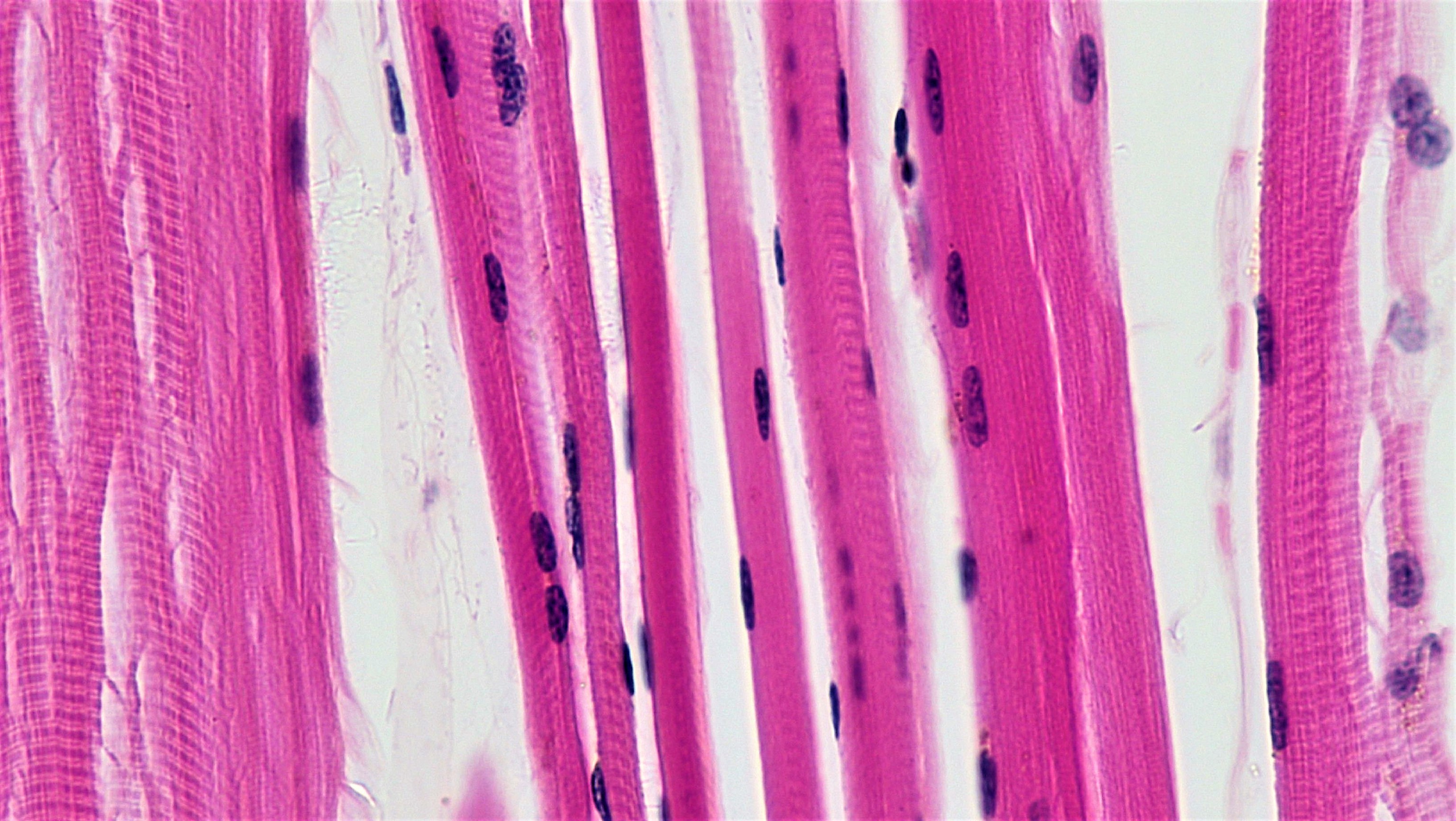
- Appearance: Under a microscope, skeletal muscle cells are striated in appearance, meaning they have visible, organized stripes due to the neat arrangement of their internal proteins.
- Structure: These fibers are long and complex, which is why skeletal muscle cells contain multiple nuclei to manage their massive cellular volume.
- Control: Crucially, skeletal muscle operates under voluntary neural control. When you consciously decide to walk or lift a syringe, you are activating these tissues.
2. Cardiac Muscle
The heart is a relentless pump that cannot afford a single misfire. Consequently, the muscle here is highly specialized.
- Location: Cardiac muscle is found exclusively in the heart.
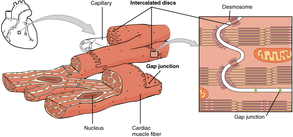
- Appearance & Structure: Like skeletal tissue, cardiac muscle cells are striated in appearance. However, unlike skeletal muscle, cardiac muscle cells typically contain a single nucleus.
- Control: You do not have to remind your heart to beat; therefore, cardiac muscle operates under involuntary neural control.
- Key Adaptation: To beat efficiently, millions of cardiac cells must fire in unison. To achieve this, cardiac muscle cells connect to each other via intercalated discs. These specialized junctions are vital because intercalated discs allow synchronized electrical signaling and contraction of cardiac tissue.
3. Smooth Muscle
If skeletal muscle moves the skeleton and cardiac muscle moves blood through the heart, smooth muscle is responsible for moving materials through the rest of the body's internal plumbing.
- Location: Smooth muscle is located in the walls of hollow internal organs. For example, the human intestines contain smooth muscle in their structural walls to push food along, and human blood vessels contain smooth muscle in their structural walls to regulate blood pressure.
- Appearance & Structure: Unlike the other two types, smooth muscle lacks visible striations. Instead of long cables, smooth muscle cells are spindle-shaped and, like cardiac cells, smooth muscle cells contain a single nucleus.
- Control: We cannot consciously control our digestion or blood pressure; thus, smooth muscle operates under involuntary neural control.
Summary Comparison Table
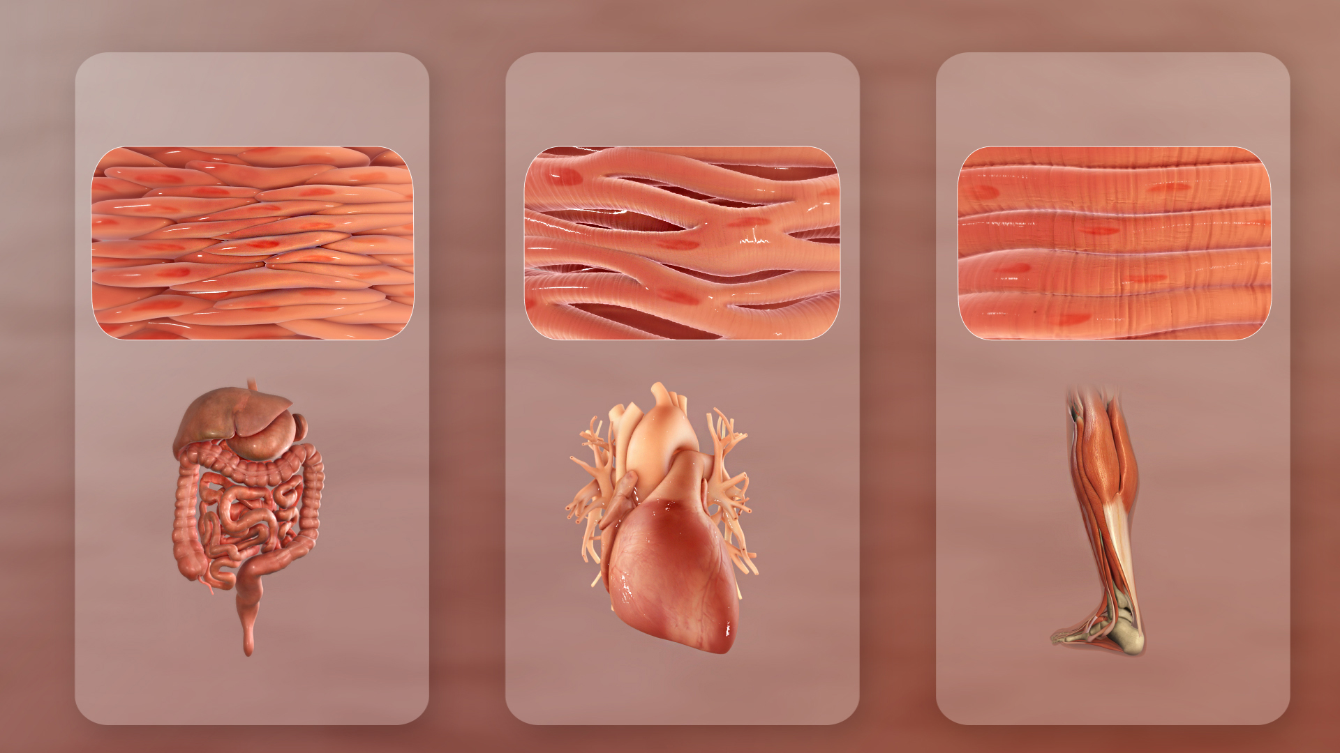
| Feature | Skeletal Muscle | Cardiac Muscle | Smooth Muscle |
|---|---|---|---|
| Striations | Yes | Yes | No |
| Control | Voluntary | Involuntary | Involuntary |
| Nuclei per cell | Multiple | Single | Single |
| Cell Shape | Long, cylindrical | Branched | Spindle-shaped |
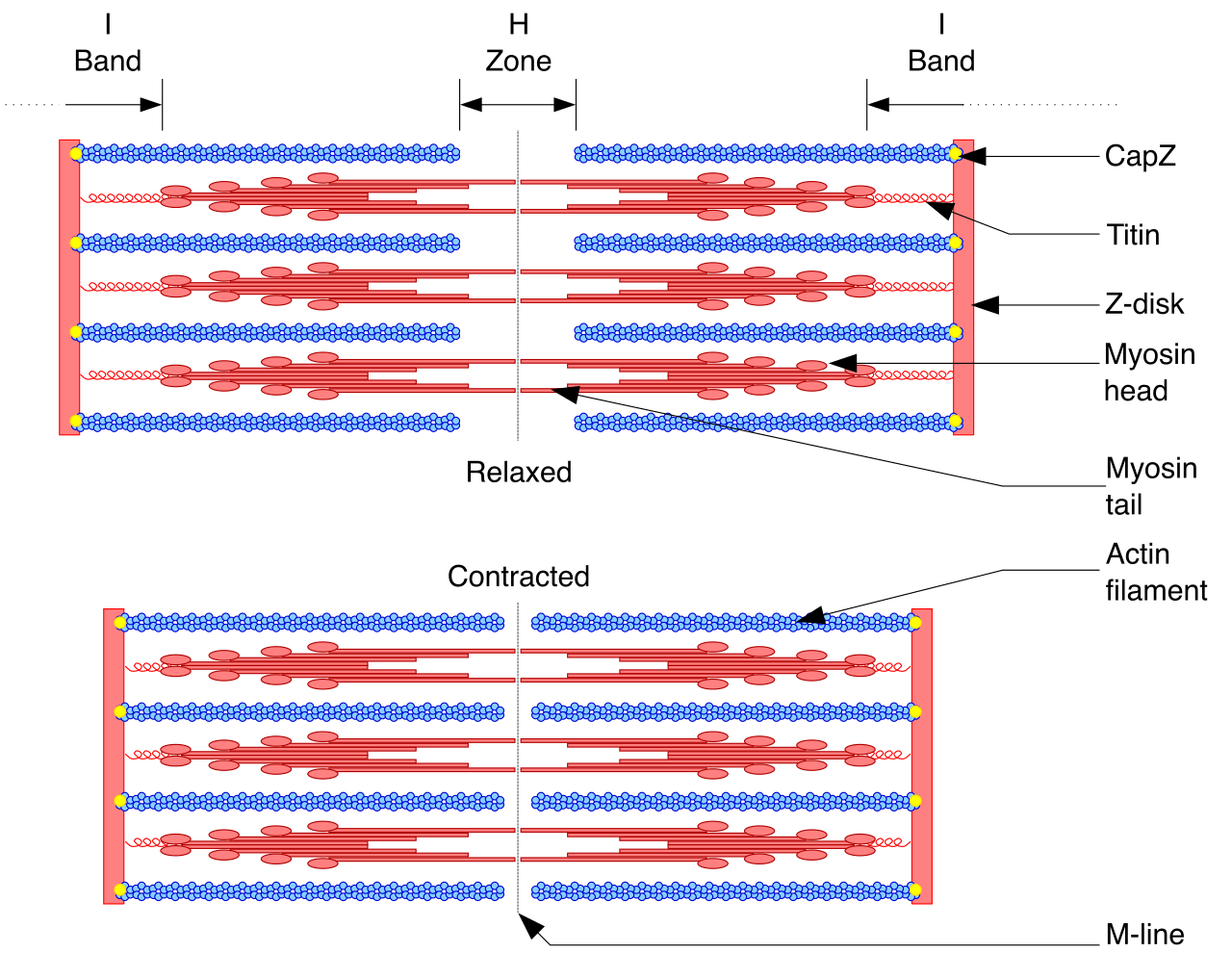
How exactly does a muscle contract? It all happens within the sarcomere, which is the basic contractile unit of a muscle fiber.
Inside the sarcomere are two crucial protein filaments:
- Actin filaments, which act as the thin filaments within a muscle sarcomere.
- Myosin filaments, which act as the thick filaments within a muscle sarcomere.
The Prime Directive of Muscle Contraction: Muscle contraction occurs when actin and myosin filaments slide past each other. It is vital to understand that actin filaments do not change their physical length during muscle contraction, nor do myosin filaments do not change their physical length during muscle contraction. The proteins themselves do not shrink; they simply overlap more tightly, like two interlacing hairbrushes.
The Lock, The Key, and The Spring
This sliding mechanism operates like an intricate molecular ratchet, heavily dependent on calcium and cellular energy.
- The Trigger: A nerve impulse signals the muscle, causing an intracellular change. Specifically, muscle contraction is initiated by the intracellular release of calcium ions.
- The Reservoir: Where does this calcium come from? The sarcoplasmic reticulum stores and releases calcium ions into the muscle cell cytoplasm.
- Unlocking the Binding Site: On the thin actin filament, the sites where myosin wants to grab hold are blocked by a protein guard called tropomyosin. To move the guard, calcium ions bind directly to the protein troponin during muscle contraction.
- The Shift: The binding of calcium to troponin causes the protein tropomyosin to shift its physical position. Because of this, the positional shift of tropomyosin exposes myosin-binding sites on the actin filament.
- The Pull (Cross-Bridge): With the sites exposed, the club-like myosin heads bind to exposed sites on the actin filament to form a cross-bridge. They then flex, pulling the actin filament inward. The repeated binding and pulling of actin by myosin heads physically shortens the sarcomere.
- The Release & Reset: To let go and pull again, the system requires energy. Adenosine triphosphate (ATP) provides the energy necessary for the myosin head to detach from actin. Once detached, adenosine triphosphate hydrolysis provides energy to re-cock the myosin head into a high-energy state, ready for the next pull.
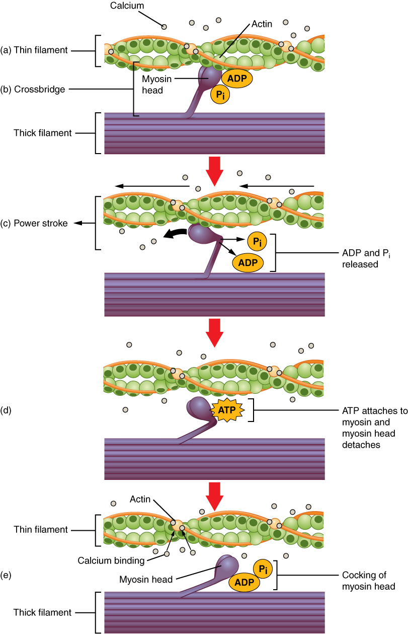
(Clinical translation: When a person dies, their body stops producing ATP. Without ATP to detach the myosin heads from the actin, the muscles lock in place—a state known as rigor mortis.)
While the muscular system focuses on mechanical work, the reproductive system is engineered for genetic continuation. Let's trace the male system chronologically, from the generation of gametes to their delivery.
1. Production
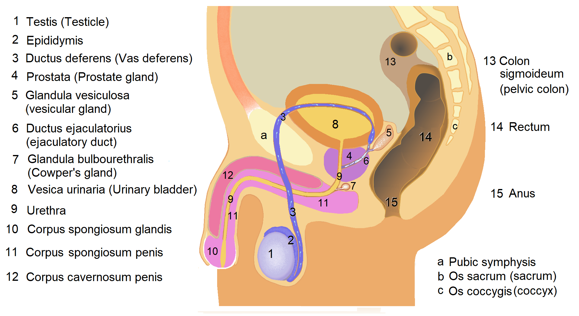
The factory floor of the male reproductive system is located in the testes, which are the primary male reproductive organs. The testes have two main jobs: the testes produce male gametes known as sperm cells, and the testes produce the steroid hormone testosterone.
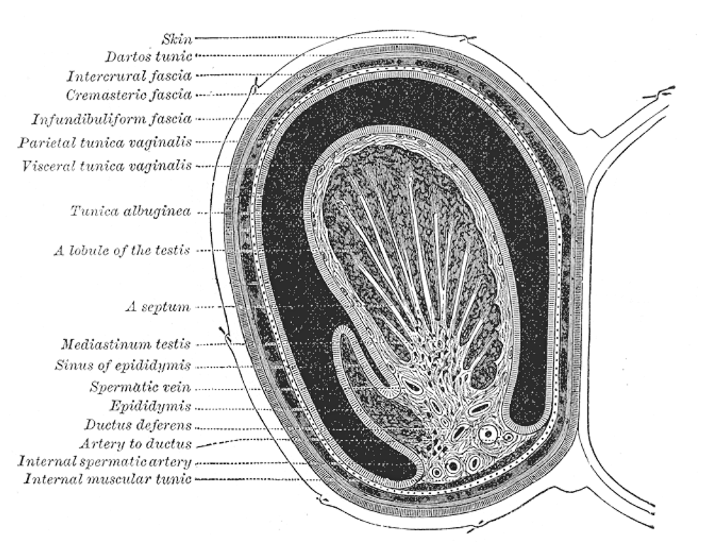
Because biological manufacturing is highly sensitive to heat, the testes cannot be kept inside the warm abdominal cavity. Instead, they reside in the scrotum, which is an external sac of skin housing the testes outside the main body cavity. The external location of the scrotum keeps the testes at a lower temperature than the core body temperature. This is an absolute physiological necessity: a sub-core body temperature is strictly required for optimal human sperm production.
2. Maturation and Storage
Once sperm are created, they are structurally complete but not yet capable of swimming. They move into the epididymis, which is a highly coiled tube located on the posterior surface of each testicle. Here, newly formed sperm cells undergo maturation within the epididymis, and these mature sperm cells are stored within the epididymis prior to ejaculation.
3. Transport and the Accessory Glands
During ejaculation, sperm travel from the epididymis through the vas deferens, which is a muscular tube transporting mature sperm away from the epididymis. The vas deferens directly carries sperm to the ejaculatory duct.
To survive the journey out of the male body and into the female body, sperm require a biological support system—semen—provided by three distinct glands:
- Seminal Vesicles: These glands produce a nutrient-rich seminal fluid. Specifically, the fluid secreted by seminal vesicles contains high concentrations of fructose. Why sugar? Because fructose from the seminal vesicles provides cellular energy required for sperm motility (powering those tiny tails).
- Prostate Gland: The female reproductive tract is naturally acidic to prevent infections. To counter this, the prostate gland secretes an alkaline fluid into the semen. The alkaline fluid from the prostate gland protects sperm from the highly acidic environment of the female reproductive tract.
- Bulbourethral Glands: Before ejaculation, the bulbourethral glands secrete a lubricating pre-ejaculate fluid. Because the male urethra serves as a common biological passage for liquid urine as well as for ejaculated semen, there may be acidic urine remnants left in the pipe. The pre-ejaculate fluid from bulbourethral glands neutralizes residual urine acidity within the male urethra so the sperm are not destroyed before they even exit the body.
While the male system is a continuous assembly line, the female system is cyclical, designed to prepare a perfectly regulated environment for a developing embryo.
1. Production
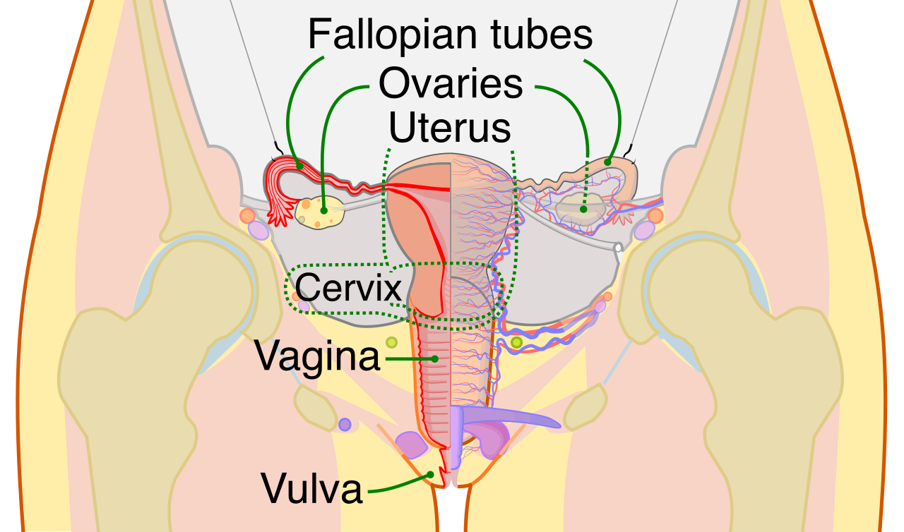
The ovaries are the primary female reproductive organs. Just as the testes have a dual role, so do the ovaries: the ovaries produce female gametes known as ova (eggs), and they act as an endocrine gland because the ovaries secrete the steroid hormone estrogen and the steroid hormone progesterone.
2. Transport and the Site of Fertilization
Once an ovum is released from an ovary, it is swept into the fallopian tubes, which are tubular structures connecting the vicinity of the ovaries to the uterus. The fallopian tubes physically transport ova from the ovaries toward the uterus.
Critically, fertilization does not typically occur in the uterus. If a woman conceives, fertilization of an ovum by a sperm cell typically occurs within the fallopian tubes.
3. Gestation and the Birth Canal
If fertilization is successful, the developing bundle of cells travels down into the uterus, which is a hollow muscular organ where a developing fetus gestates. The inner lining of this organ is the endometrium, which is the inner mucosal lining of the uterus. A successful pregnancy relies on the fact that a fertilized egg implants directly into the endometrium to establish a pregnancy.
At the base of the uterus is the cervix, which constitutes the lower narrow portion of the uterus. The cervix opens directly into the superior end of the vagina. Finally, the vagina serves as the muscular birth canal during parturition (childbirth) and serves the dual purpose of being the receptacle that receives sperm during human sexual intercourse.
To truly grasp reproduction, we must look at the genetic mathematics and the chemical messengers (hormones) that orchestrate it all.
The Math of Meiosis
Normal human body cells (somatic cells) have 46 chromosomes. But if a sperm with 46 chromosomes fused with an egg with 46 chromosomes, the resulting baby would have 92 chromosomes—a biological impossibility.
Therefore, gametes (sperm and eggs) undergo a special type of cell division called meiosis. Meiosis during gametogenesis produces haploid gamete cells. "Haploid" means half, so haploid gamete cells contain exactly half the normal human somatic number of chromosomes. To be precise, human haploid gametes contain exactly twenty-three chromosomes.
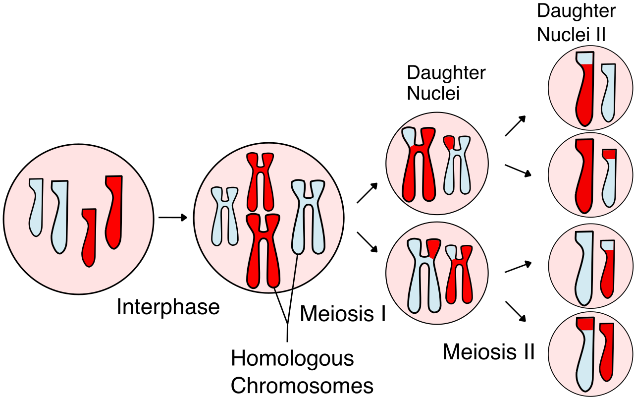
- Spermatogenesis is the biological process of sperm cell development, and it occurs continuously within the seminiferous tubules of the testes.
- Oogenesis is the biological process of egg cell development, and unlike the continuous male process, oogenesis occurs exclusively within the ovarian follicles.
The Hormonal Control Room
The reproductive systems are controlled by a brilliant feedback loop initiated in the brain. You must know these specific hormonal triggers for the TEAS exam:
Follicle-Stimulating Hormone (FSH):
- In males: Follicle-stimulating hormone directly stimulates the production of sperm in human males.
- In females: Follicle-stimulating hormone directly stimulates the maturation of ovarian follicles in human females. (The name gives it away!)
Luteinizing Hormone (LH):
- In males: Luteinizing hormone stimulates Leydig cells in the testes to produce testosterone.
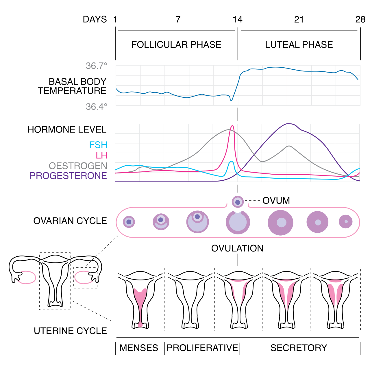
- In females: A sudden systemic surge in luteinizing hormone rapidly triggers ovulation in human females.
The Female Cycle: Ovulation & Uterine Preparation
- Estrogen: As the follicle matures under the influence of FSH, it secretes estrogen. Estrogen heavily regulates the physiological development of female secondary sexual characteristics and estrogen triggers the initial proliferative thickening of the endometrium during the menstrual cycle, preparing the "soil" of the uterus.
- Ovulation: Initiated by the LH surge, ovulation involves the physical release of a mature egg from a ruptured ovarian follicle.
- Progesterone: Once the egg bursts free, the remnants of that ruptured follicle transform into a new structure. The corpus luteum forms from the ruptured ovarian follicle immediately following ovulation. This new structure takes on a critical job: the corpus luteum actively secretes the hormone progesterone. Why does this matter? Because progesterone maintains the structural integrity of the uterine endometrial lining for potential pregnancy. If progesterone drops, the lining sheds (menstruation).
The Climax: Fertilization
The culmination of this vast anatomical and hormonal preparation is a microscopic event. Fertilization is the biological fusion of a haploid sperm cell and a haploid egg cell.
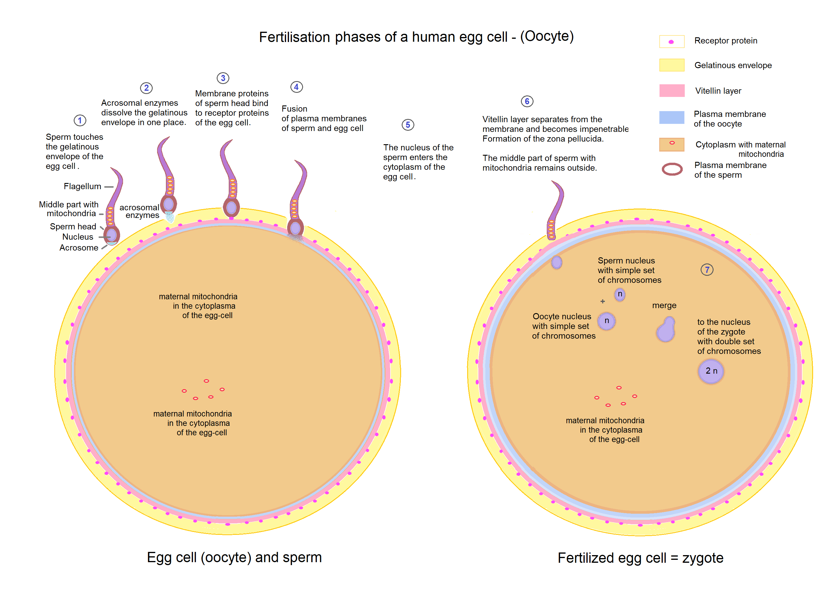
When 23 chromosomes from the father meet 23 chromosomes from the mother, the fusion of a sperm and an egg produces a single unified cell called a zygote. Through this elegant fusion, the math is restored: a normal human zygote is a diploid cell containing exactly forty-six chromosomes, containing the entire architectural blueprint necessary to build a new human being.