Integumentary and Endocrine Systems
Not sure you’re ready?
Take the ~3-minute readiness diagnostic and see where you stand.
When a trauma patient arrives in the emergency department, their immediate survival heavily depends on two complementary organ systems: a physical barrier that has kept out pathogens and prevented massive fluid loss, and an internal signaling network actively adjusting their heart rate, blood pressure, and metabolism to cope with shock. The physical barrier is the integumentary system; the signaling network is the endocrine system. Understanding how human anatomy defends its borders and communicates across distances is not just textbook trivia. It is the mechanistic foundation of every vital sign, wound assessment, and medication response you will manage as a clinician.
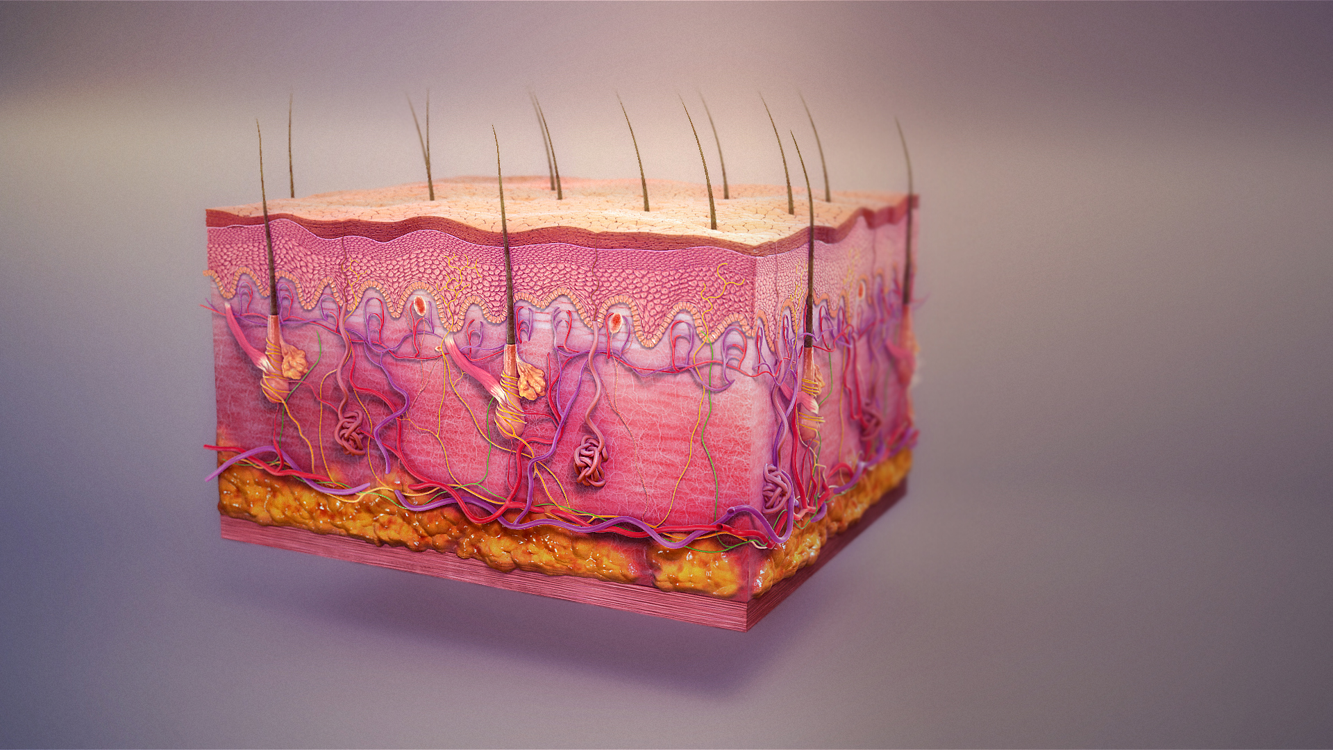
The integumentary system is your body’s primary defense against the outside world. To understand how it works, imagine a medieval fortress. It has an outer stone wall to take the beating of the elements, a bustling courtyard beneath it supplying resources, and a deep foundation anchoring the whole structure. In the skin, these three layers are the epidermis, the dermis, and the hypodermis.
1. The Epidermis (The Outer Wall)
The epidermis is the outermost layer of the skin. Because it serves as the frontline shield, the epidermis is an avascular tissue layer—meaning it lacks its own blood supply. It cannot bleed, and it must rely entirely on diffusion from the underlying dermis for oxygen and nutrients.
The cellular makeup of this wall is highly specialized:
- Keratinocytes: These are the most abundant cell type in the epidermis. They are the "bricklayers," constantly producing the fibrous protein keratin, which provides a tough, water-resistant protective layer for the skin surface.
- Melanocytes: Specialized cells found in the basal layer of the epidermis. They synthesize the pigment melanin. Melanin acts as microscopic sunglasses for your skin; it protects the DNA of skin cells from ultraviolet radiation damage.
- Langerhans cells: These are the patrolling guards—immune cells residing within the epidermis to intercept invading microbes.
- Merkel cells: Specialized touch receptors located in the epidermis that allow you to feel delicate textures.
The Stratified Layers of the Epidermis
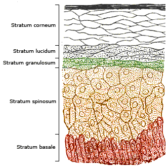
The epidermis is constructed in distinct strata (layers), progressing from deep to superficial:
| Layer | Structure and Function |
|---|---|
| Stratum Basale | The deepest layer of the epidermis. Because it is closest to the dermal blood supply, the stratum basale continuously generates new skin cells through mitotic division. |
| Stratum Spinosum | An epidermal layer just above the basale that provides strength and flexibility to the skin. |
| Stratum Granulosum | An epidermal layer where migrating cells accumulate massive amounts of keratin and begin to die as they move further from the blood supply. |
| Stratum Lucidum | A unique, clear layer of the epidermis found only in the thick skin of the palms and soles. |
| Stratum Corneum | The most superficial layer of the epidermis. It consists entirely of dead, flattened, keratin-filled cells that you constantly shed. |
2. The Dermis (The Infrastructure)
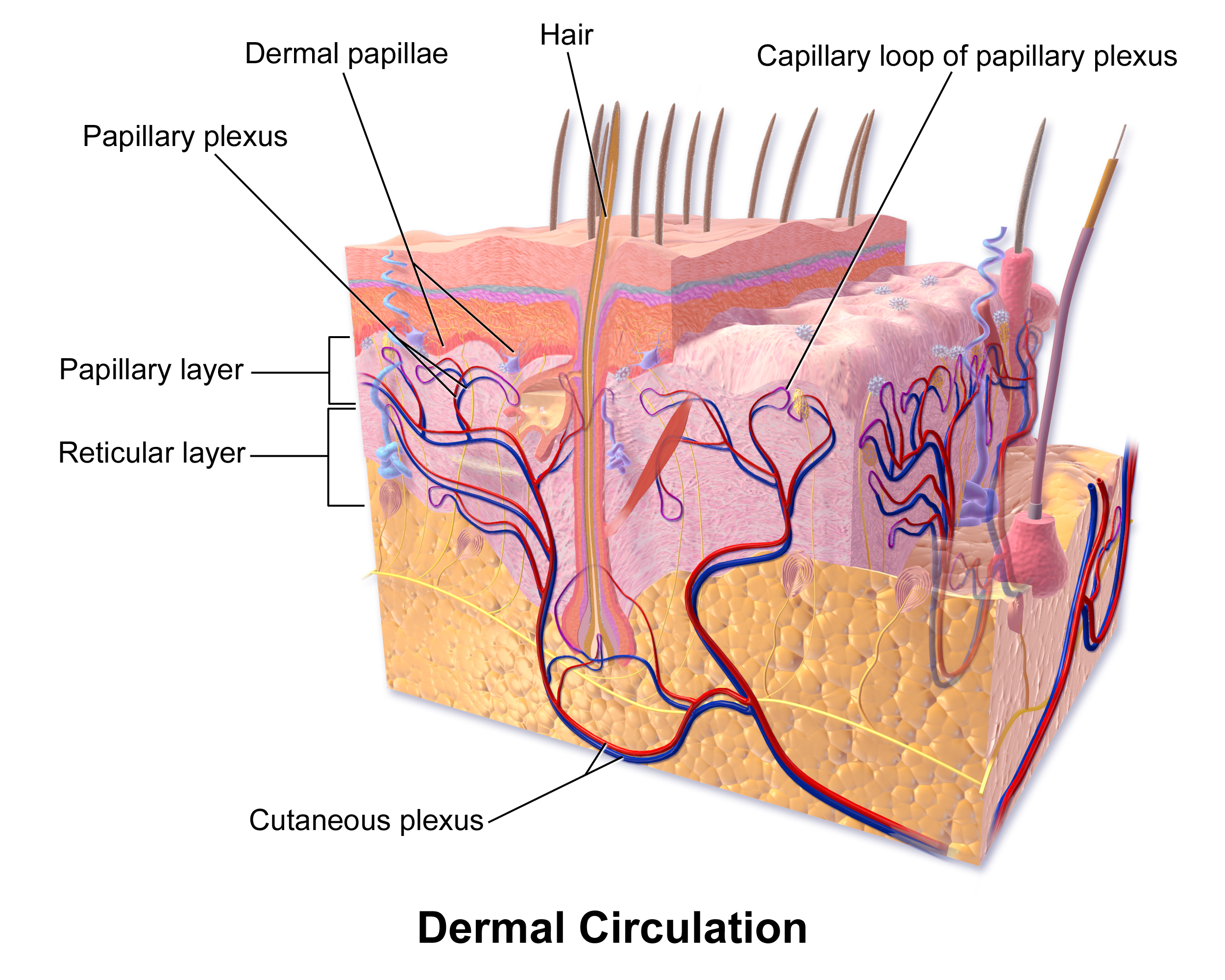
Directly beneath the epidermis lies the dermis, the middle layer of the skin. If the epidermis is the brick wall, the dermis is the living infrastructure.
The dermis contains a rich network of blood vessels that nourish the skin and help regulate heat. It is heavily wired, containing sensory nerve endings for temperature, pain, and touch. The dermis is also the manufacturing center for your accessory structures: it is where hair follicles are situated, and it houses both sweat glands and sebaceous glands.
3. The Hypodermis (The Foundation)
The hypodermis is the deepest layer of the integumentary system. Clinically, it is interchangeably referred to as the subcutaneous layer (where you will administer "sub-Q" injections like insulin).
The hypodermis consists primarily of adipose tissue (fat) and loose connective tissue. This composition allows it to serve two major purposes:
- The adipose tissue in the hypodermis provides crucial thermal insulation for the body.
- The hypodermis acts as an energy reservoir due to its high concentration of fat cells.
Thermoregulation
A primary role of the integumentary system is to regulate body temperature. It achieves this dynamically through blood flow and moisture:
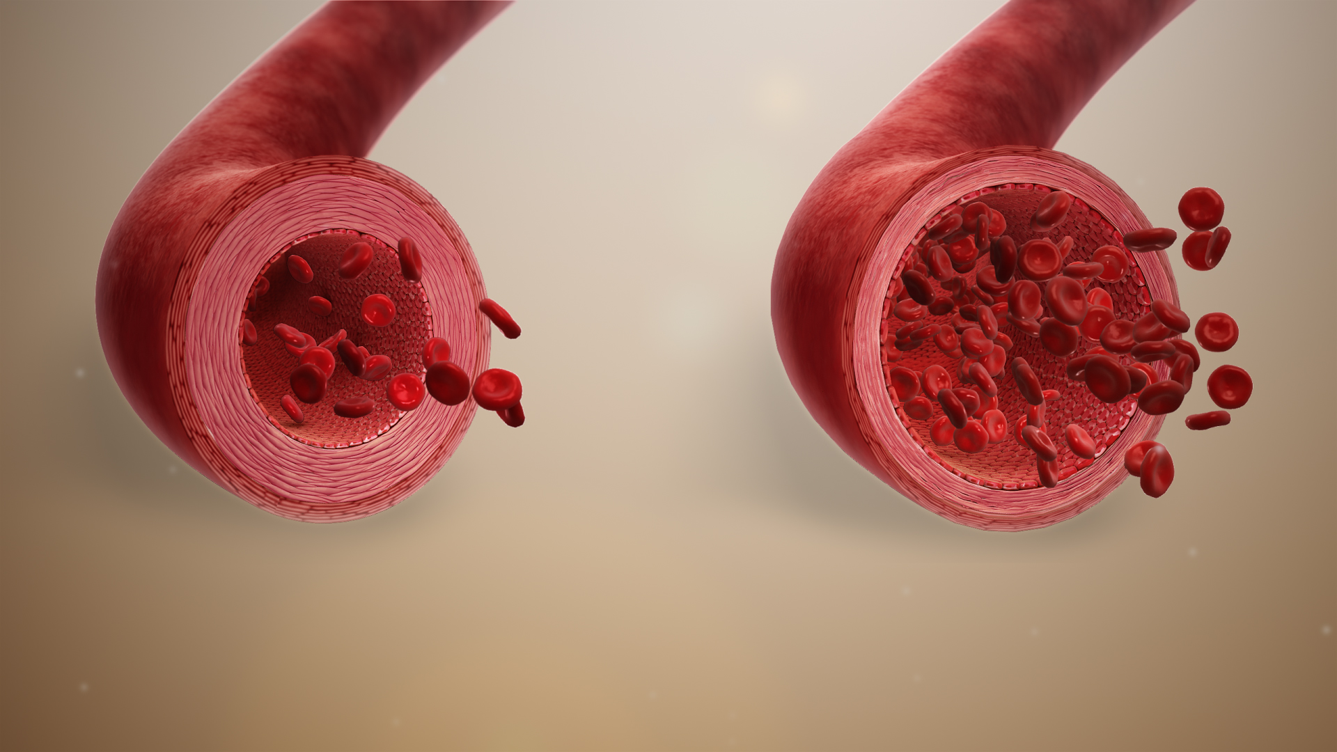
- Heat Loss: When core temperature spikes, the nervous system triggers vasodilation of dermal blood vessels. This increases blood flow to the skin surface to facilitate heat loss. Simultaneously, the integumentary system regulates body temperature through the evaporation of sweat, which pulls heat away from the skin.
- Heat Conservation: In freezing environments, the body triggers vasoconstriction of dermal blood vessels, which restricts blood flow to the skin to conserve internal body heat for vital organs.
Glands of the Skin
Core Concept: Exocrine Glands Before diving into skin glands, we must classify them. Exocrine glands secrete substances onto epithelial surfaces or into body cavities via tubular ducts. Salivary glands are classified as exocrine glands, and sweat glands are classified as exocrine glands.
There are three key exocrine glands in the skin:
- Eccrine sweat glands: These secrete a watery fluid directly onto the skin surface. The primary physiological function of eccrine sweat glands is to lower body temperature.
- Apocrine sweat glands: These secrete a thicker, protein-rich sweat directly into hair follicles. They are highly concentrated in the axillary (armpit) and genital regions and are responsible for body odor when local bacteria break down the proteins.
- Sebaceous glands: These secrete a lipid-rich substance called sebum. Sebum naturally lubricates the skin and hair. More importantly, sebum creates a hydrophobic barrier that helps prevent excessive water loss from the skin.
Hair and Cellular Byproducts
Hair is primarily composed of dead, tightly packed keratinized cells—very similar to the stratum corneum. Attached to individual hair follicles are arrector pili, which are tiny smooth muscles. When you get a chill or a fright, the contraction of arrector pili muscles causes hair to stand upright (goosebumps).

The skin also performs vital biochemical tasks. For instance, the skin synthesizes the inactive precursor of vitamin D when exposed to ultraviolet light. This is clinically paramount because vitamin D synthesized by the skin is essential for adequate calcium absorption in the digestive tract. Additionally, the skin functions as a minor excretory organ; it excretes minor amounts of metabolic wastes such as urea and lactic acid through sweat.
While the integumentary system physically shields the body, the endocrine system controls the internal environment through chemical messaging.
Unlike exocrine glands (which have ducts), endocrine glands are entirely ductless structures. Because they lack ducts, endocrine glands secrete chemical messengers called hormones directly into the surrounding interstitial fluid and bloodstream. The bloodstream then broadcasts these hormones to target cells across the entire body.
Hormone Chemistry: How Messages Enter the Cell
The way a hormone interacts with a cell depends entirely on its chemical structure. Think of cell membranes as heavily guarded bouncers made of fat (the phospholipid bilayer).
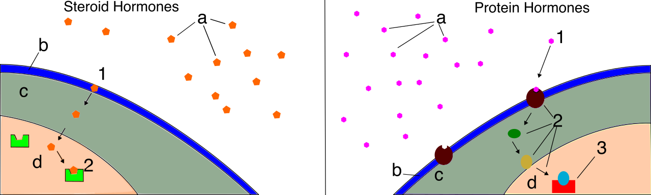
- Steroid hormones: These are fundamentally lipid-soluble molecules. Because "like dissolves like," lipid-soluble hormones can freely diffuse directly through the phospholipid bilayer of a target cell membrane. Once inside, lipid-soluble hormones typically bind to intracellular receptors to directly alter cellular gene expression.
- Non-steroid hormones: These are fundamentally water-soluble molecules. Consequently, water-soluble hormones cannot pass through the lipid core of a target cell membrane. To deliver their message, water-soluble hormones must bind to surface receptors to activate secondary messenger systems inside the target cell.
The Feedback Loops
To maintain homeostasis, hormone levels must be tightly controlled.
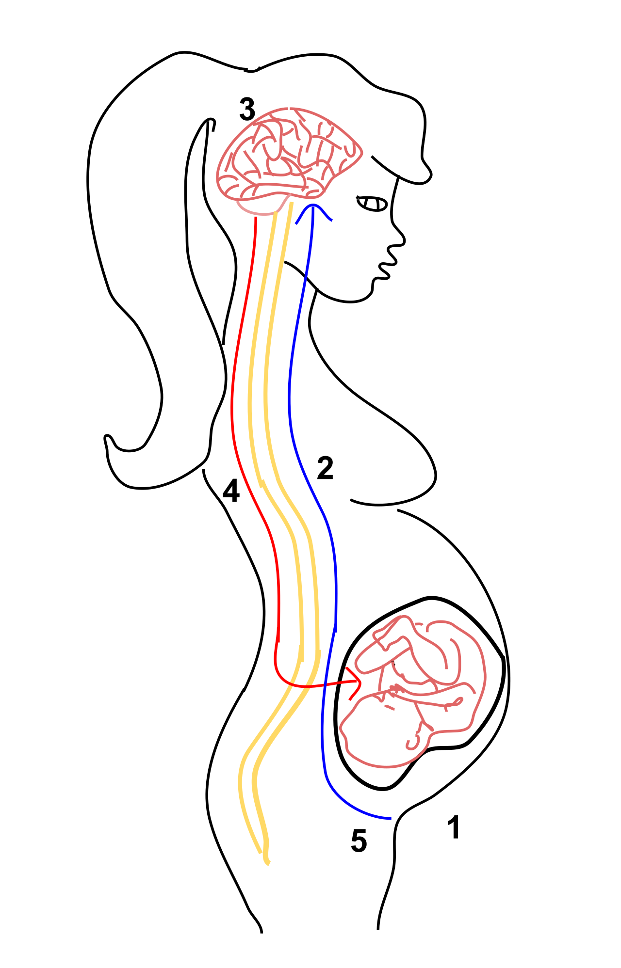
- Negative Feedback: This is a biological regulatory mechanism that reverses a deviation from a physiological set point. The vast majority of endocrine hormones are regulated by negative feedback loops. In a typical endocrine negative feedback loop, an elevated hormone level inhibits the further secretion of that same hormone (like a thermostat turning off the heat once the room is warm).
- Positive Feedback: This is a regulatory mechanism that amplifies a physiological change until a specific endpoint is reached. For example, the release of oxytocin during childbirth is controlled by a positive feedback loop, driving stronger and stronger contractions until the baby is delivered.
To master the endocrine system, you must know the major glands, the hormones they produce, and their effects.
The Master Axis: Hypothalamus and Pituitary Gland
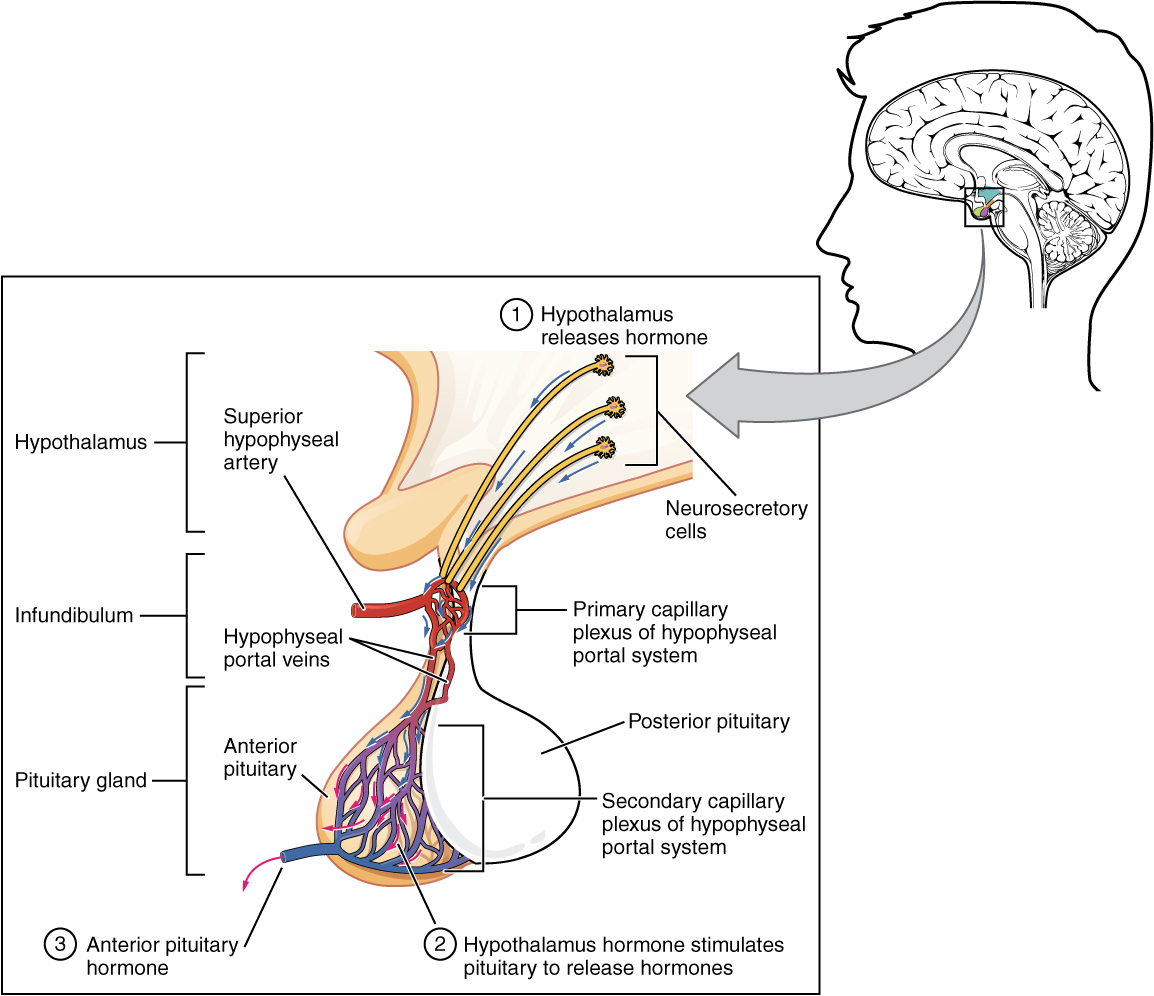
The hypothalamus is a brain structure that serves as the primary link between the nervous and endocrine systems. The hypothalamus produces releasing and inhibiting hormones. Interestingly, these hypothalamic releasing hormones exclusively target the anterior pituitary gland, dictating its activity.
Furthermore, the hypothalamus synthesizes two vital hormones of its own: antidiuretic hormone (ADH) and oxytocin.
However, the hypothalamus doesn't release ADH and oxytocin directly into the broader bloodstream. Instead, it sends them to the posterior pituitary gland. The posterior pituitary gland stores and releases hormones that were originally produced by the hypothalamus. Therefore, the posterior pituitary gland does not synthesize its own hormones.
- Antidiuretic hormone (ADH) is released into the bloodstream from the posterior pituitary gland. ADH targets the kidneys to increase water reabsorption into the blood, effectively preventing dehydration.
- Oxytocin is also released into the bloodstream from the posterior pituitary gland. Oxytocin stimulates intense uterine muscle contractions during labor, and later, oxytocin stimulates milk ejection from the mammary glands during breastfeeding.
In contrast, the anterior pituitary gland is a true glandular factory that synthesizes and secretes a multitude of its own hormones:
- Growth hormone (GH): Primarily targets bone and skeletal muscle tissues to stimulate overall cellular growth and division.
- Thyroid-stimulating hormone (TSH): Commands the thyroid gland to produce and release its own metabolic hormones.
- Adrenocorticotropic hormone (ACTH): Targets the adrenal cortex to stimulate the release of corticosteroids such as cortisol.
- Follicle-stimulating hormone (FSH) and Luteinizing hormone (LH): Both are synthesized and secreted here. Follicle-stimulating hormone and luteinizing hormone target the male and female gonads to regulate reproductive functions.
- Prolactin: Synthesized and secreted to stimulate the production of milk within the mammary glands.
The Pineal and Thymus Glands
- Pineal Gland: A small endocrine structure located deep within the brain. The pineal gland secretes the hormone melatonin, which regulates the body's circadian rhythms and the sleep-wake cycle.
- Thymus: An endocrine gland located in the upper chest immediately behind the sternum. The thymus secretes a group of hormones called thymosins. Thymosins stimulate the maturation of T-lymphocytes for the adaptive immune system.
Metabolism and Calcium Control: Thyroid and Parathyroids
The thyroid gland is located in the anterior neck just below the larynx. It is central to metabolic control.
- The thyroid gland secretes triiodothyronine (T3) and thyroxine (T4). Triiodothyronine and thyroxine regulate the basal metabolic rate of virtually all body cells. Note that dietary iodine is a strictly required mineral for the synthesis of thyroid hormones.
- The thyroid gland also secretes the hormone calcitonin. Calcitonin lowers elevated blood calcium levels by stimulating bone-building osteoblast activity (taking calcium from the blood and putting it into bone).
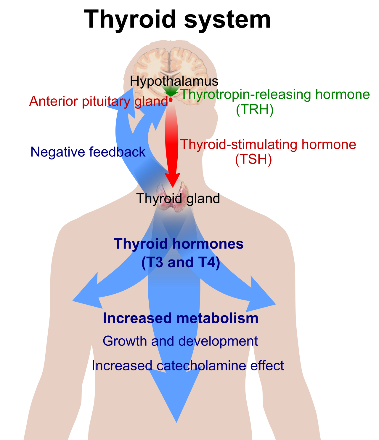
Tucked onto the posterior surface of the thyroid gland are four small structures known as the parathyroid glands.
- The parathyroid glands secrete parathyroid hormone (PTH). Parathyroid hormone does the exact opposite of calcitonin: it raises low blood calcium levels by stimulating bone-resorbing osteoclast activity (breaking down bone to release calcium into the blood).
The Pancreas: Blood Glucose Control
The pancreas is unique because it exhibits dual functionality as both an endocrine gland and an exocrine gland (secreting digestive enzymes into ducts).
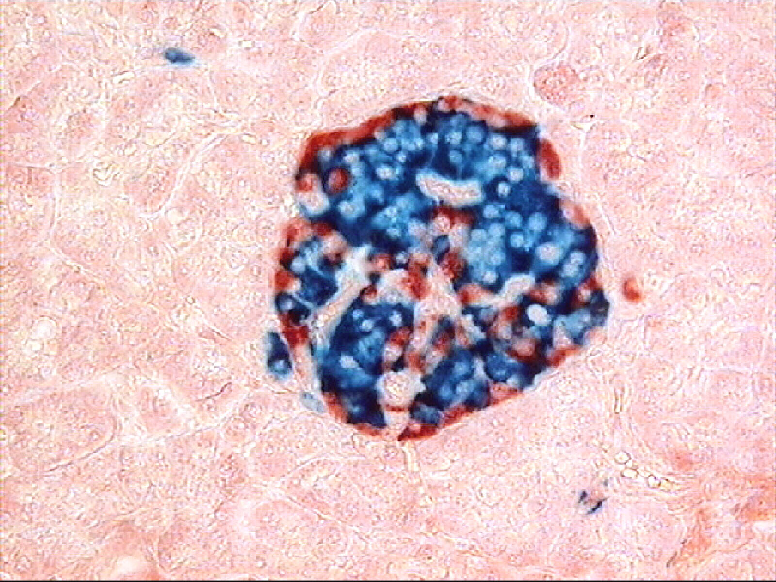
The specific endocrine cells of the pancreas are clustered in regions called the Islets of Langerhans.
- Alpha cells within the pancreatic Islets of Langerhans secrete the hormone glucagon. Glucagon raises blood glucose levels by stimulating the liver to break down stored glycogen into glucose.
- Beta cells within the pancreatic Islets of Langerhans secrete the hormone insulin. Insulin lowers blood glucose levels by facilitating the transport of glucose from the blood into target cells.
Stress and Survival: The Adrenal Glands
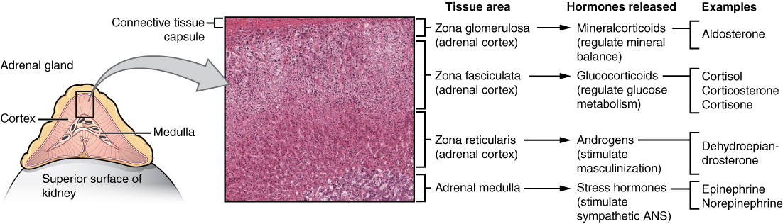
The adrenal glands are structurally located on the superior pole of each kidney. They are separated into two distinct regions:
- The Adrenal Cortex: The outer glandular region of the adrenal gland. It produces slow, long-term steroid hormones.
- The adrenal cortex secretes the hormone cortisol. Cortisol is a glucocorticoid that increases blood glucose levels to assist the body during long-term stress.
- The adrenal cortex also secretes the hormone aldosterone. Aldosterone is a mineralocorticoid that targets the kidneys to heavily increase sodium reabsorption. Because water follows salt, the increased sodium reabsorption driven by aldosterone indirectly causes water retention and elevated blood pressure.
- The Adrenal Medulla: The inner neural region of the adrenal gland.
- The adrenal medulla secretes epinephrine and norepinephrine. These hormones initiate the rapid, short-term fight-or-flight response, causing immediate spikes in heart rate and airway dilation.
The Reproductive Gonads
The gonads respond to pituitary signals (FSH and LH) to drive reproduction.
- Ovaries: The primary reproductive gonads in biological females. The ovaries secrete the steroid hormones estrogen and progesterone. Estrogen actively promotes the development and maintenance of female secondary sex characteristics. Progesterone primarily acts to prepare and maintain the inner uterine lining for potential pregnancy.
- Testes: The primary reproductive gonads in biological males. The testes secrete the steroid hormone testosterone. Testosterone promotes the development of male secondary sex characteristics and regulates continuous sperm production.