Respiratory and Cardiovascular Systems
Not sure you’re ready?
Take the ~3-minute readiness diagnostic and see where you stand.
The human body operates on a continuous, inescapable demand for oxygen and the rapid elimination of cellular exhaust. At the center of this operation are two intimately linked systems: the respiratory system, which procures the oxygen and vents the carbon dioxide, and the cardiovascular system, a high-pressure logistical network that transports these gases to and from trillions of cells. For a clinician, mastering the mechanics of these systems is not mere anatomical trivia; it is the foundation of sustaining life. When a patient gasps for air or presents with a fading pulse, the margin between recovery and collapse is dictated by the physics of pressure gradients, the chemistry of gas diffusion, and the electrical rhythm of a beating heart.
To understand ventilation, we must trace the path of a single breath from the atmosphere to the microscopic depths of the lungs. The upper and lower respiratory tracts are engineered to prepare and deliver air with maximum efficiency while defending the delicate internal tissues from environmental hazards.
The Upper Airway: Filtration and Preparation
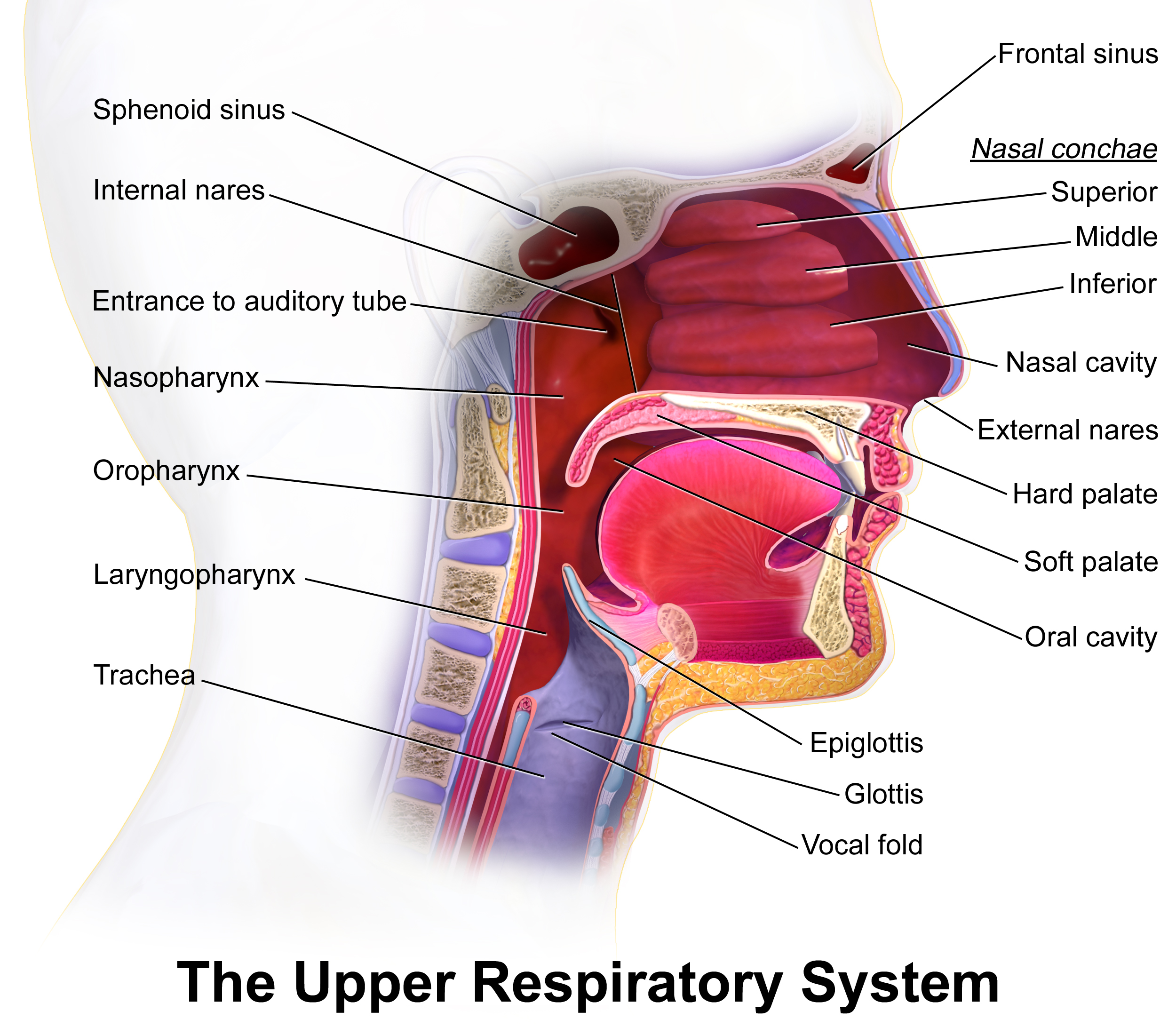
When atmospheric air enters the body, it is entirely unfit for the lungs. It is cold, dry, and laden with microscopic debris. The nasal cavity warms incoming inhaled air and simultaneously moistens incoming inhaled air, bringing it to body temperature and 100% humidity so it does not shock the delicate lung tissue. Furthermore, the nasal cavity filters incoming air using mucus and filters incoming air using nasal hairs, trapping dust, pathogens, and particulate matter before they can penetrate deeper.
From the nasal cavity, air descends into the pharynx (the throat). The pharynx serves as a shared passageway for air moving to the lungs as well as a shared passageway for food moving to the esophagus. Because this single corridor handles both air and solid matter, the body requires a mechanical sorting mechanism to prevent choking. This sorting is achieved by the epiglottis, which is a flap composed of elastic cartilage. The epiglottis covers the larynx during swallowing to prevent food from entering the airway.
Safely past the epiglottis, air enters the larynx (the voice box). The larynx contains the vocal cords, which vibrate to produce sound as air is expelled.
The Lower Airway: The Tracheobronchial Tree
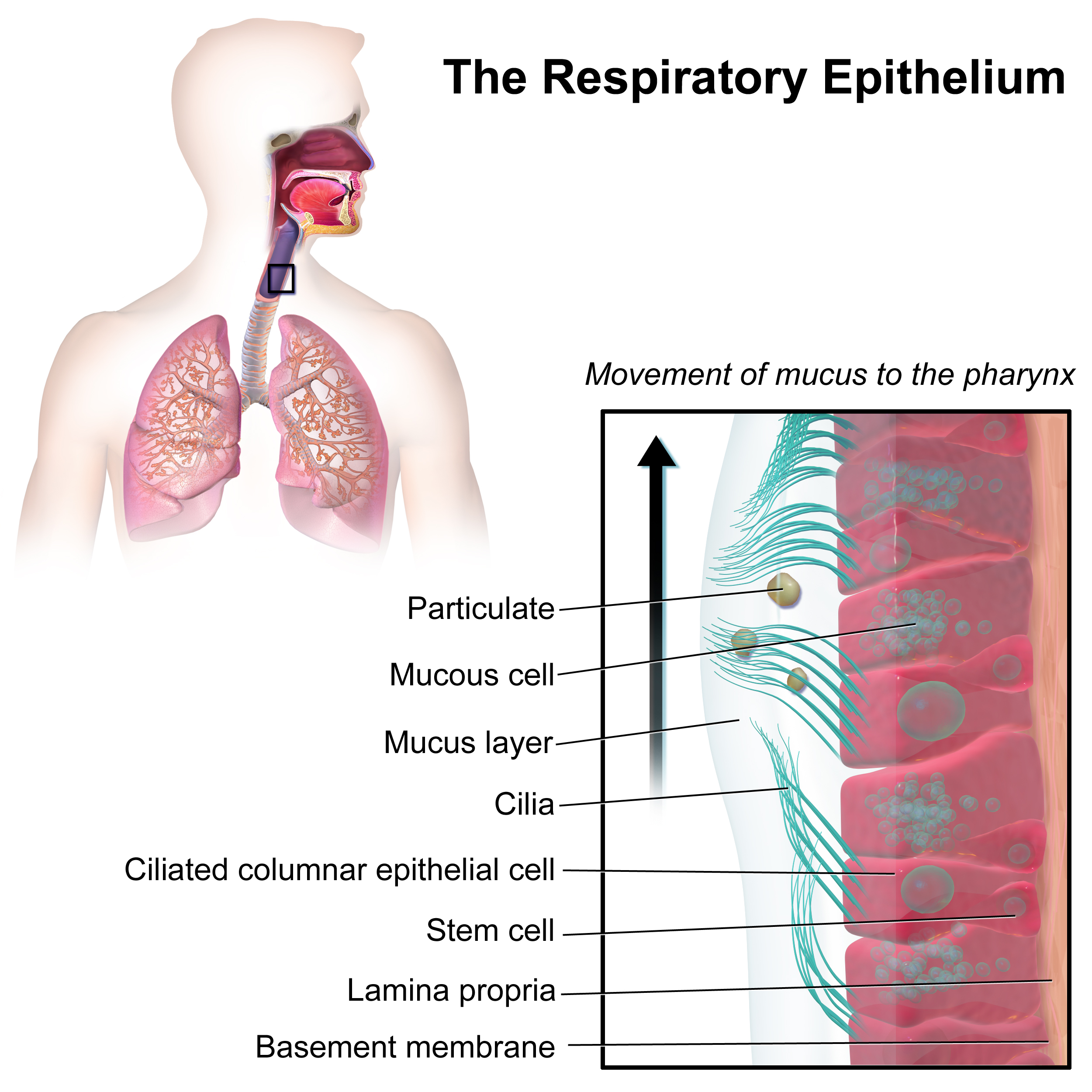
Below the larynx, the respiratory tract becomes dedicated entirely to air transport. The primary conduit is the trachea, a rigid tube that is maintained in an open position by C-shaped rings of cartilage. These rings prevent the airway from collapsing under negative pressure during a sharp inhalation. The inner walls of the trachea possess their own defense mechanism: the inner lining of the trachea contains ciliated epithelial cells. Like thousands of microscopic brooms, ciliated epithelial cells in the respiratory tract sweep mucus and trapped debris upward away from the lungs toward the pharynx, where it can be swallowed or expelled.
At its base, the trachea divides into the right primary bronchus and the left primary bronchus, directing air into the two lungs.
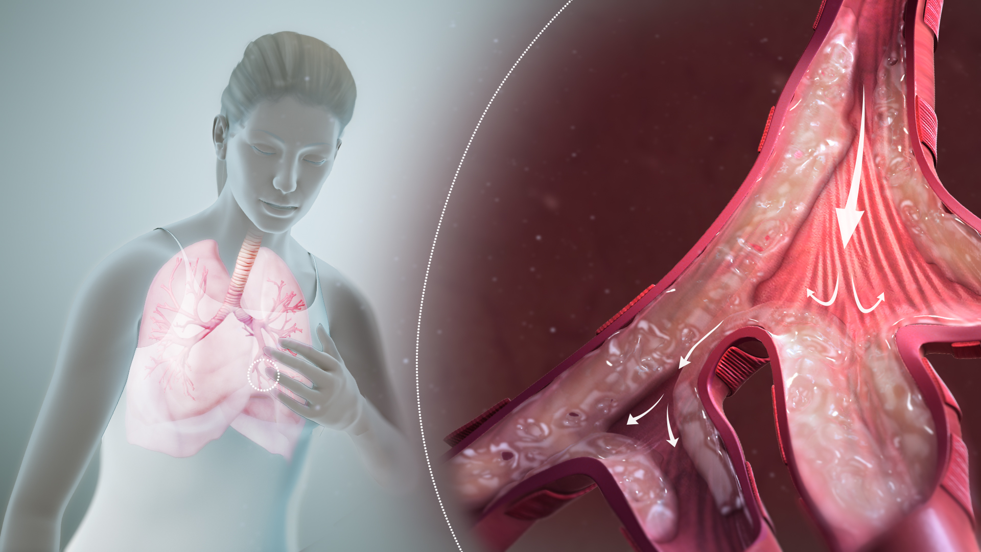
The lungs themselves are asymmetrical to accommodate the heart. The right lung possesses three distinct lobes, whereas the left lung possesses two distinct lobes. Inside the lungs, the primary bronchi branch into progressively smaller airways called bronchioles. Unlike the larger trachea, bronchioles lack cartilaginous rings. Instead, bronchioles are surrounded by smooth muscle, which can contract or relax to regulate airflow.
Clinical Correlate: This smooth muscle is the primary battleground in asthmatic patients. Asthma is a respiratory disorder characterized by spastic constriction of the bronchiolar smooth muscle, severely restricting airflow and making exhalation labored.
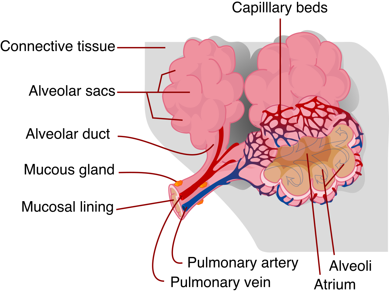
Eventually, the bronchioles terminate in microscopic air sacs called alveoli.
The Alveoli and The Pleura
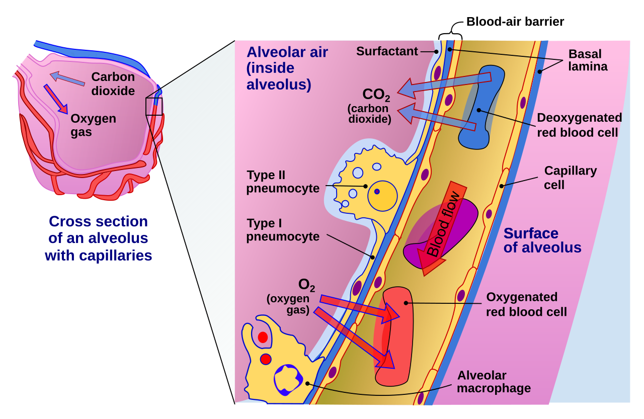
Alveoli are the primary functional sites of gas exchange in the respiratory system. To facilitate rapid chemical transfer, the wall of an alveolus consists of a single layer of simple squamous epithelium. Simple squamous epithelium allows for the rapid diffusion of respiratory gases because the physical barrier between the air and the bloodstream is exceptionally thin.
Inside these air sacs, moisture creates surface tension, which threatens to pull the delicate walls inward and collapse the sac. To counteract this, Type II alveolar cells secrete a lipoprotein fluid called surfactant. Surfactant reduces surface tension within the fluid lining of the alveoli, and this reduced surface tension from surfactant prevents the alveoli from collapsing during exhalation.
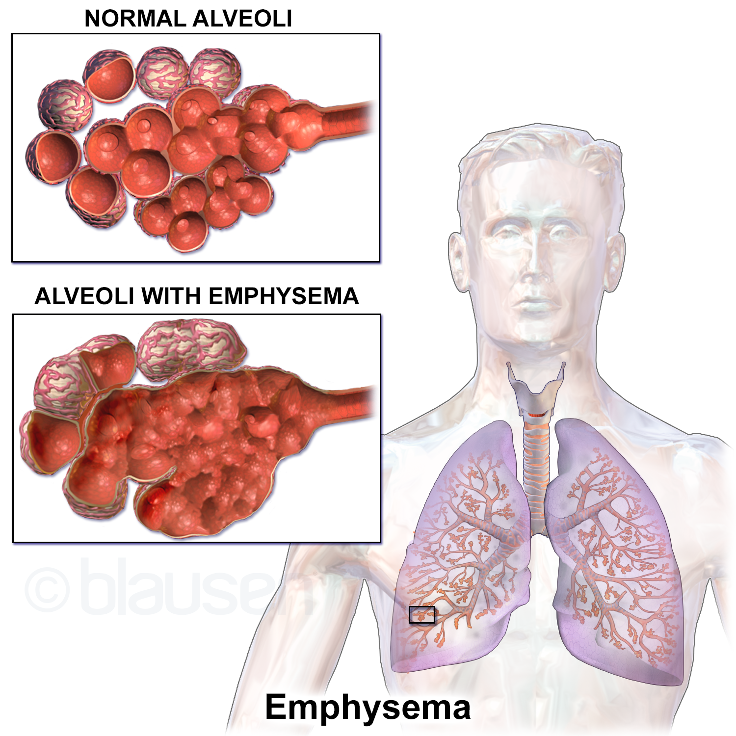
Clinical Correlate: Emphysema is a disease that causes the progressive destruction of alveolar walls. As the walls break down, multiple small sacs merge into larger, floppy spaces. The destruction of alveolar walls significantly decreases the total surface area available for gas exchange, permanently depriving the patient's blood of adequate oxygen.
Surrounding the entire lung structure is a protective casing known as the pleura. The pleura is a double-layered serous membrane surrounding the lungs. The inner layer, known as the visceral pleura, firmly attaches to the outer surface of the lungs. The outer layer, the parietal pleura, lines the inner wall of the thoracic cavity. A vital lubricant, pleural fluid between the visceral and parietal pleura minimizes friction during lung expansion, allowing the lungs to glide smoothly against the ribs with every breath.
Ventilation is the mechanical process of moving atmospheric air into and out of the lungs. It is governed strictly by Boyle's Law of physics: pressure and volume are inversely related. To move air, the body changes the volume of the chest cavity to create pressure gradients.
Inhalation and Exhalation
Inhalation is initiated by the contraction of the diaphragm, a dome-shaped muscle at the base of the chest. The diaphragm moves downward toward the abdominal cavity during contraction. Simultaneously, the contraction of the external intercostal muscles elevates the rib cage.
By dropping the floor and expanding the walls, the expansion of the thoracic cavity increases the internal volume of the lungs. Because the space is larger, an increase in lung volume decreases intrapulmonary air pressure. Nature abhors a vacuum; therefore, air flows into the lungs when intrapulmonary pressure drops below atmospheric pressure.
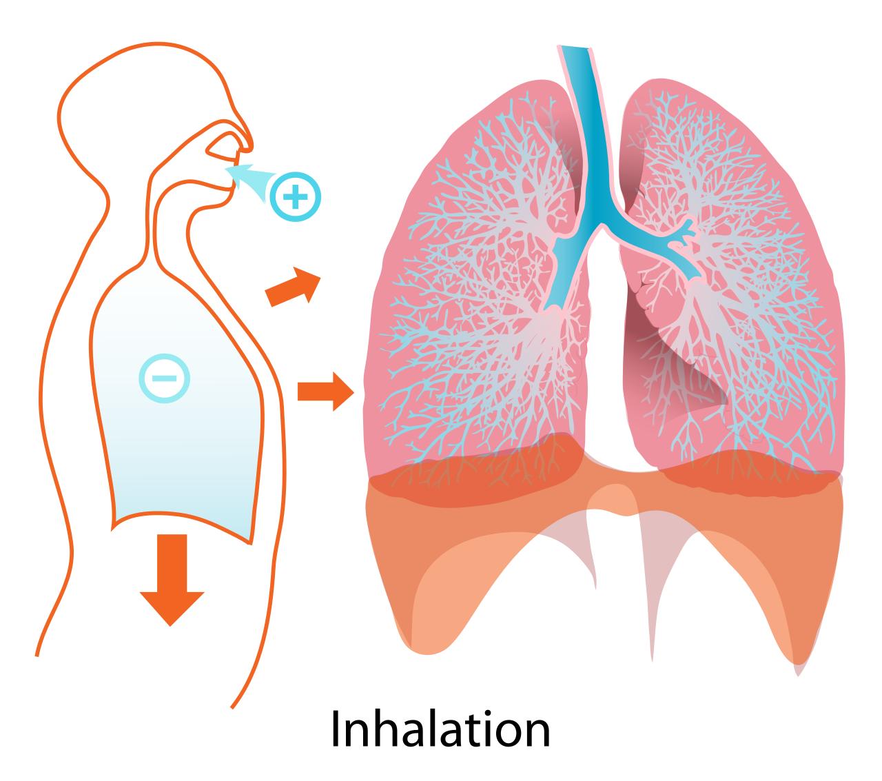
Conversely, exhalation is typically a passive process driven by the relaxation of respiratory muscles. Relaxation of the diaphragm decreases the volume of the thoracic cavity. As the walls close in, a decrease in thoracic volume increases intrapulmonary air pressure. Consequently, air is forced out of the lungs when intrapulmonary pressure exceeds atmospheric pressure.
We quantify this airflow using specific respiratory metrics:
- Tidal volume is the amount of air inhaled or exhaled during a normal resting breath.
- Residual volume is the amount of air remaining in the lungs following a maximal forceful exhalation. (This residual air keeps the alveoli permanently inflated and prevents lung collapse.)
The Chemistry of Diffusion and Neurological Control
Once air is in the alveoli, physics hands the baton to chemistry. Gas exchange across the alveolar membrane occurs via passive diffusion—gases move naturally from areas of high concentration to low concentration. Oxygen diffuses from the air in the alveoli into the blood of the pulmonary capillaries, while carbon dioxide diffuses from the blood of the pulmonary capillaries into the air of the alveoli to be exhaled.
But how does the body know when and how fast to breathe? The brain's autonomic systems run the operation. The medulla oblongata of the brainstem controls the basic rhythm of breathing. Counterintuitively, the body is less concerned with oxygen levels than it is with cellular exhaust. The respiratory center in the brain monitors the concentration of carbon dioxide in the blood.
When you exercise or hold your breath, carbon dioxide builds up. Because CO2 forms carbonic acid in aqueous solutions, an increase in blood carbon dioxide concentration lowers the pH of the blood, making it more acidic. A drop in blood pH stimulates the medulla oblongata to increase the breathing rate, driving you to exhale faster to blow off the excess CO2 and restore blood pH balance.
If the lungs are the intake manifold, the heart is the master pump, pushing a specialized fluid matrix through thousands of miles of vascular tubing.
Composition of Blood
Blood is not a uniform red liquid; it is a complex, multi-part connective tissue.
| Blood Component | Scientific Name | Primary Characteristics & Functions |
|---|---|---|
| Plasma | Plasma | Plasma constitutes the liquid extracellular matrix of whole blood. Blood plasma is composed predominantly of water. It serves as the great biological solvent: blood plasma transports dissolved proteins, transports circulating endocrine hormones, and transports metabolic waste products to the kidneys for excretion. |
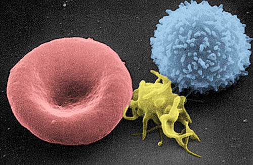
| Red Blood Cells | Erythrocytes | Erythrocytes are commonly known as red blood cells. Unique among cells, mature human erythrocytes lack a cellular nucleus. The absence of a nucleus in erythrocytes maximizes internal cellular volume for hemoglobin storage. They are packed with it: erythrocytes contain the iron-rich protein hemoglobin, and hemoglobin binds reversibly to oxygen molecules for transport in the bloodstream. |
| White Blood Cells | Leukocytes | Leukocytes are commonly known as white blood cells. Their role is defense; leukocytes protect the body against invading pathogens. |
| Platelets | Thrombocytes | Thrombocytes are commonly known as platelets. Platelets are cellular fragments that initiate the blood clotting cascade, plugging leaks in damaged vessels. |
The Chambers and Vessels
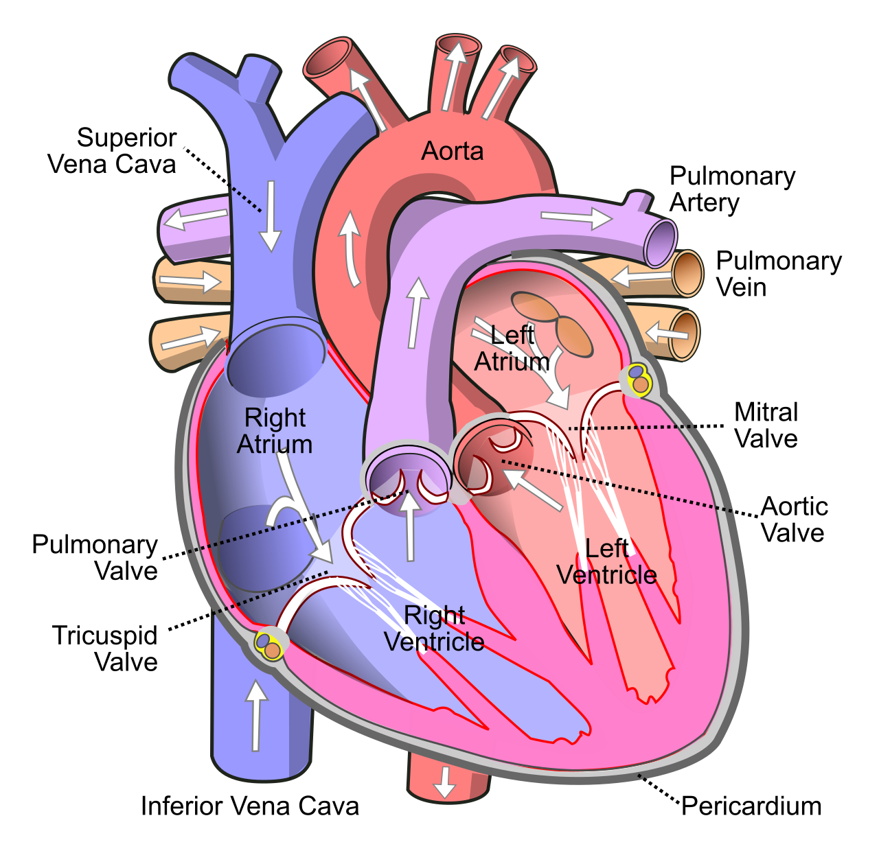
The human heart contains four chambers. The upper chambers are for receiving, and the lower chambers are built for immense physical labor.
- The right atrium is an upper receiving chamber of the heart.
- The left atrium is an upper receiving chamber of the heart.
- The right ventricle is a lower pumping chamber of the heart.
- The left ventricle is a lower pumping chamber of the heart.
To organize the massive task of circulating blood, the cardiovascular system is divided into two distinct loops:
- The pulmonary circuit comprises the flow of blood between the heart and the lungs. (Low pressure, designed strictly for gas exchange).
- The systemic circuit comprises the flow of blood between the heart and the peripheral body tissues. (High pressure, designed to deliver nutrients everywhere else).
Tracing the Blood Flow
Understanding hemodynamics requires tracing a drop of blood exactly as it navigates the heart's anatomy.
- Return from the body (Deoxygenated): Exhausted, oxygen-poor blood returns to the heart. The superior vena cava delivers deoxygenated blood from the upper body directly into the right atrium, while the inferior vena cava delivers deoxygenated blood from the lower body directly into the right atrium.
- Into the Right Ventricle: Blood flows from the right atrium into the right ventricle through the tricuspid valve. The tricuspid valve prevents the backward flow of blood into the right atrium during ventricular contraction.
- To the Lungs (Pulmonary Circuit): The right ventricle pumps deoxygenated blood toward the lungs. Blood exits the right ventricle by passing through the pulmonary semilunar valve. From there, the pulmonary trunk carries blood from the right ventricle to the pulmonary arteries, and these pulmonary arteries transport deoxygenated blood away from the heart to the lungs. (Note: This is the rare instance in human anatomy where an artery carries deoxygenated blood).
- Return from the Lungs (Oxygenated): After picking up oxygen at the alveoli, oxygenated blood returns from the lungs to the heart via the pulmonary veins. (Again, a rare instance of veins carrying highly oxygenated blood). The left atrium receives oxygenated blood from the pulmonary veins.
- Into the Left Ventricle: Blood flows from the left atrium into the left ventricle through the bicuspid valve. In medical practice, the bicuspid valve is clinically referred to as the mitral valve.
- Out to the Body (Systemic Circuit): The left ventricle pumps oxygenated blood throughout the systemic circulation. Because it must push blood all the way to the top of the brain and the tips of the toes against high resistance, the myocardium of the left ventricle is significantly thicker than the myocardium of the right ventricle. Blood exits the left ventricle by passing through the aortic semilunar valve. From there, it enters the systemic highway: the aorta is the largest artery in the human cardiovascular system.
The Vascular Highway
Vessels change dramatically depending on the pressure they handle and their distance from the heart.
- Arteries: Arteries are defined as blood vessels that carry blood away from the heart. Because they face the massive brunt of ventricular pressure, arterial walls contain a thick layer of smooth muscle and contain an abundant layer of elastic connective tissue to stretch and recoil with every heartbeat.
- Capillaries: As arteries branch and shrink, they become arterioles, which then feed the capillaries. Capillaries are the microscopic blood vessels connecting arterioles to venules. At the tissue level, the walls of capillaries consist of a single layer of endothelial cells. The extreme thinness of capillary walls facilitates the exchange of nutrients and gases with surrounding tissues.
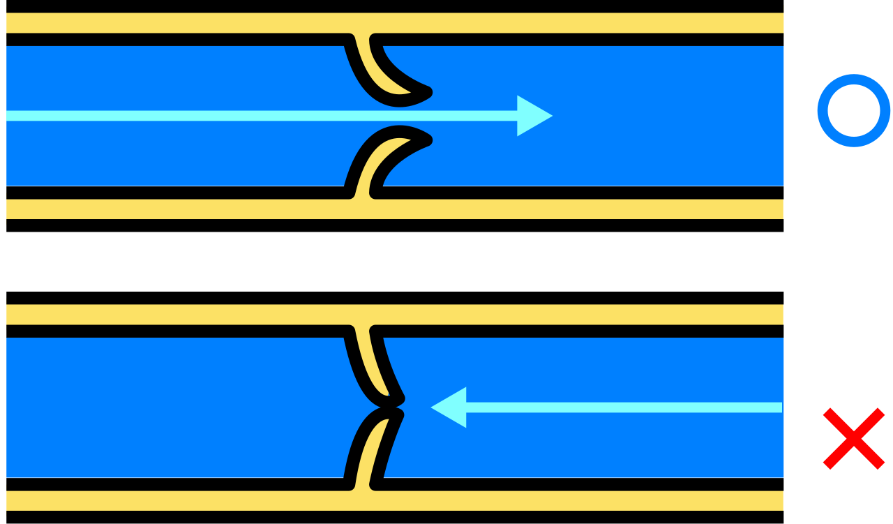
- Veins: Exiting the tissues, capillaries merge into venules, which merge into veins. Veins are defined as blood vessels that transport blood toward the heart. Because venous blood has lost most of the pressure generated by the heart, it struggles to climb back up the legs. To solve this, many systemic veins contain internal one-way valves. Venous valves prevent the backward pooling of blood due to gravity, ensuring blood only flows homeward.
The cardiac cycle consists of alternating periods of muscular contraction and relaxation. We define these phases clinically:
- Systole refers to the contraction phase of the cardiac cycle. During this explosive phase, ventricular systole forces blood into the aorta and ventricular systole forces blood into the pulmonary trunk.
- Diastole refers to the relaxation phase of the cardiac cycle. During this quiet period, the chambers of the heart passively fill with blood during diastole.
The Heart's Electrical Conduction System
The heart does not require a signal from the brain to beat; it generates its own electricity.
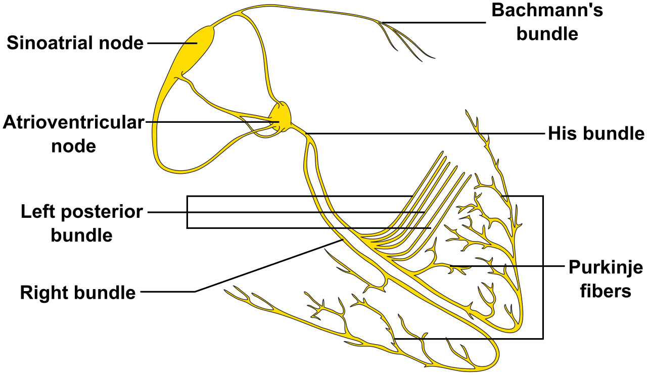
The spark begins at the sinoatrial node, which is located in the upper wall of the right atrium. The sinoatrial node serves as the primary natural pacemaker of the heart, and generates the initial electrical impulses that trigger atrial contraction.
This electrical wave sweeps over the atria and hits a designated checkpoint: the atrioventricular node, which is located at the junction of the atria and ventricles. Why have a checkpoint? The atrioventricular node briefly delays the electrical impulse traveling from the atria to the ventricles. The delay at the atrioventricular node allows the atria to fully empty blood into the ventricles prior to ventricular contraction. Without this fractional delay, the upper and lower chambers would squeeze simultaneously, fighting each other and drastically reducing cardiac output.
After the delay, the signal races down the interventricular septum and into the outer ventricular walls via specialized conductive wiring: Purkinje fibers distribute electrical action potentials rapidly throughout the ventricular myocardium, causing a synchronized, forceful upward squeeze.
Hemodynamics: Blood Pressure and Pathology
All of this mechanical pumping generates fluid tension against the vessel walls. Blood pressure is the hydrostatic force exerted by circulating blood against the inner walls of blood vessels.
When you measure blood pressure, you are quantifying the force during the two phases of the cardiac cycle:
- Systolic blood pressure is the maximum arterial pressure recorded during ventricular contraction.
- Diastolic blood pressure is the minimum arterial pressure recorded during ventricular relaxation.
As clinicians, we measure these forces externally. A sphygmomanometer is the medical device used to measure blood pressure.
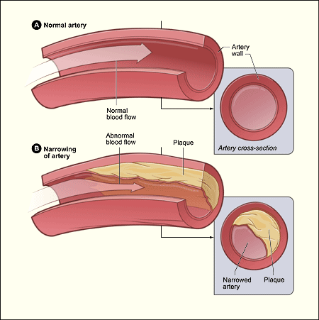
When the cardiovascular system breaks down, the physical dynamics of this pressure radically change. Atherosclerosis is characterized by the accumulation of lipid plaques within the inner walls of arteries, which narrows the physical diameter of the tubing. If you force the same volume of fluid through a narrower tube, pressure inevitably skyrockets. This leads directly to hypertension, which is the clinical term for chronic abnormally high blood pressure. Left unchecked, hypertension steadily destroys the delicate capillary beds in the kidneys, eyes, and brain, illustrating perfectly why understanding these pressure dynamics is the cornerstone of clinical vigilance.