Human Anatomy - General Orientation
Not sure you’re ready?
Take the ~3-minute readiness diagnostic and see where you stand.
To treat the human body, you must first understand how to navigate it. Before a surgeon can operate or a nurse can triage a trauma patient, they must rely on a universally agreed-upon coordinate system and structural hierarchy. The human body is not a random assortment of tissues; it is a masterpiece of biological engineering, governed by strict physical laws and geographic coordinates. Just as an architect must understand how a building is constructed from raw materials to a finished structure, a healthcare professional must understand how the body organizes itself, from microscopic particles to the dynamic equilibrium that keeps the organism alive.
The human body is built on a strict, additive hierarchy. Each level of complexity is constructed entirely from the components of the level below it. To understand a patient’s illness, you must often trace the symptom back to its smallest structural root.
At the foundation of this hierarchy, atoms combine to form molecules. These invisible chemical building blocks dictate the physical properties of everything in the universe. In the human body, these molecules combine to form cellular organelles, which are the specialized, microscopic machines—like mitochondria or nuclei—that drive life.
These organelles are housed within the cell, which is the basic structural and functional unit of all living organisms. A single cell is the smallest independent unit capable of life. However, cells rarely work alone.
When you have groups of similar cells performing a common, specialized function, they form tissues. You can think of tissues as the "fabrics" of the human body. There are only four basic tissue types in the human body: epithelial, connective, muscle, and nervous tissue. Every structure inside you is woven from these four materials.
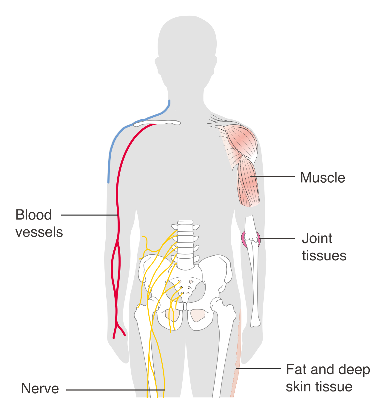
When the body needs to perform complex, localized tasks, it builds organs. Organs are discrete structures composed of at least two different tissue types working together to perform specific functions. For example, your stomach contains epithelial tissue to line its inner surface, muscle tissue to churn food, and nervous tissue to regulate digestion.
Stepping further out, organ systems are groups of organs that cooperate to accomplish a common physiological purpose. The digestive system, for instance, requires the coordinated effort of the esophagus, stomach, and intestines.
Finally, the organism is the highest level of structural organization representing the sum total of all interacting organ systems. You are an organism, kept alive by the flawless, continuous collaboration of your cellular machinery and macroscopic organs.
If a patient is lying on their stomach, is their kneecap pointing "forward" or "down"? If they are upside down, where is "up"? In medicine, ambiguity is dangerous. To ensure that "left" always means left and "front" always means front, anatomists developed a universal reference point.
Standard anatomical position requires the human body to be standing erect. Every medical diagram, chart, and directional term assumes the body is exactly in this posture, regardless of how the patient is actually situated in a hospital bed.
In this standard anatomical position:
- The feet are parallel and flat on the floor.
- The eyes gaze forward.
- The arms hang at the sides of the body.
- The palms of the hands face forward.
- The thumbs point away from the body.
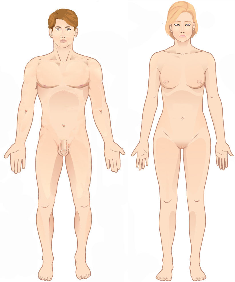
Because the palms are facing forward, the forearms are uncrossed. If a surgeon refers to a structure on the "front" of the forearm, they are always referring to the palm-side, even if the patient's arms are currently crossed over their chest.
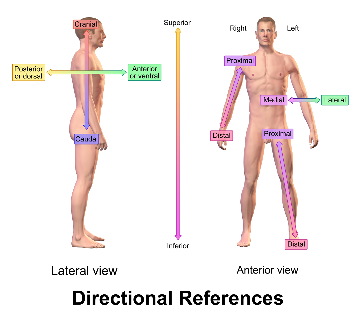
With the map oriented, we can use precise vocabulary to describe where one body part is located relative to another.
| Term | Definition | Clinical Context / Equivalence |
|---|---|---|
| Superior / Inferior | Superior describes a position toward the head or upper part of a structure.<br><br>Inferior describes a position away from the head or toward the lower part of a structure. | The heart is superior to the stomach. The pelvis is inferior to the ribcage. |
| Anterior / Posterior | Anterior describes a position toward the front of the body.<br><br>Posterior describes a position toward the back of the body. | Ventral is used interchangeably with anterior in human anatomy.<br><br>Dorsal is used interchangeably with posterior in human anatomy. |
| Medial / Lateral | Medial describes a position toward or at the midline of the body.<br><br>Lateral describes a position away from the midline of the body. | The nose is medial to the ears. The arms are lateral to the chest. |
| Proximal / Distal | Proximal describes a position closer to the origin of the body part or the point of attachment of a limb to the body trunk.<br><br>Distal describes a position farther from the origin of a body part or the point of attachment of a limb to the body trunk. | Crucial for limbs. The elbow is proximal to the wrist. The fingers are distal to the palm. |
| Superficial / Deep | Superficial describes a position toward or at the surface of the body.<br><br>Deep describes a position away from the surface of the body. | Skin is superficial to the skeletal muscles. Bones are deep to the skin. |
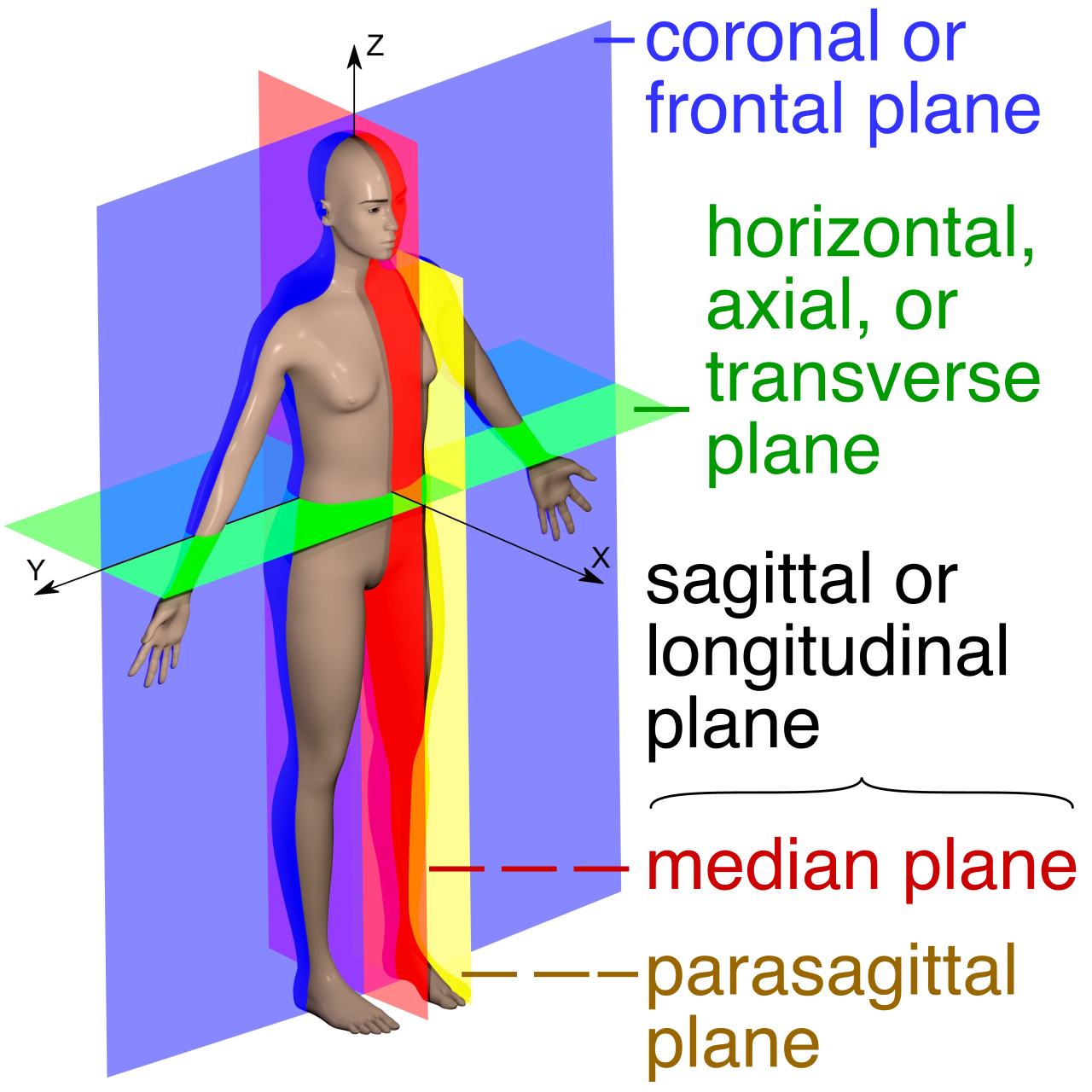
When viewing an MRI, a CT scan, or a cross-section of tissue under a microscope, you are looking at a slice of the body. A body plane is an imaginary flat surface that passes through the body to provide a frame of reference.
There are three primary planes:
- The Sagittal Plane: A vertical plane that divides the body into right and left portions.
- If the cut goes perfectly down the center, it is a midsagittal plane, which divides the body exactly at the midline into equal right and left halves.
- If the cut is offset, it is a parasagittal plane, which divides the body into unequal right and left portions.
- The Frontal Plane: This plane divides the body into anterior and posterior portions (front and back). In clinical settings, the frontal plane is synonymous with the coronal plane.
- The Transverse Plane: This horizontal slice divides the body into superior and inferior portions (top and bottom). Accordingly, the transverse plane is synonymous with the horizontal plane.
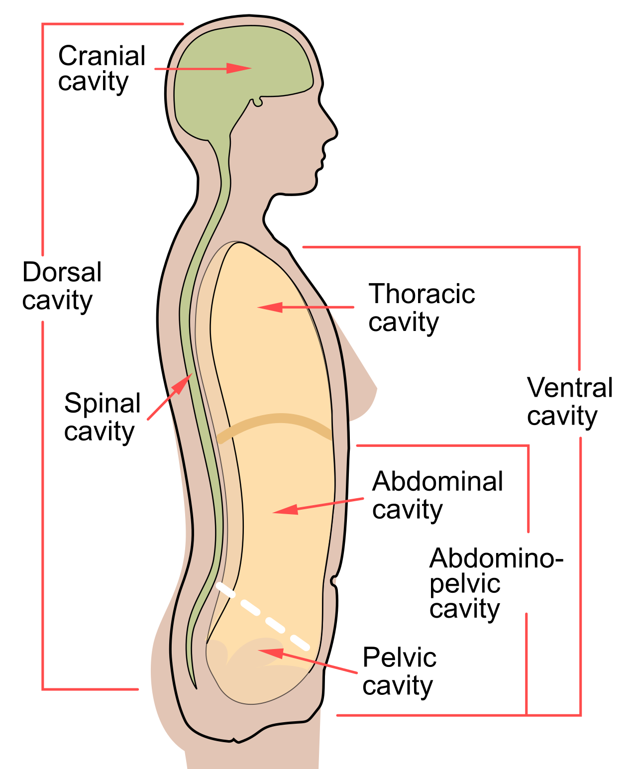
The human organism is not solid; it is built with internal spaces that house, protect, and permit the expansion of organs. There are two major sets of cavities: the dorsal and the ventral.
The Dorsal Body Cavity
Tucked along the posterior (back) of the body, the dorsal body cavity protects the central nervous system organs. It is entirely encased in bone and consists of two continuous subdivisions:
- The dorsal body cavity contains the cranial cavity and the spinal cavity.
- The cranial cavity encases the brain within the rigid skull.
- The spinal cavity encloses the spinal cord within the vertebral column.
The Ventral Body Cavity
Located anteriorly, the ventral body cavity houses the internal organs collectively called viscera. Unlike the dorsal cavity, the ventral cavity is largely unprotected by complete bony enclosures, allowing organs like the stomach and lungs to expand and contract.
The ventral body cavity is subdivided into the thoracic cavity and the abdominopelvic cavity. What separates them? The diaphragm, a dome-shaped muscle that separates the thoracic cavity from the abdominopelvic cavity.
- The thoracic cavity contains the heart and lungs.
- Below the diaphragm, the abdominopelvic cavity is artificially divided into the abdominal cavity and the pelvic cavity (there is no physical barrier separating them).
- The abdominal cavity contains the stomach, intestines, spleen, and liver.
- The pelvic cavity contains the urinary bladder, reproductive organs, and rectum.
Mapping the Abdomen
Because the abdominopelvic cavity is so large and contains so many overlapping organs, healthcare providers need a way to pinpoint pain or anomalies.
For quick triage and clinical localization, medical personnel divide the abdominopelvic cavity into four quadrants. For example, if a patient presents with sharp pain in the right upper quadrant of the abdominopelvic cavity, which contains the liver and gallbladder, the triage nurse immediately suspects gallstones.
However, for more granular, academic detail, anatomists divide the abdominopelvic cavity into nine distinct regions for precise anatomical studies (such as the epigastric, umbilical, and hypogastric regions).
Structural anatomy provides the map, but physiology provides the engine. The overarching goal of the human machine is survival, which requires maintaining a hyper-specific internal environment despite a chaotic external world.
Homeostasis is the physiological process of maintaining a relatively stable internal environment despite continuous external changes.
Crucially, homeostasis represents a dynamic state of equilibrium rather than an unchanging, static state. Your blood pressure, temperature, and glucose levels fluctuate constantly, but they are tightly constrained within a narrow, life-sustaining range.
The Control Mechanism
To maintain this dynamic equilibrium, the body relies on rapid communication loops, typically via the nervous or endocrine systems. Every homeostatic control mechanism consists of three interdependent components:
- The Receptor: This is a sensor that monitors the environment and responds to specific stimuli (such as thermoreceptors in your skin detecting a drop in temperature).
- The Control Center: Usually the brain or an endocrine gland, the control center determines the set point at which the variable is maintained, analyzes the input from the receptor, and determines the appropriate response.
- The Effector: This component provides the means for the control center's response to the stimulus (such as muscles shivering to generate heat).
Negative Feedback: The Braking System
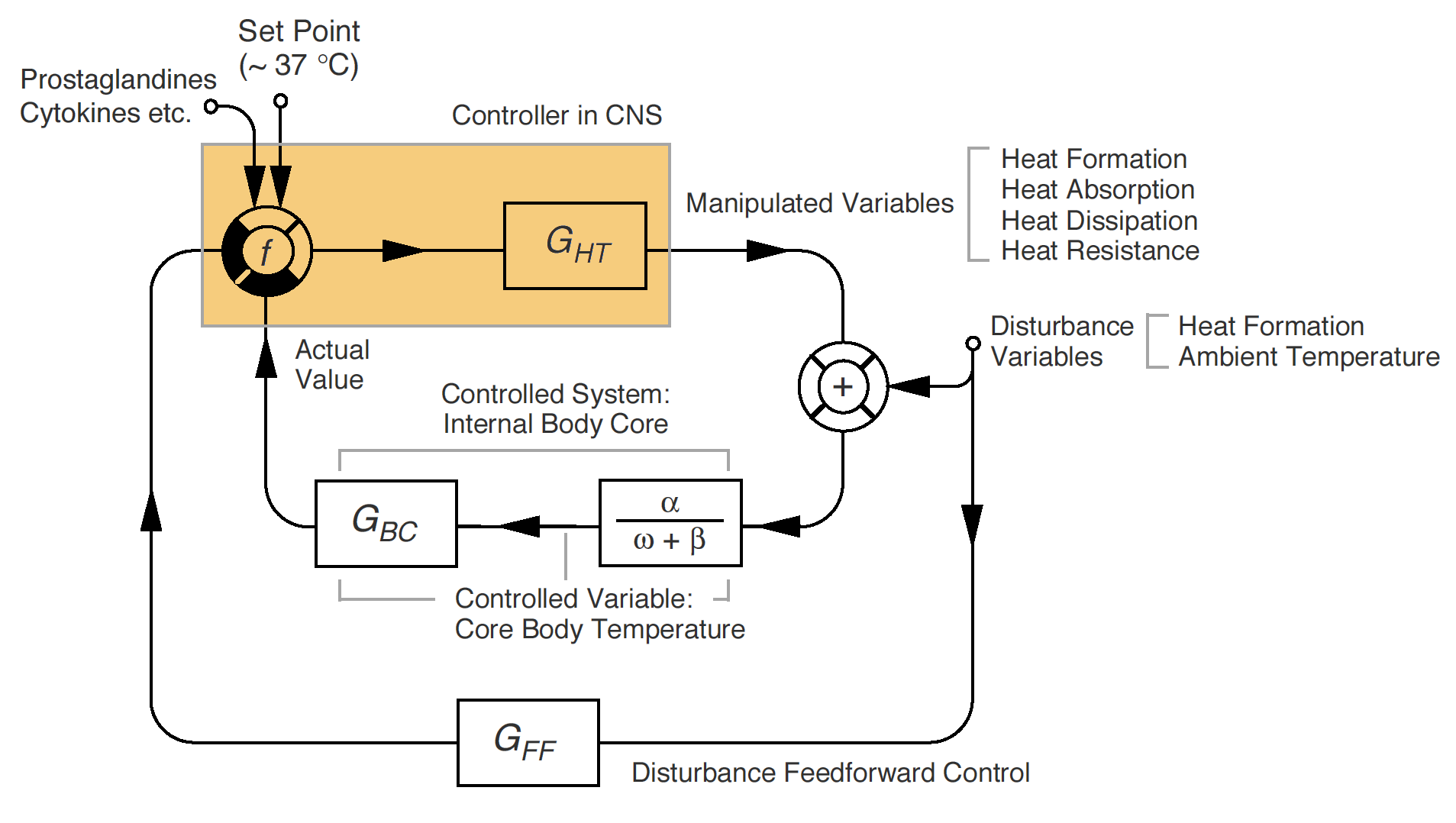
The vast majority of homeostatic mechanisms in the body are negative feedback loops. Negative feedback mechanisms act to reduce or completely shut off the original stimulus. They function exactly like the thermostat in your house. If it gets too hot, the AC kicks on to bring the temperature down, opposing the initial change.
- Human body temperature regulation is an example of a negative feedback mechanism.
- The regulation of blood glucose levels by insulin is an example of a negative feedback mechanism. (When blood sugar rises, insulin forces cells to absorb the glucose, driving the blood sugar back down).
Positive Feedback: The Accelerator
While negative feedback seeks stability, positive feedback seeks completion. Positive feedback mechanisms act to enhance or exaggerate the original stimulus. Instead of pulling the body back to a baseline, in a positive feedback mechanism, the physiological response accelerates in the same direction as the initial change.
Because positive feedback loops are explosive and inherently destabilizing, they control infrequent, episodic events that need a definitive conclusion:
- Blood clotting is an example of a positive feedback mechanism. When a vessel tears, platelets rush in and release chemicals that attract more platelets, escalating rapidly until the clot seals the tear.
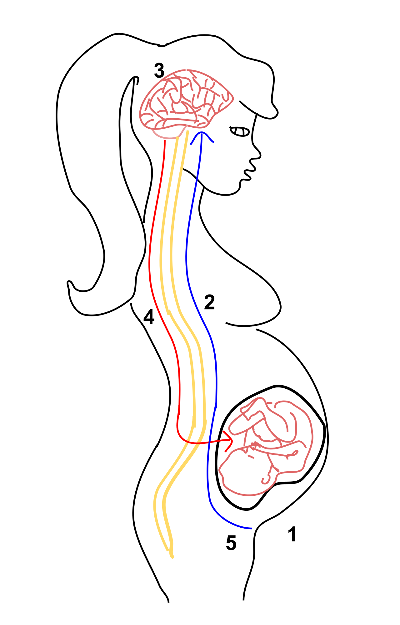
- Uterine contractions during childbirth represent an example of a positive feedback mechanism. The hormone oxytocin causes the uterus to contract, pushing the baby against the cervix. This stretching triggers the release of more oxytocin, causing stronger contractions, a loop that accelerates until the baby is delivered.