Cardiovascular and Respiratory Systems
Not sure you’re ready?
Take the ~3-minute readiness diagnostic and see where you stand.
The human body is fundamentally a fluid-driven machine designed to solve a single, relentless problem: how to deliver a continuous supply of microscopic oxygen molecules to trillions of demanding cells while instantly removing their toxic carbon dioxide exhaust. Every breath drawn and every pulse palpated represents the coordinated mechanical and fluid dynamics of two inseparable loops: the cardiovascular and respiratory systems. In clinical practice, measuring a patient’s vital signs—blood pressure, heart rate, respiratory rate, and oxygen saturation—is simply observing the real-time efficiency of these twin engines. Understanding how they structurally interlock, pump, and diffuse gases is the absolute foundation of all clinical assessment.
To understand circulation, you must first understand the medium being circulated. Whole blood is not a simple red liquid; it is a complex, living tissue composed of specialized cells suspended in a dynamic fluid matrix.
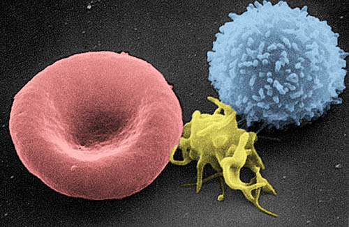
Blood plasma constitutes the extracellular liquid matrix component of whole blood. Because it must carry dissolved nutrients, hormones, and cellular waste, blood plasma is composed of approximately 90 percent water. Flowing within this watery matrix are three primary formed elements, each engineered for a specific physiological task:
| Cellular Element | Common Name | Primary Function & Characteristics |
|---|---|---|
| Erythrocytes | Red Blood Cells (RBCs) | The primary physiological function of erythrocytes is the transport of oxygen to bodily tissues. Remarkably, mature human erythrocytes completely lack a nucleus, trading their genetic material for more internal cargo space. They contain abundant molecules of an iron-bearing protein called hemoglobin. These hemoglobin molecules bind reversibly to oxygen in the lungs for transportation through the bloodstream, holding on to it tightly until they reach oxygen-starved tissues. |
| Leukocytes | White Blood Cells (WBCs) | Leukocytes function primarily as active components of the body's immune system to identify and destroy pathogens. They are the mobile infantry of your bloodstream. |
| Thrombocytes | Blood Platelets | Thrombocytes initiate and facilitate the physiological blood clotting process. When a vessel is breached, these fragments adhere together to plug the leak. |
Blood is channeled throughout the body in a closed network of tubes designed to handle varying degrees of pressure and flow.
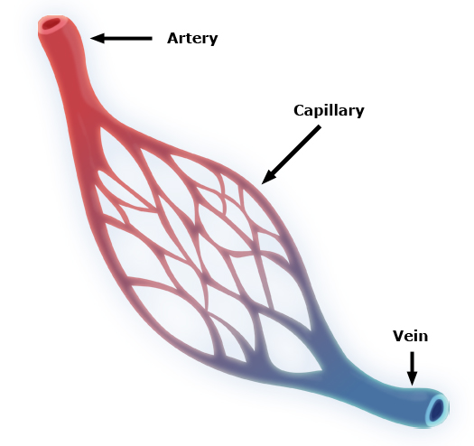
- Arteries are defined functionally as blood vessels that carry blood away from the heart. Because they receive blood directly from the heart's forceful pumping, their walls are thick and elastic.
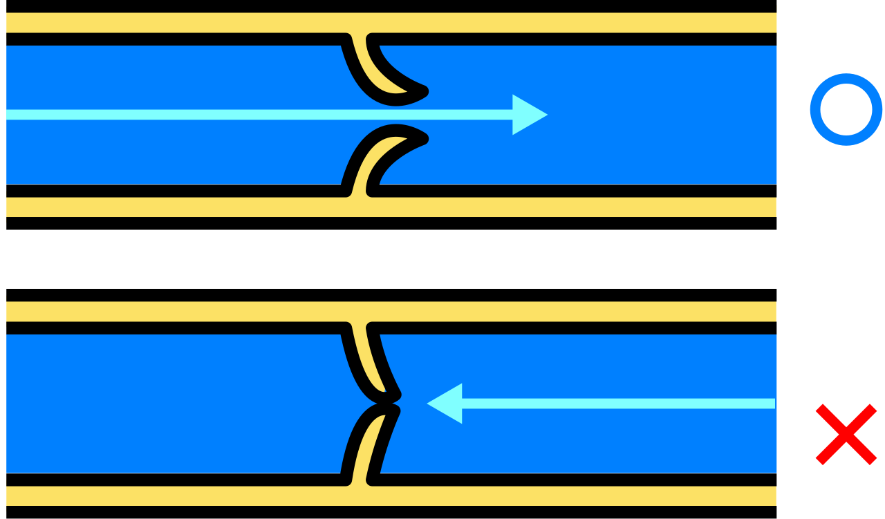
- Veins are defined functionally as blood vessels that carry blood toward the heart. Because venous blood is under low pressure and must often travel upward from the lower extremities, many veins in the human body contain internal one-way valves. The one-way valves located inside veins prevent the backward flow of blood against the pull of gravity. (When these valves fail, blood pools, resulting in varicose veins).
- Capillaries are the microscopic bridge between arteries and veins. Capillaries are microscopic blood vessels featuring walls that are precisely one cell thick. The extreme thinness of capillary walls enables the rapid exchange of gases and nutrients between circulating blood and surrounding tissues.
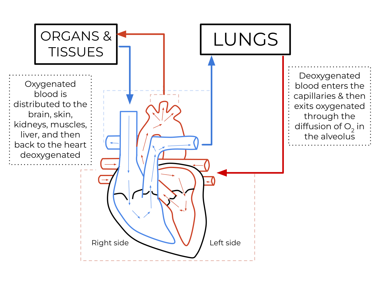
The human heart is not a single pump; it is two distinct pumps acting simultaneously side-by-side. The right side handles the low-pressure pulmonary circuit (blood going to the lungs), while the left side handles the high-pressure systemic circuit (blood going to the entire body).
The Cardiac Cycle The pumping action of the heart muscle operates in two alternating phases:
Tracing the Pathway of Blood
To master cardiac anatomy, you must be able to trace a single drop of blood as it completes a full loop through both circuits.
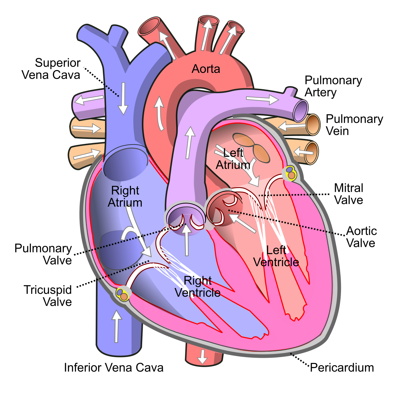
- Return from the Body: Deoxygenated blood from the upper and lower body enters the heart through the superior and inferior vena cava.
- Right Atrium: The superior and inferior vena cava empty deoxygenated blood directly into the right atrium.
- Right Ventricle: Blood passes from the right atrium into the right ventricle through the tricuspid valve (a mechanical set of "doors" preventing backflow).
- To the Lungs: The right ventricle pumps deoxygenated blood outward through the pulmonary valve. Blood exiting the pulmonary valve enters the pulmonary artery.
- Crucial Exception: The pulmonary artery is the only artery in the adult human body that carries deoxygenated blood. The pulmonary artery transports deoxygenated blood to the lungs for oxygenation.
- Return from the Lungs: After picking up oxygen at the capillary beds of the lungs, oxygenated blood returns from the lungs to the heart via the pulmonary veins.
- Crucial Exception: The pulmonary veins are the only veins in the adult human body that carry oxygenated blood.
- Left Atrium: The pulmonary veins empty oxygenated blood directly into the left atrium.
- Left Ventricle: Blood flows from the left atrium into the left ventricle through the mitral valve. (Note: The mitral valve is interchangeably referred to as the bicuspid valve).
- Out to the Body: The left ventricle pumps oxygenated blood outward through the aortic valve. Blood exiting the aortic valve immediately enters the aorta. The aorta distributes oxygenated blood to the entire systemic circulation.
Because of its heavy workload, the left ventricle contains the thickest muscular wall of all the heart chambers. The thick muscular wall of the left ventricle generates the high pressure required to pump blood throughout the entire systemic circuit, reaching all the way from the crown of the head to the tips of the toes.
For the heart chambers to pump blood effectively, their muscular contractions (systole) must be perfectly timed. If the atria and ventricles squeezed at the exact same time, blood would simply smash together. This timing is controlled by an intrinsic electrical wiring system.
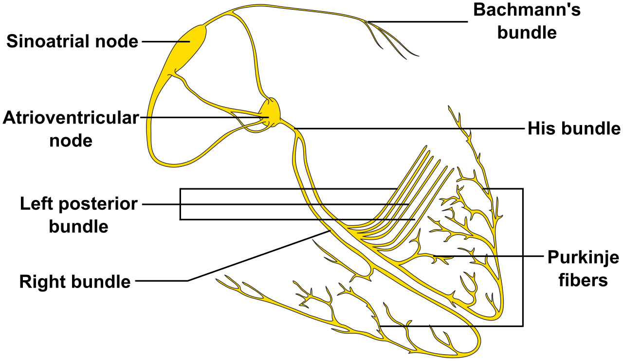
- The Sinoatrial (SA) Node: The sinoatrial node is located in the upper wall of the right atrium. The sinoatrial node functions as the primary natural pacemaker of the human heart, continuously firing rhythmic electrical impulses.
- The Atrioventricular (AV) Node: The electrical impulse generated by the sinoatrial node travels directly to the atrioventricular node. The atrioventricular node performs a critical mechanical function: it briefly delays the cardiac electrical impulse to allow the atria to completely empty blood into the ventricles before the ventricles are triggered to squeeze.
- The Bundle of His and Purkinje Fibers: Cardiac electrical signals travel from the atrioventricular node down the bundle of His. Finally, Purkinje fibers distribute the cardiac electrical impulse throughout the right and left ventricular muscle walls, triggering a powerful, coordinated upward contraction.
While the heart pumps blood, the respiratory tract is responsible for capturing ambient oxygen and conditioning it before it meets the bloodstream.
The pathway of air follows a precise anatomical route:
- Nasal Cavity & Pharynx: Inhaled atmospheric air passes from the nasal cavity directly into the pharynx (the throat). The pharynx functions as a shared anatomical passageway for both respiratory air and ingested food.
- The Epiglottis: Because the pharynx is a shared pathway, the body needs a way to separate food from air. The epiglottis is a distinct flap of elastic cartilage positioned superior to the larynx. When you swallow, the epiglottis reflexively folds downward to prevent food and liquid from entering the lower respiratory tract. (Failure of this mechanism results in aspiration).
- The Larynx: Air passes next to the larynx, which is situated between the pharynx and the trachea. The larynx is informally called the voice box.
- The Trachea: The air then enters the windpipe, or trachea. The trachea is supported structurally by a series of C-shaped rings composed of hyaline cartilage. The rigid cartilaginous rings of the trachea prevent the airway tube from collapsing during rapid pressure changes.
- The Bronchial Tree: The inferior end of the trachea bifurcates (splits) into the right primary bronchus and the left primary bronchus. These primary bronchi branch continuously into progressively smaller secondary and tertiary bronchi. Tertiary bronchi branch repeatedly into very narrow tubes known as bronchioles.
- The Terminal End: Finally, the smallest of these branches, the terminal bronchioles, connect directly to clusters of microscopic air sacs called alveoli.
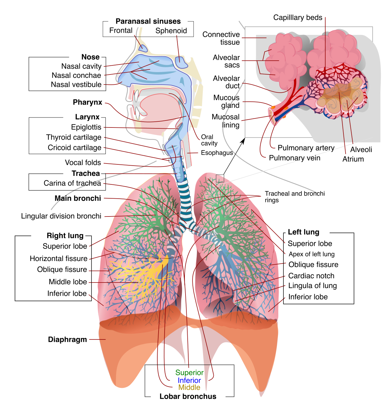
Alveoli serve as the fundamental functional units of the respiratory system. Everything else in the respiratory tract is just plumbing designed to get air to these microscopic balloons.
Each alveolus is surrounded intimately by an extensive network of pulmonary capillaries. Pulmonary gas exchange occurs directly across the thin respiratory membrane of the alveoli. This exchange does not require the cell to burn energy; rather, pulmonary gas exchange is driven entirely by the physical process of simple passive diffusion. Gases naturally move down their concentration gradients:
- During alveolar gas exchange, oxygen diffuses from a higher concentration in the alveoli to a lower concentration in the capillary blood.
- During alveolar gas exchange, carbon dioxide diffuses from a higher concentration in the capillary blood to a lower concentration in the alveoli, where it can be exhaled.
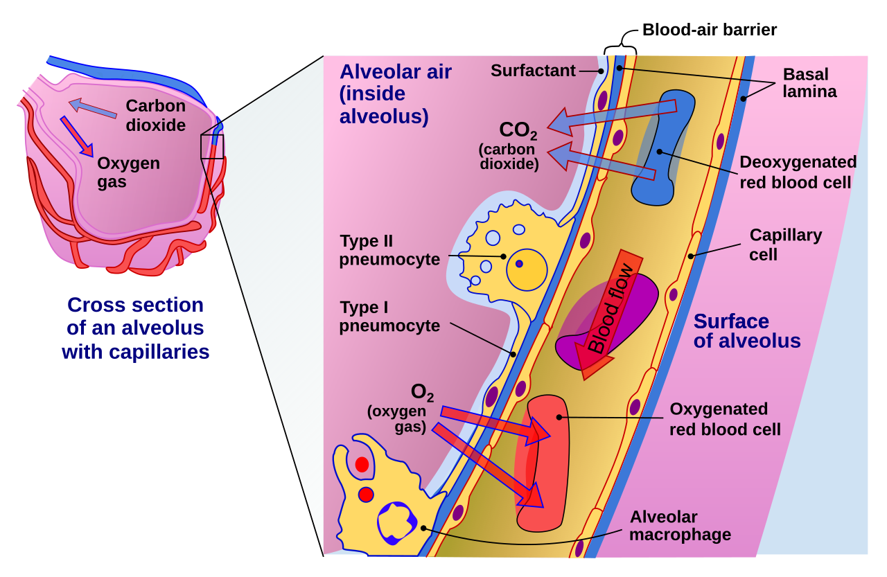
Because the walls of the alveoli are exceptionally thin and coated in a layer of water, surface tension naturally makes them want to collapse inward and stick shut, much like the wet inside of a deflated balloon. To counteract this, Type II alveolar cells secrete a specialized lipoprotein mixture known as pulmonary surfactant. Pulmonary surfactant significantly lowers the surface tension of the fluid lining the interior of the alveoli. By actively reducing surface tension, pulmonary surfactant prevents the fragile alveoli from collapsing completely during exhalation.
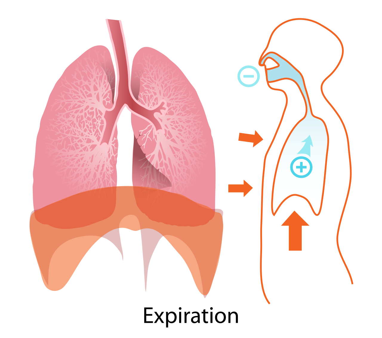
Air does not move into the lungs on its own; it requires a pressure gradient. This gradient is manufactured by changes in the volume of your chest cavity, governed by Boyle’s Law (which dictates that volume and pressure are inversely related).
The primary engine of this system is the diaphragm, a dome-shaped sheet of skeletal muscle that physically separates the thoracic cavity (containing the heart and lungs) from the abdominal cavity (containing the stomach, liver, and intestines).
Inhalation (Active)
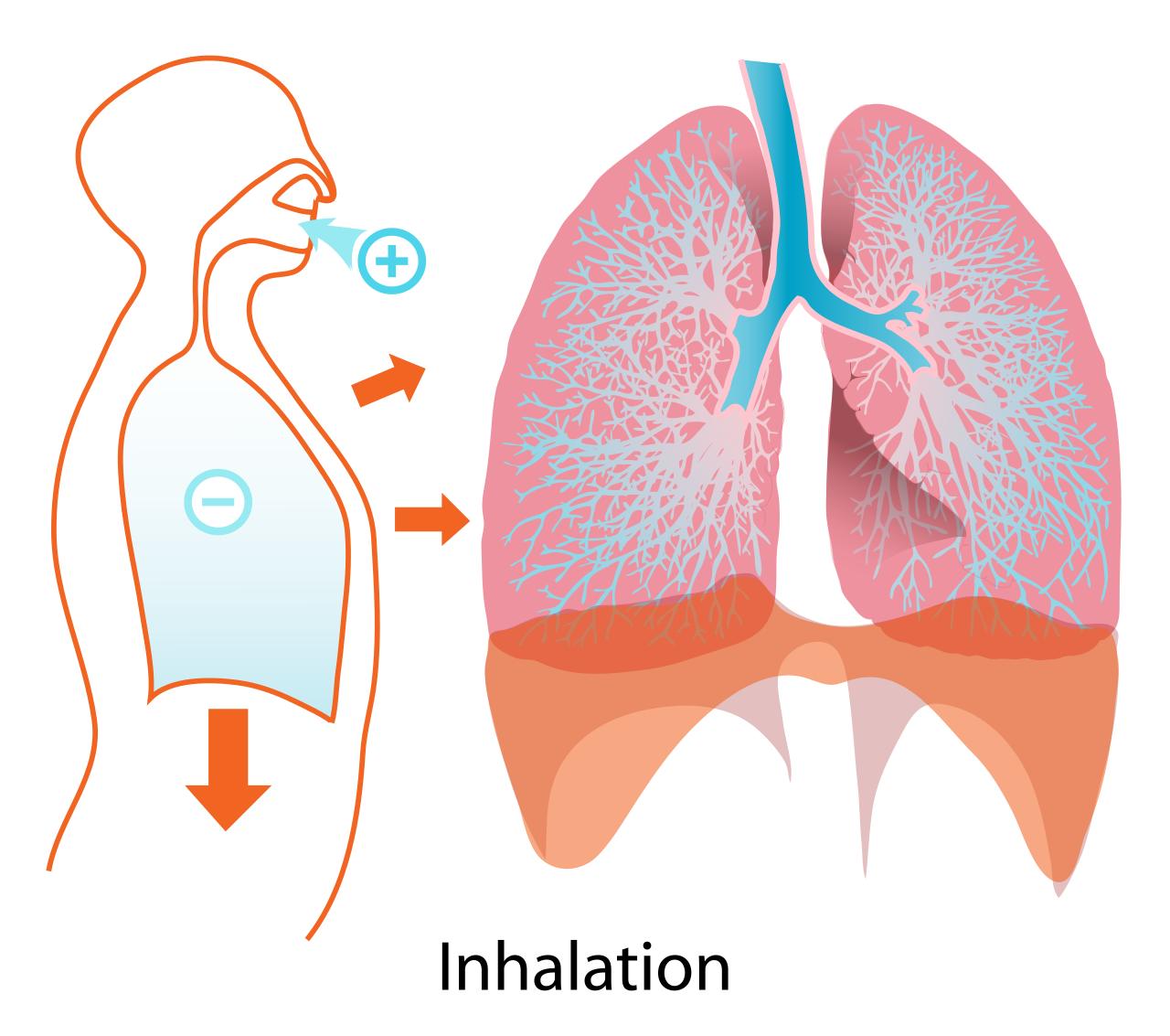
The active contraction of the diaphragm muscle initiates pulmonary inhalation. During inhalation, the contracting diaphragm muscle pulls downward toward the abdominal cavity.
The downward movement of the contracting diaphragm physically increases the total volume of the thoracic cavity. According to the laws of physics, an expansion in thoracic cavity volume causes a proportional decrease in internal pulmonary pressure relative to the outside atmosphere. Atmospheric air flows inward into the lungs whenever atmospheric pressure exceeds the internal pressure of the thoracic cavity.
Exhalation (Passive)
During passive normal exhalation, the diaphragm and external intercostal (rib) muscles relax simultaneously. The relaxation of respiratory muscles decreases overall thoracic volume to force residual air out of the lungs. The internal pressure briefly rises above atmospheric pressure, pushing the carbon-dioxide-rich air back up the bronchial tree, out the trachea, and out of the body, completing the cycle.