Skeletal and Muscular Systems
Not sure you’re ready?
Take the ~3-minute readiness diagnostic and see where you stand.
The human body is a marvel of biomechanical engineering, functioning much like a dynamic, high-rise hospital building. The structural steel and concrete—the skeletal system—do far more than prevent gravity from crushing the fragile internal machinery; they actively manufacture the very cells that keep the system alive and stockpile the mineral currency required to keep the lights on. Interlocking with this physical scaffolding is an intricate array of biological motors—the muscular system—that translates chemical energy and electrical impulses into precise kinetic force. For the future healthcare professional, mastering the mechanics of bones, joints, and muscles is not merely an exercise in anatomical memorization. When you are transferring a bedbound patient, assessing a rapid, irregular heartbeat, or explaining why a frail patient's fracture is healing poorly, you are directly interacting with the sliding filaments, cellular matrices, and anatomical levers defined within these biological systems.
It is easy to view bones as inert, lifeless calcified sticks. In reality, bone is a highly vascular, intensely active organ.
Core Functions of the Skeletal System
At its most fundamental level, the skeletal system provides structural physical support for the human body and structurally protects internal human organs from physical damage (such as the ribs shielding the lungs, or the skull housing the brain). By partnering with the muscular system, bones act as physical levers to facilitate body movement.
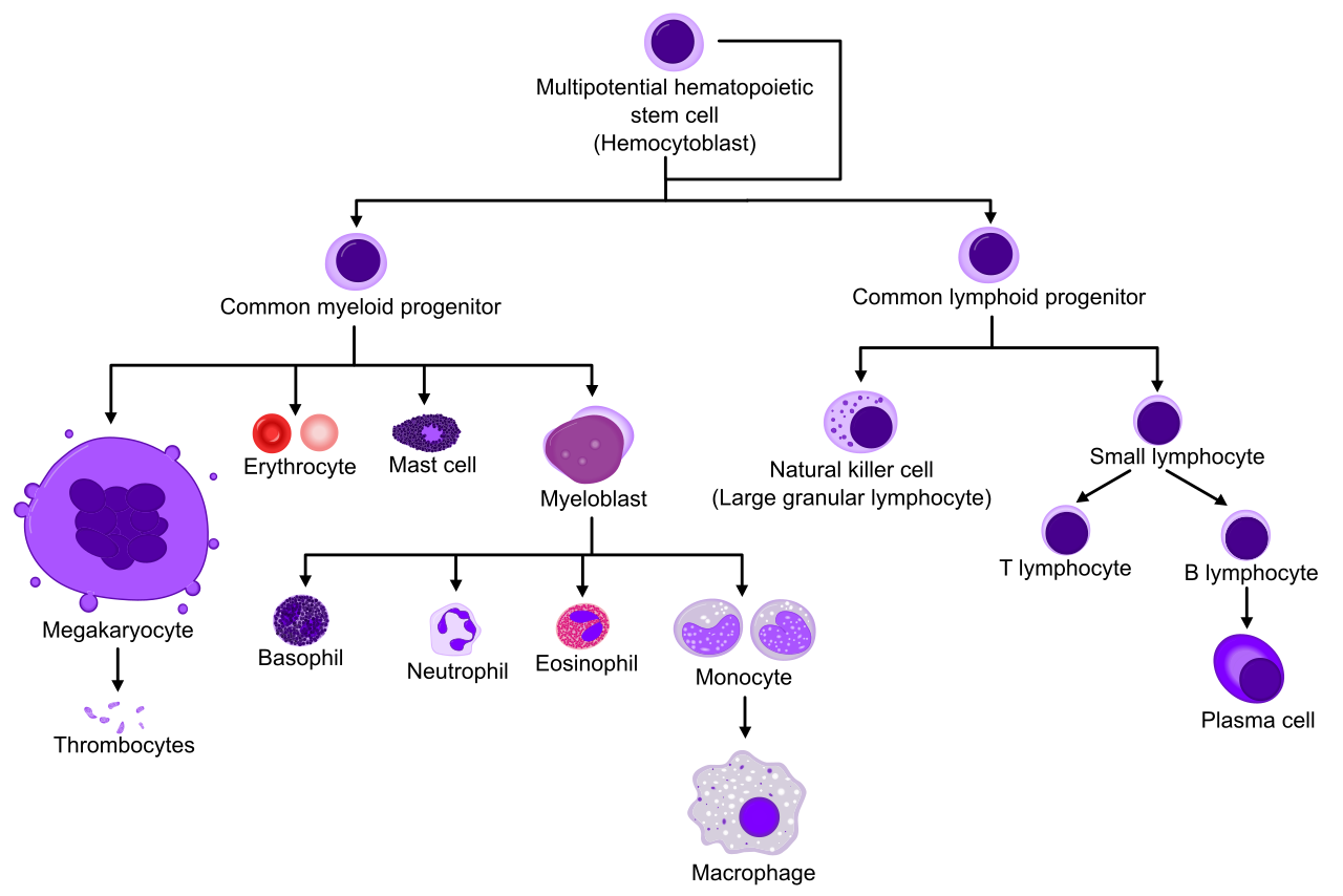
Beyond biomechanics, bones serve critical metabolic roles. Bones chemically store essential minerals such as calcium and phosphorus, releasing them into the bloodstream to maintain homeostatic balance. Furthermore, bones are the factories of the blood. Hematopoiesis is the biological process of blood cell formation, and hematopoiesis occurs specifically within the red bone marrow of certain bones. As we age, some marrow changes function; yellow bone marrow primarily stores adipose tissue as an internal energy reserve.
Macroscopic and Microscopic Anatomy
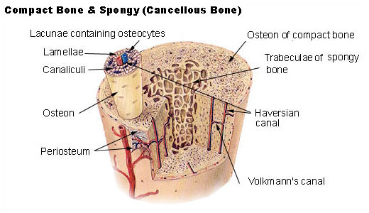
Consider the architecture of a bone from the outside in. The periosteum is a dense fibrous connective tissue membrane covering the outer surface of bones, serving as the critical anchor point for nerves, blood vessels, and tendons.
Beneath this lies the hard shell. Compact bone forms the dense, solid outer layer of all human bones. If you look at compact bone under a microscope, it looks like a cross-section of tightly packed tree trunks. The osteon is the basic microscopic cylindrical unit of compact bone.
Deeper inside, the architecture changes to save weight without sacrificing strength. Spongy bone contains a highly porous internal network of structural units called trabeculae. Spongy bone is commonly found within the interior regions of human flat bones, and spongy bone is typically located at the anatomical ends of human long bones.
In a long bone, these macroscopic regions have specific names:
- The diaphysis is the main central tubular shaft of a human long bone (primarily compact bone).
- The epiphysis is the expanded rounded end section of a human long bone (primarily spongy bone housing red marrow).
The Cellular Workforce
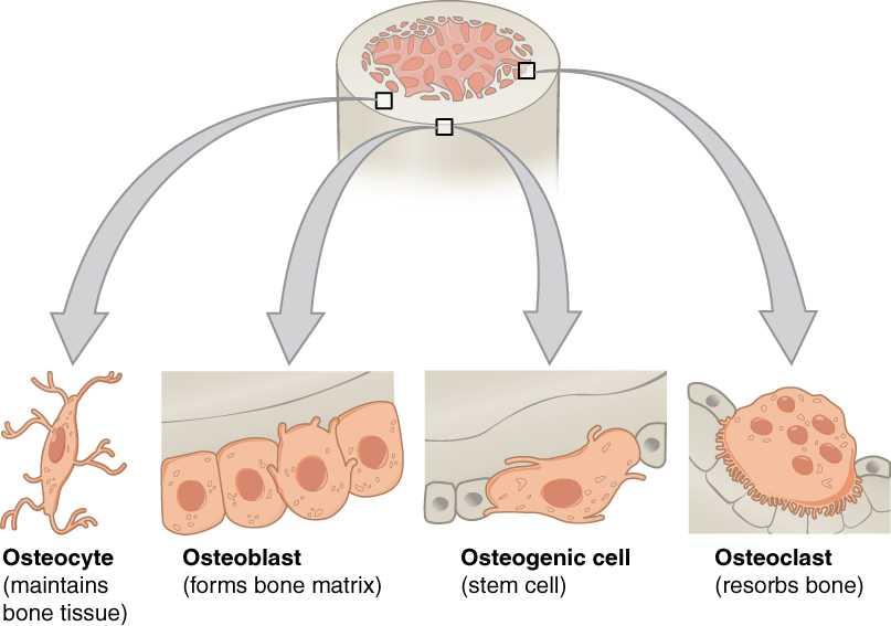
Bone tissue is constantly being demolished and rebuilt in response to physical stress and metabolic needs. Three primary cells manage this:
- Osteoblasts are specialized bone-forming cells responsible for synthesizing new bone matrix.
- Osteoclasts are large multinucleated cells responsible for breaking down bone tissue during bone resorption, liberating stored calcium back into the blood.
- Osteocytes are mature bone cells permanently embedded within the mineralized bone matrix, acting as stress sensors.
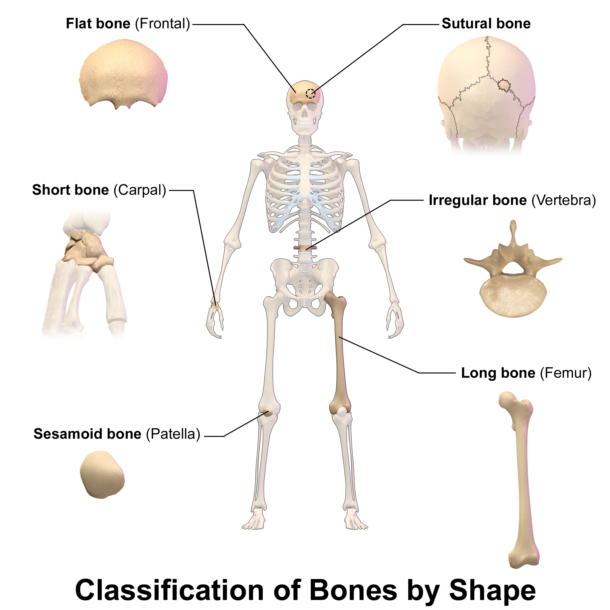
Form dictates function. Human bones are anatomically categorized by their unique shapes, which perfectly match their mechanical roles.
- Long bones possess an anatomical structure that is significantly longer than it is wide. The human femur (thigh bone) is an anatomical example of a long bone, designed to handle immense compressive weight and act as a large lever.
- Short bones feature a roughly cube-shaped physical anatomical structure. The carpal bones located in the human wrist are anatomical examples of short bones, allowing for intricate, multi-directional gliding movements.
- Flat bones feature a thin, flattened, and often slightly curved physical structure. The human sternum (breastbone) is an anatomical example of a flat bone, serving as a broad shield for the heart.
- Irregular bones possess complex shapes that prevent classification in other standard structural categories. Human vertebrae are anatomical examples of irregular bones, intricately shaped to protect the spinal cord while allowing flexibility.
- Sesamoid bones are small independent bones fully embedded within a muscle tendon. The human patella (kneecap) is an anatomical example of a sesamoid bone; it acts as a specialized fulcrum to improve the leverage of the thigh muscles.
Joints (Articulations)
Where two bones meet, we have a joint. Joints are functionally classified by how much movement they allow.
- Synarthroses are classified as joints that permit zero physical movement. For example, the cranial fibrous sutures of the human skull are functionally classified as synarthroses.
- Amphiarthroses are classified as joints that permit a slight degree of physical movement. The pubic symphysis located in the human pelvis is functionally classified as an amphiarthrosis, stretching just enough during childbirth to allow passage.
- Diarthroses are classified as joints that are freely movable in multiple directions. All human synovial joints fall under the functional classification of diarthroses (like the knee, shoulder, and elbow).
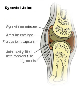
The Synovial Joint Apparatus
Because diarthroses move so freely, they require specialized hardware to prevent the bones from grinding themselves to dust:
- Articular cartilage completely covers the ends of articulating bones within a synovial joint to reduce friction.
- Synovial fluid chemically and physically lubricates the internal joint cavity of a synovial joint, acting much like motor oil.
- Bursae are enclosed fluid-filled sacs that reduce physical friction between moving tissues near functional joints.
- Ligaments are rigid bands of dense connective tissue physically connecting one bone to another bone (stabilizing the joint).
- Tendons are thick bands of dense fibrous connective tissue physically connecting a muscle to a bone (transferring the pulling force of the muscle).
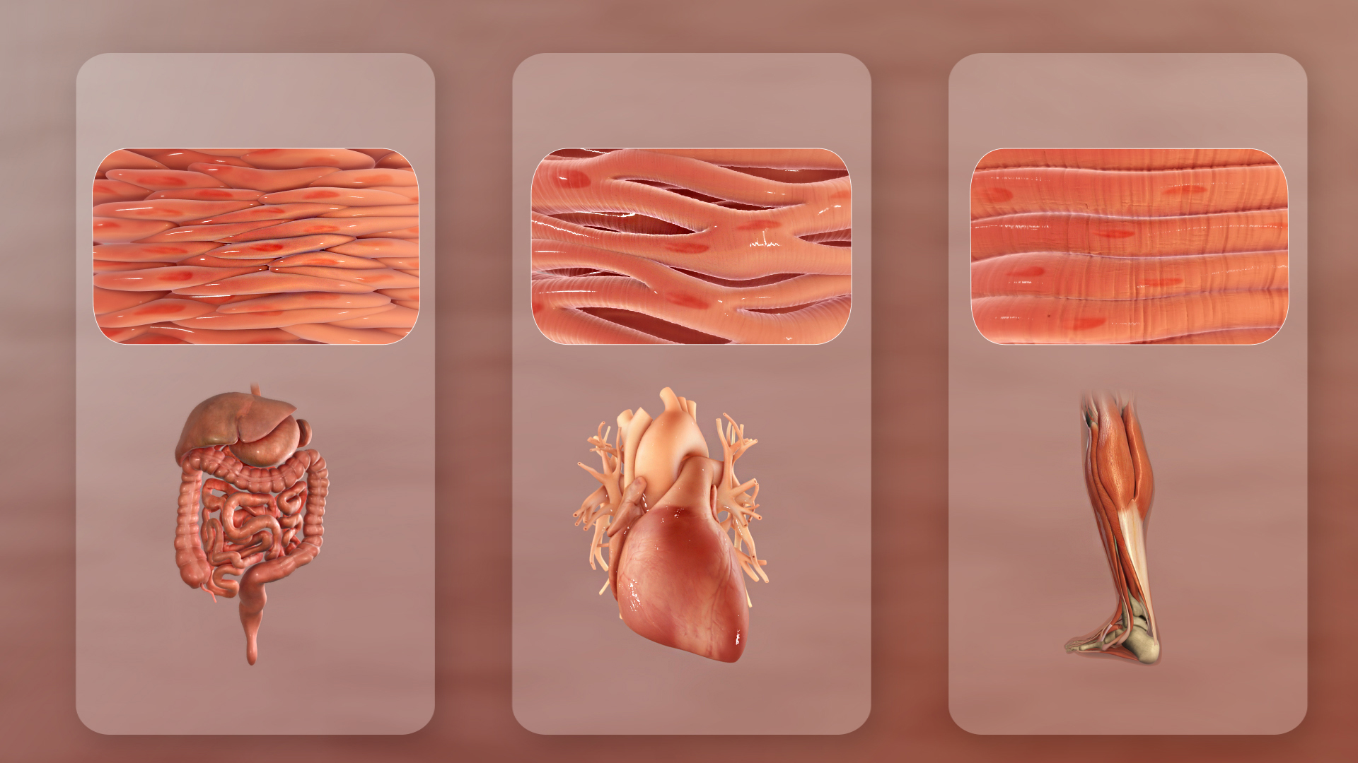
Not all muscle tissue is the same. The body utilizes three distinct types of muscle tissue to accomplish different physiological tasks. Understanding these differences is critical for pharmacology and pathology.
| Feature | Skeletal Muscle | Cardiac Muscle | Smooth Muscle |
|---|---|---|---|
| Location | Attached to bones via tendons. | Exclusively located within the muscular wall of the human heart. | Physically lines the interior functional walls of hollow human organs like the intestines, and is located within the structural walls of human blood vessels. |
| Control | Subject to direct conscious control by the somatic nervous system. Anatomically classified as voluntary muscle. | Functions entirely independently of conscious nervous system control. Anatomically classified as involuntary muscle. | Functions entirely independently of conscious human control. Anatomically classified as involuntary muscle. |
| Microscopic Appearance | Consistently exhibits a highly striated (striped) appearance under a light microscope. | Possesses a clearly striated visual appearance under microscopic examination. | Completely lacks any microscopic physical striations. |
| Cellular Structure | Individual skeletal muscle cells contain multiple cell nuclei situated near the cell periphery. | Individual cardiac muscle cells typically contain a single centrally located nucleus. | Individual smooth muscle cells are entirely spindle-shaped and contain a single central nucleus. |
| Unique Features | Capable of rapid, forceful contractions but fatigues easily. | Intercalated discs are specialized physical junctions directly connecting individual cardiac muscle cells, allowing electrical signals to pass instantly across the heart. | Sustains long, slow contractions (peristalsis) without fatiguing. |
How does an electrical thought in your brain become the physical reality of picking up a cup of coffee? The answer lies in microscopic protein filaments dragging themselves across one another.
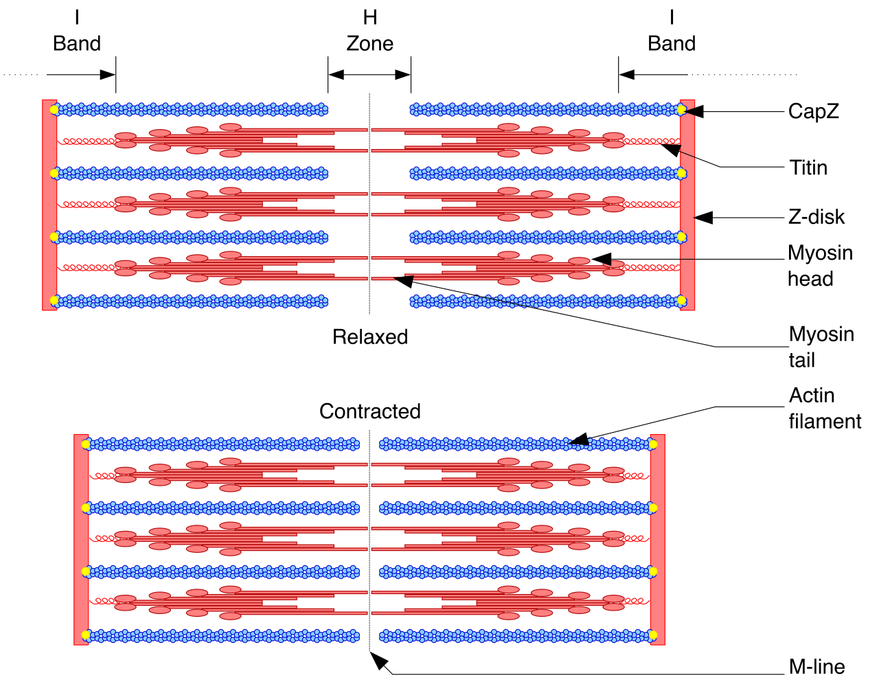
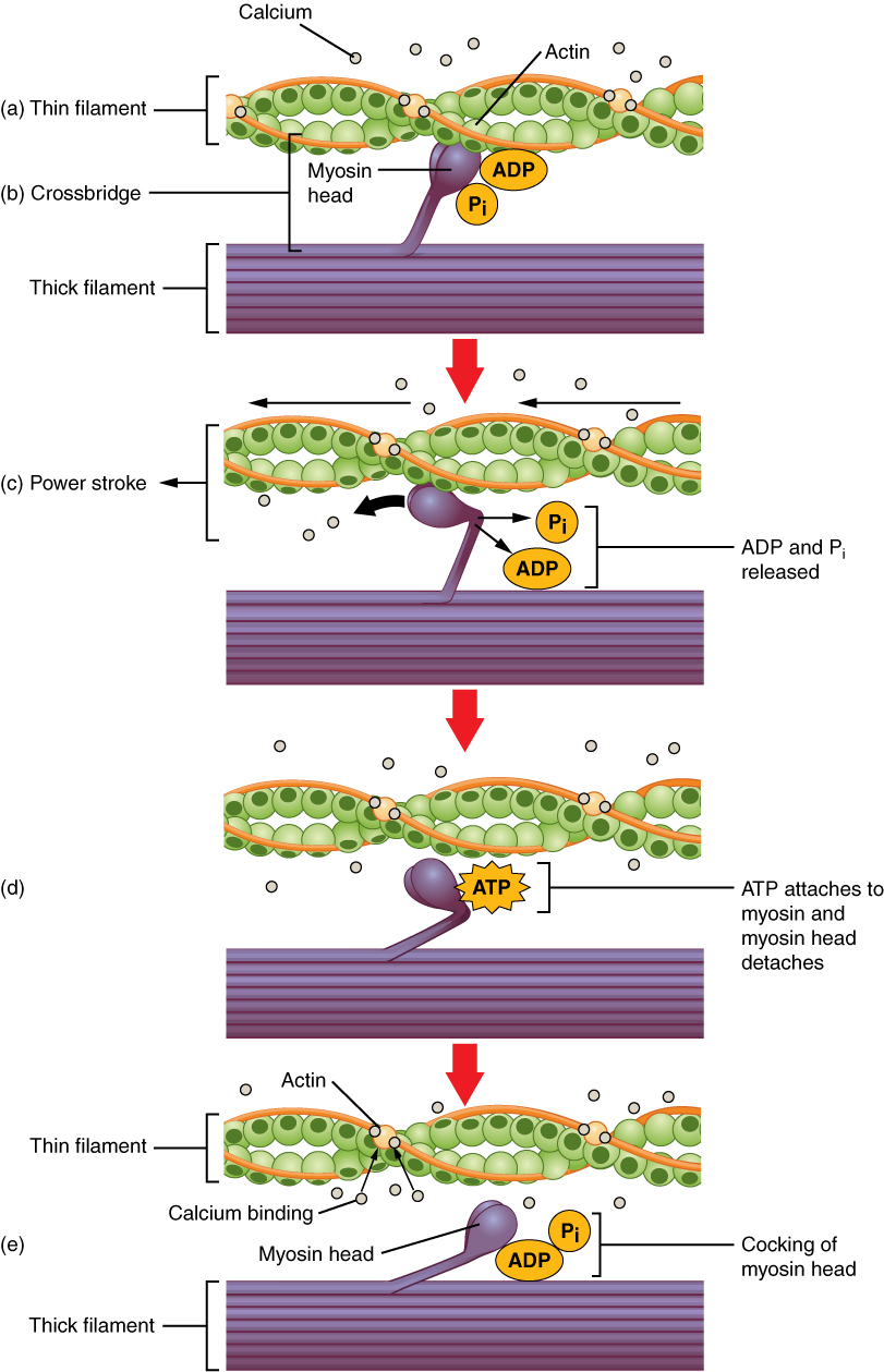
The Sliding Filament Theory describes muscle contraction as the physical sliding of actin filaments directly past myosin filaments.
To grasp this, we must zoom in on a muscle cell. Inside the cell are long protein cords. Actin protein molecules aggregate to form the thin filaments found within functional muscle fibers. Myosin protein molecules aggregate to form the thick filaments found within functional muscle fibers. These organized filaments are packed into repetitive units. The sarcomere serves as the fundamental contractile physical unit of a single striated muscle fiber.
The Contraction Cycle: Step-by-Step
- The Signal: The nervous system sends a signal to the muscle. The neuromuscular junction is the specialized chemical synapse connecting a motor neuron directly to a muscle fiber.
- The Trigger: Acetylcholine acts as the primary chemical neurotransmitter released at the human neuromuscular junction. Acetylcholine release at the neuromuscular junction chemically triggers the initial start of a muscle contraction.
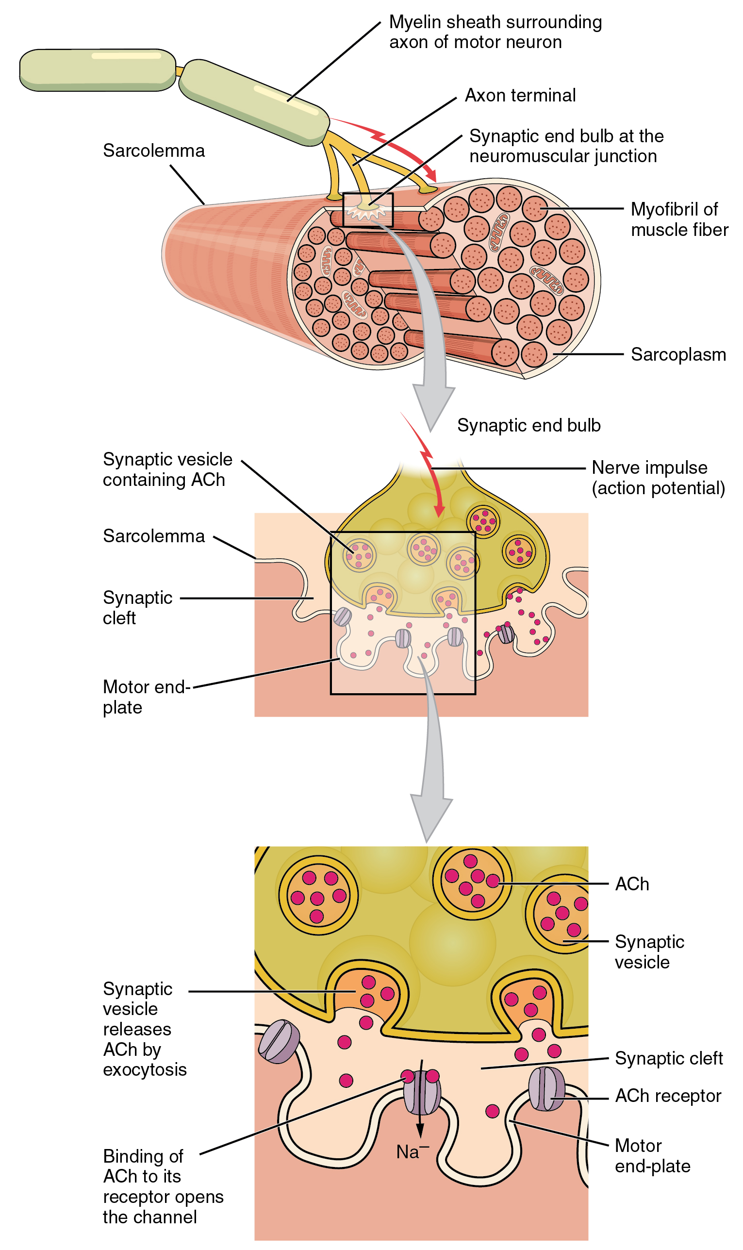
- Calcium Release: The acetylcholine sparks an electrical charge. An action potential traveling down a transverse tubule forces the sarcoplasmic reticulum to release internal calcium ions.
- Unlocking the Binding Sites: Calcium ions initiate the muscle contraction cycle by chemically binding to specific troponin molecules on the actin filament. The chemical binding of calcium to troponin physically forces the attached tropomyosin protein to shift its position.
- The Cross-Bridge: The physical shifting of tropomyosin mechanically exposes specific myosin-binding active sites on the actin filament. A chemical cross-bridge successfully forms when a single myosin head firmly binds to an exposed active site on an actin filament.
- The Power Stroke: The mechanical power stroke directly occurs when a bound myosin head pivots to pull the actin filament toward the geometric center of the sarcomere. This microscopic pull, multiplied across millions of sarcomeres, physically shortens the entire muscle.
- Detachment and Reset: Adenosine triphosphate (ATP) provides the crucial cellular energy required for the bound myosin head to physically detach from the actin filament. Clinical note: When a person dies, ATP production stops. Because ATP is required to detach the myosin heads, the muscles become chemically locked in a contracted state—a phenomenon known as rigor mortis.
Nurses and allied-health professionals must instantly identify major muscle groups to administer intramuscular injections, assess stroke deficits, and guide physical rehabilitation. Muscles generally work in opposing pairs: flexors decrease the angle of a joint, while extensors increase it. Abductors move limbs away from the midline; adductors pull them back.
Head and Neck
- Masseter: The masseter muscle elevates the human mandible to mechanically close the jaw during the chewing process. It is one of the strongest muscles in the body relative to its size.
- Sternocleidomastoid (SCM): The sternocleidomastoid muscle facilitates the physical rotation and forward flexion of the human head.
The Torso
- Pectoralis Major: The pectoralis major is a massive superficial muscle situated completely on the anterior human chest. The pectoralis major muscle continually generates the physical force needed to adduct and flex the human arm.
- Latissimus Dorsi: The latissimus dorsi is a massive broad muscle situated completely on the human back. The latissimus dorsi muscle generates the physical force needed to completely extend and adduct the arm at the shoulder joint.
- Rectus Abdominis: The rectus abdominis is a paired vertical muscle located directly on the anterior wall of the human abdomen (commonly known as the "six-pack").
The Upper Extremities
- Deltoid: The deltoid muscle anatomically covers the entire exterior structure of the human shoulder joint. The deltoid muscle primarily acts as the main mechanical abductor of the human arm (and is a prime site for intramuscular vaccinations).
- Biceps Brachii: The biceps brachii muscle functions as the primary mechanical flexor of the human forearm at the elbow joint.
- Triceps Brachii: Operating opposite the biceps, the triceps brachii muscle functions as the primary mechanical extensor of the human forearm at the elbow joint.
The Lower Extremities
- Gluteus Maximus: The gluteus maximus is structurally the largest individual muscle located in the human buttocks. The gluteus maximus muscle primarily acts as the major mechanical extensor of the human hip joint, vital for climbing stairs or standing up from a chair.
- Quadriceps Femoris: The quadriceps femoris is a specific functional group of four distinct muscles located on the anterior human thigh. Working together, the quadriceps femoris muscle group mechanically extends the lower human leg directly at the knee joint.
- Hamstring Group: Antagonistic to the quadriceps, the hamstring group consists of three anatomically distinct muscles located completely on the posterior human thigh. The hamstring muscle group mechanically flexes the lower human leg directly at the knee joint.
- Gastrocnemius: The gastrocnemius is the largest major superficial muscle situated on the posterior human calf. The gastrocnemius muscle directly facilitates plantar flexion of the human foot at the ankle joint (the action of pointing your toes downward or standing on your tiptoes).