Digestive and Urinary Systems
Not sure you’re ready?
Take the ~3-minute readiness diagnostic and see where you stand.
The human body operates as an exquisite thermodynamic machine, demanding a continuous supply of chemical energy and a relentless, highly calibrated mechanism for waste removal. To maintain homeostasis, the body relies on two massive physiological operations: the extraction of nutrients from the external environment and the meticulous filtration of the blood to remove metabolic byproducts. The digestive system dismantles raw, complex organic matter into usable molecular building blocks, while the urinary system acts as the ultimate chemical arbiter, deciding exactly what remains in the bloodstream and what is expelled. For any clinician, understanding this interface is not merely academic; it is the foundation of patient care. When a patient presents with a soaring potassium level, severe dehydration, or an inability to absorb a crucial vitamin, the answers always trace back to the mechanics of these two systems.
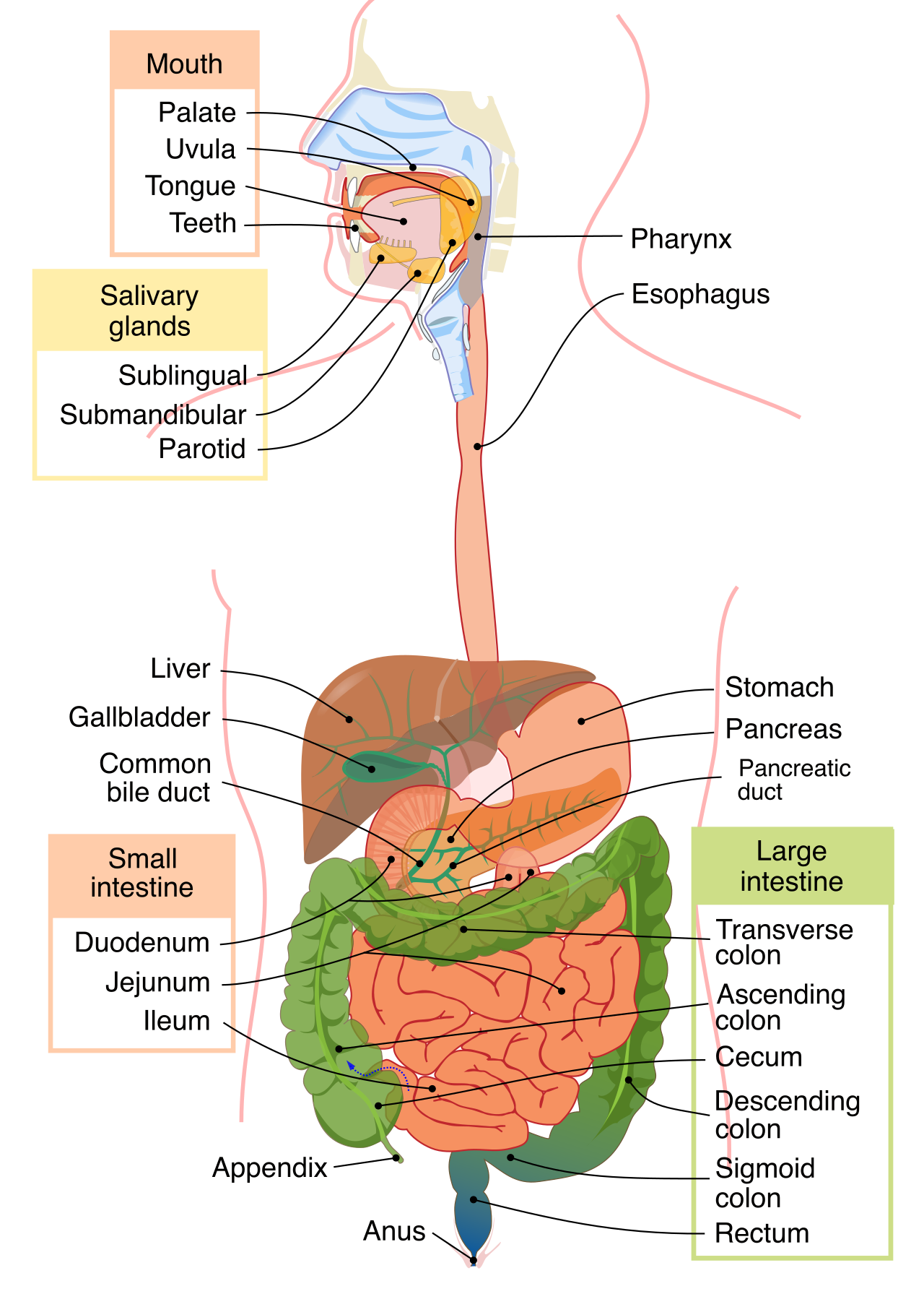
To understand digestion is to understand a biological disassembly line. The gastrointestinal tract is a continuous tube extending from the mouth to the anus. Everything inside this tube is technically "outside" the body's internal environment until it crosses the cellular barrier of the intestinal lining.
The Upper Tract: Mechanical Breakdown and Initial Chemistry
The disassembly process begins immediately in the oral cavity, where mechanical digestion begins in the mouth through the physical breakdown of food by chewing. But mechanical breakdown is only half the battle. As you chew, salivary glands secrete saliva into the oral cavity.
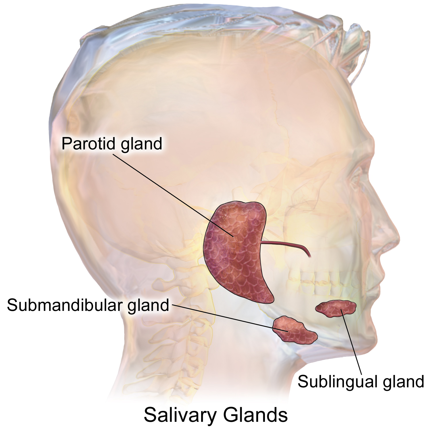
Clinical Insight: Saliva contains the digestive enzyme salivary amylase. Because of this, salivary amylase begins the chemical digestion of carbohydrates in the mouth. If you chew a piece of plain bread long enough, it begins to taste sweet. This is the amylase actively cleaving complex starches into simple sugars.
Once chewed, the food must navigate the throat. The pharynx serves as a common passageway for both food and air. To ensure the food travels down the digestive tract and not into the respiratory system, a crucial mechanical trapdoor engages: the epiglottis folds over the glottis to prevent food from entering the trachea during swallowing.
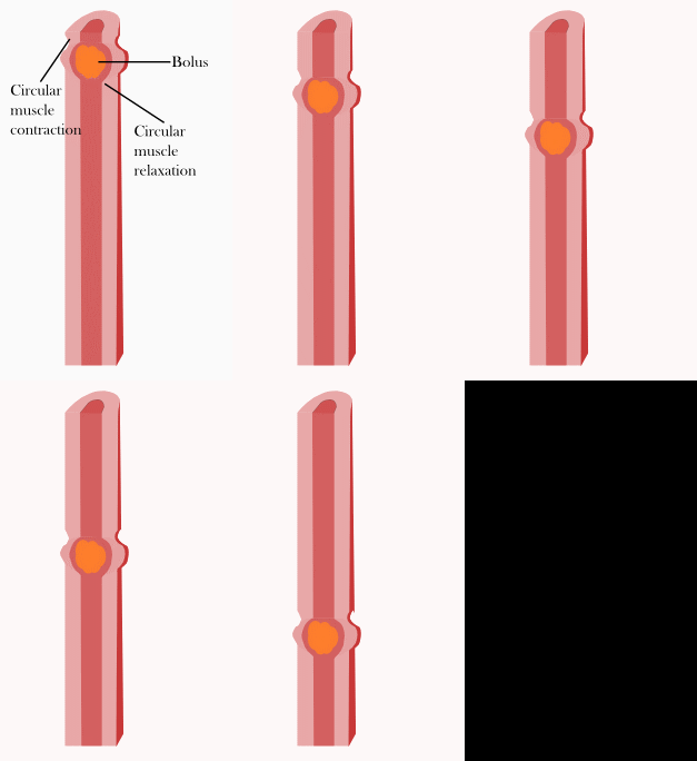
The food bolus then enters the esophagus. The esophagus is a muscular tube connecting the pharynx to the stomach. It does not rely on gravity to move food downward. Instead, peristalsis consists of rhythmic, wave-like muscular contractions propelling food through the digestive tract. As the food reaches the end of the esophagus, it passes through a muscular valve. The lower esophageal sphincter prevents acidic stomach contents from backing up into the esophagus. When this sphincter fails, patients experience gastroesophageal reflux disease (GERD).
The Stomach: The Chemical Cauldron
The stomach is essentially an acidic holding tank. Functionally, the stomach temporarily stores ingested food. To accommodate a massive influx of volume, folds in the inner lining of the stomach wall are called rugae. These rugae allow the stomach to stretch and expand significantly after a large meal.
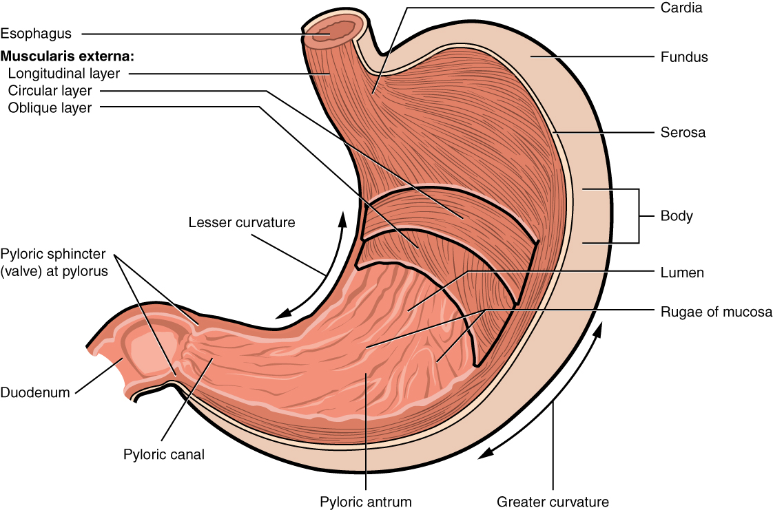
Inside, the stomach actively mixes food with gastric juices to form a semi-fluid mass called chyme. This mixing requires a potent chemical environment, managed by two highly specialized types of cells in the stomach lining:
- Parietal Cells: Parietal cells in the stomach lining secrete hydrochloric acid. This acid is so strong that it drops the gastric pH to nearly 2.0. Hydrochloric acid in the stomach creates a highly acidic environment to destroy ingested pathogens. Additionally, parietal cells in the stomach secrete a glycoprotein called intrinsic factor. This is critically important for clinicians: intrinsic factor is required for the intestinal absorption of vitamin B12. Patients who undergo gastric bypass or lose parietal cell function cannot absorb B12 and will develop pernicious anemia.
- Chief Cells: Chief cells in the stomach lining secrete pepsinogen. Pepsinogen is an inactive enzyme (a zymogen). Why inactive? Because if the cells synthesized active protein-destroying enzymes, they would digest themselves. Instead, they release the safe precursor. Once in the stomach lumen, hydrochloric acid in the stomach converts the inactive precursor pepsinogen into the active enzyme pepsin. Once activated, pepsin initiates the chemical digestion of dietary proteins within the stomach.
The Small Intestine & Accessory Organs: The Hub of Absorption
The stomach simply preps the meal. The small intestine is the primary site of nutrient digestion and absorption in the human body.
Anatomically, the small intestine is anatomically divided into the duodenum, jejunum, and ileum.
- The duodenum is the first and shortest section of the small intestine.
- The jejunum is the middle section of the small intestine.
- The ileum is the final and longest section of the small intestine.
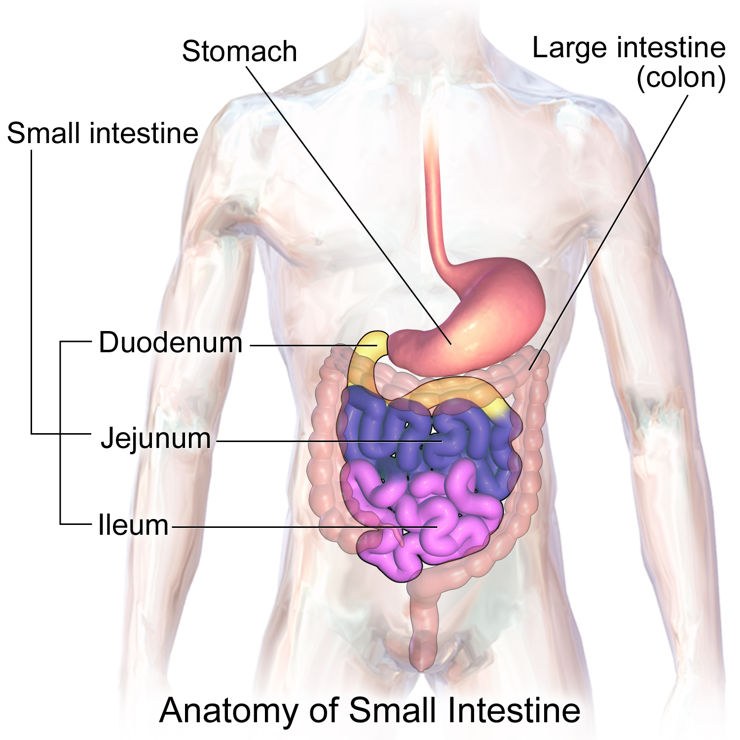
The real chemical magic happens in the short, crucial duodenum. The duodenum receives acidic chyme directly from the stomach. Because this chyme is highly acidic, it would burn the intestinal lining if not immediately neutralized. Therefore, the duodenum relies heavily on the accessory digestive organs.
The Pancreas
The pancreas functions as both an endocrine gland and an exocrine gland. While its endocrine function regulates blood sugar (insulin), its digestive role relies on exocrine tissue. Exocrine cells of the pancreas secrete pancreatic juice into the duodenum. Pancreatic juice contains a high concentration of bicarbonate ions. These basic bicarbonate ions from the pancreas neutralize the highly acidic chyme entering the duodenum.
Simultaneously, the pancreas delivers a full suite of digestive enzymes:
- The pancreas secretes the enzyme pancreatic amylase to digest complex carbohydrates.
- The pancreas secretes the enzyme pancreatic lipase to digest triglycerides into fatty acids and glycerol.
- The pancreas secretes protease enzymes like trypsin and chymotrypsin to digest proteins.
The Liver and Gallbladder
The duodenum receives bile from the gallbladder and liver. The liver continuously produces a greenish-yellow fluid called bile. This bile is essential for handling dietary fats, which do not mix with the watery environment of the gut. Bile emulsifies large dietary fat globules into smaller droplets to increase the surface area for fat-digesting enzymes.
When we aren't eating, the gallbladder—a small muscular pouch located on the inferior surface of the liver—stores excess bile produced by the liver. To save space, the gallbladder concentrates stored bile by actively removing water. Later, when you eat a rich meal, the gallbladder contracts and releases bile into the duodenum upon the arrival of fatty foods.
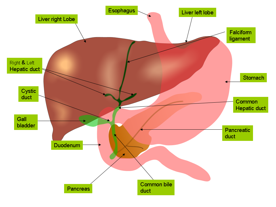
Beyond Digestion: The liver is the body's primary biochemical plant. Beyond producing bile, the liver stores excess systemic glucose in the form of glycogen. It also acts as a security checkpoint: the liver detoxifies blood by chemically altering and removing potentially harmful substances.
Absorbing the Nutrients
With carbohydrates, fats, and proteins fully broken down, absorption begins. The inner wall of the small intestine is engineered purely to maximize surface area. Villi are tiny, finger-like projections lining the mucosal wall of the small intestine. Under a microscope, the cells making up the villi have their own projections: microvilli are microscopic cellular membrane projections on the epithelial cells of the small intestine. Together, intestinal villi and microvilli significantly increase the internal surface area to maximize nutrient absorption.
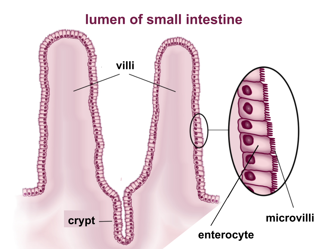
Nutrients are absorbed via two different circulatory pathways in the villi:
- Blood capillaries within the intestinal villi absorb amino acids and monosaccharides (sugars and proteins go straight to the bloodstream).
- Lymphatic vessels called lacteals within the intestinal villi absorb dietary fats (fats are too large for capillaries and must travel through the lymphatic system first).
The Large Intestine: Water Reclamation
By the time the remains of a meal exit the ileum, almost all usable nutrients have been extracted. What is left is mostly water and indigestible fiber. The large intestine primarily absorbs water and electrolytes from indigestible food matter.
Anatomically, the large intestine includes the cecum, colon, rectum, and anal canal. The colon is subdivided into the ascending, transverse, descending, and sigmoid sections.
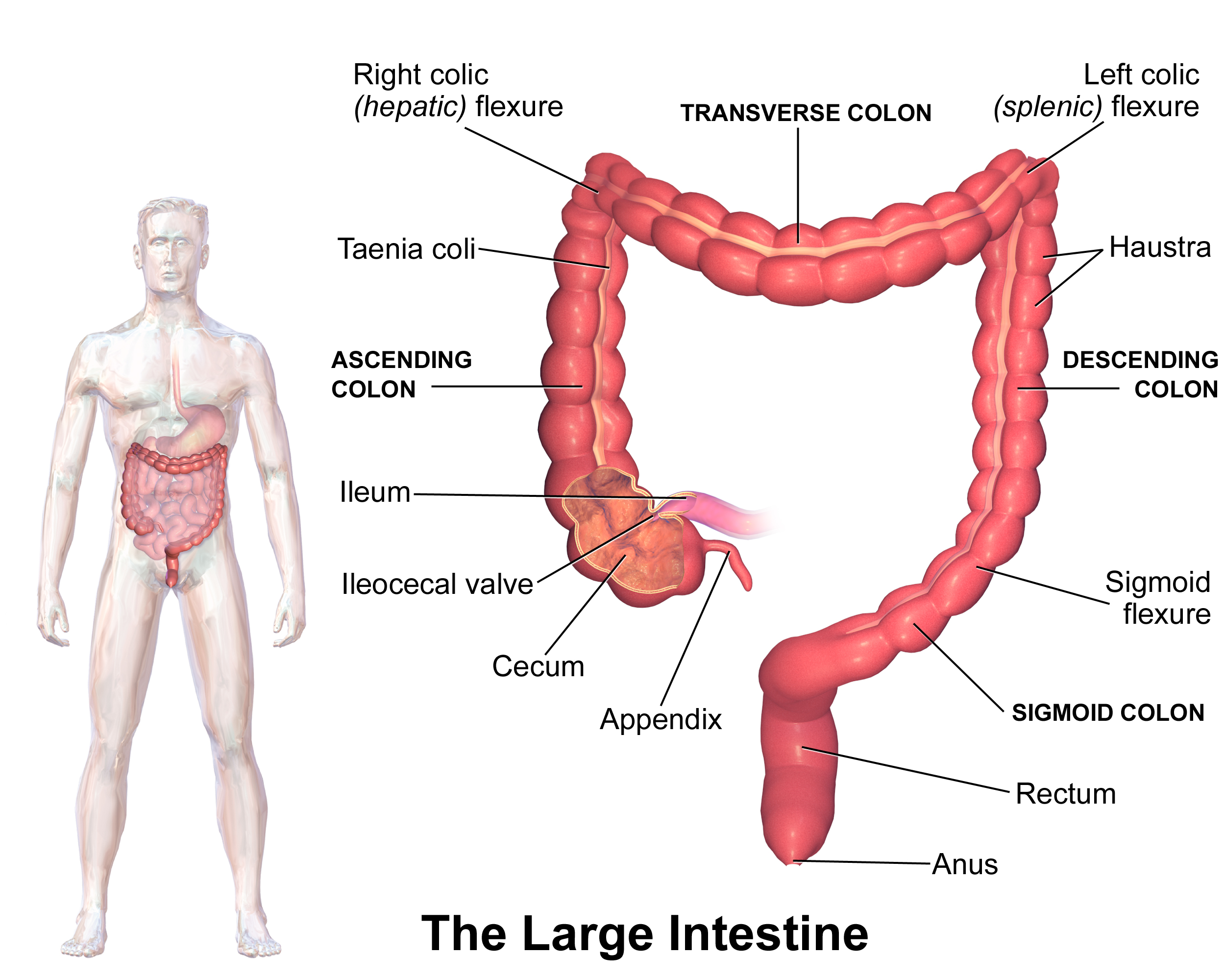
The large intestine is not sterile; it hosts a massive microbiome. Normal bacterial flora residing in the large intestine synthesize vitamin K, which is vital for blood clotting. Finally, the rectum stores solid feces prior to elimination from the body.
While the digestive system brings raw materials into the blood, the urinary system ensures the blood's chemical composition remains perfectly balanced. The urinary system consists of two kidneys, two ureters, one urinary bladder, and one urethra.
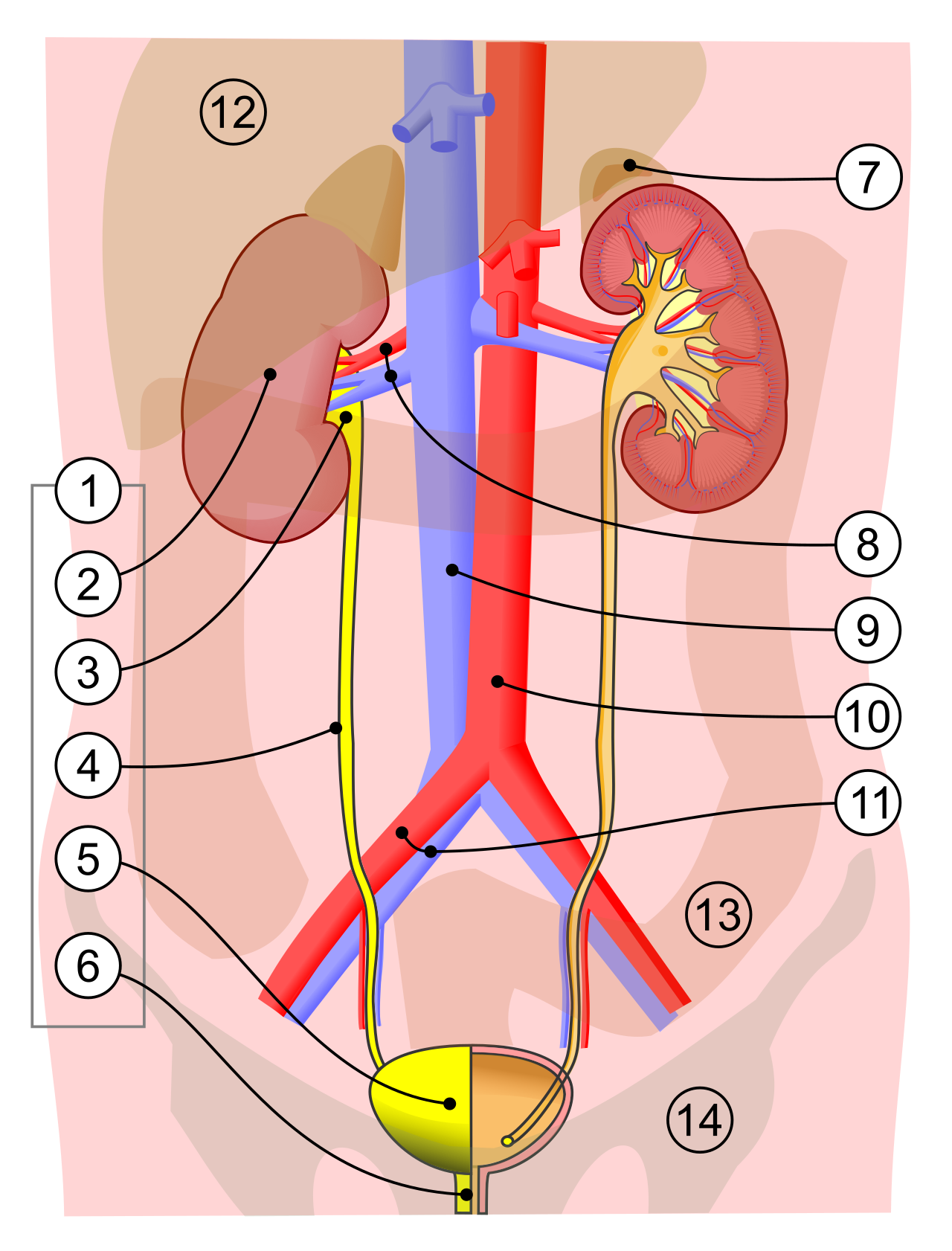
Macro-Anatomy of the Plumbing
The primary function of the kidneys is to filter blood and excrete metabolic wastes in the form of urine.
If you slice a kidney in half, you will see distinct zones. The renal cortex is the lighter, outermost layer of the kidney. Deeper inside, the renal medulla is the darker, inner portion of the kidney. The renal medulla contains striated, cone-shaped structures called renal pyramids.
As urine is formed in these tissues, it drains toward the center. The renal pelvis is a funnel-like dilated anatomical space inside the kidney. The renal pelvis collects newly formed urine and channels the fluid into the ureter.
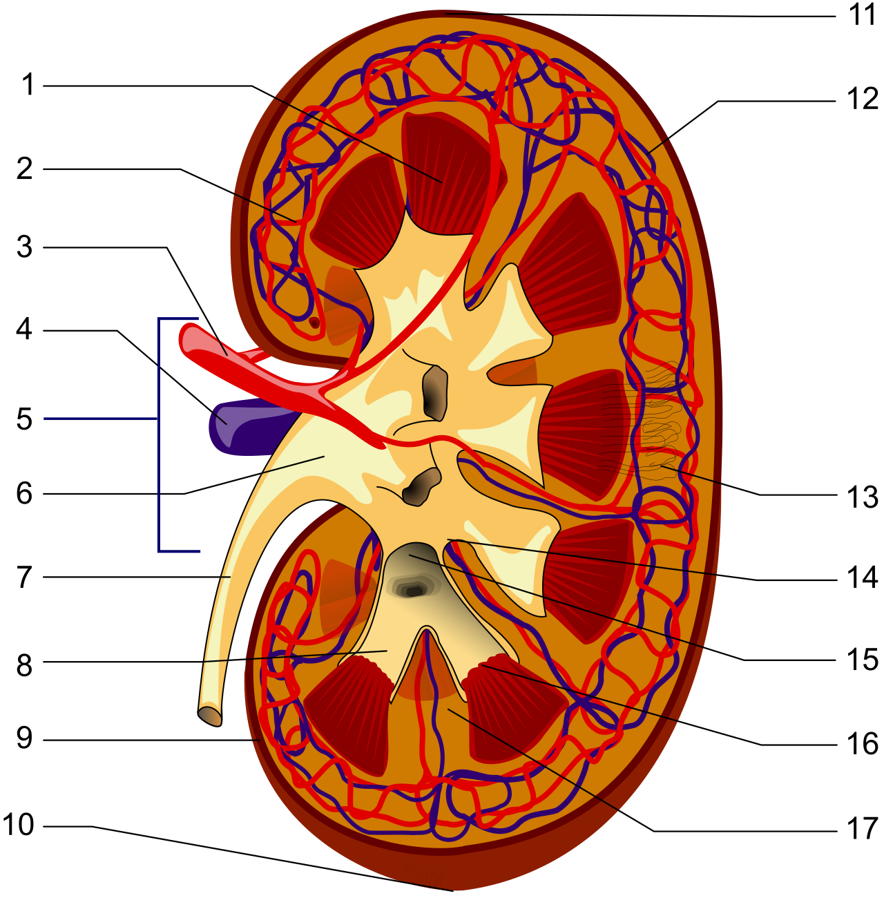
From there, the plumbing takes over: ureters are muscular tubes responsible for transporting urine from the kidneys to the urinary bladder. The urinary bladder is an expandable, hollow muscular organ that stores urine, and finally, the urethra is a thin tube conveying urine from the urinary bladder to the external environment.
The Nephron: The Microscopic Filter
The massive job of blood filtration is carried out by millions of microscopic workers. The nephron is the microscopic structural and functional unit of the kidney. Astonishingly, each human kidney contains approximately one million individual nephrons.
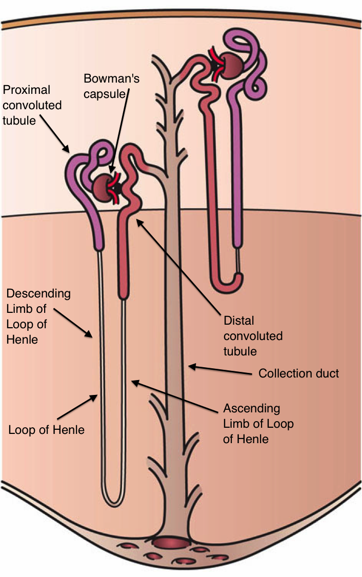
Each nephron features a standard anatomical sequence:
- A renal corpuscle consists of a glomerulus surrounded by a Bowman's capsule.
- The glomerulus is a specialized spherical network of blood capillaries.
- Bowman's capsule is a double-walled cup-like sac enclosing the glomerulus.
- The proximal convoluted tubule is the highly coiled segment of the nephron immediately following Bowman's capsule.
- The Loop of Henle is a U-shaped nephron tubule extending downward into the renal medulla.
- The distal convoluted tubule connects the Loop of Henle to the collecting duct.
- Finally, multiple individual nephrons empty finished urine into a single shared collecting duct.
The Three Steps of Urine Formation
Urine formation involves three sequential processes: glomerular filtration, tubular reabsorption, and tubular secretion.
1. Glomerular Filtration (The Bulk Dump)
Think of glomerular filtration as emptying your entire desk drawer onto the floor before deciding what to keep. Glomerular filtration is a passive process driven by hydrostatic blood pressure pushing water and small solutes out of capillaries.
The initial glomerular filtrate contains water, glucose, amino acids, urea, and various ions. However, the glomerulus is a selective physical filter based on size. Red blood cells and white blood cells are too large to pass through a healthy glomerular filtration membrane. Likewise, large plasma proteins are prevented from entering the glomerular filtrate under normal physiological conditions.
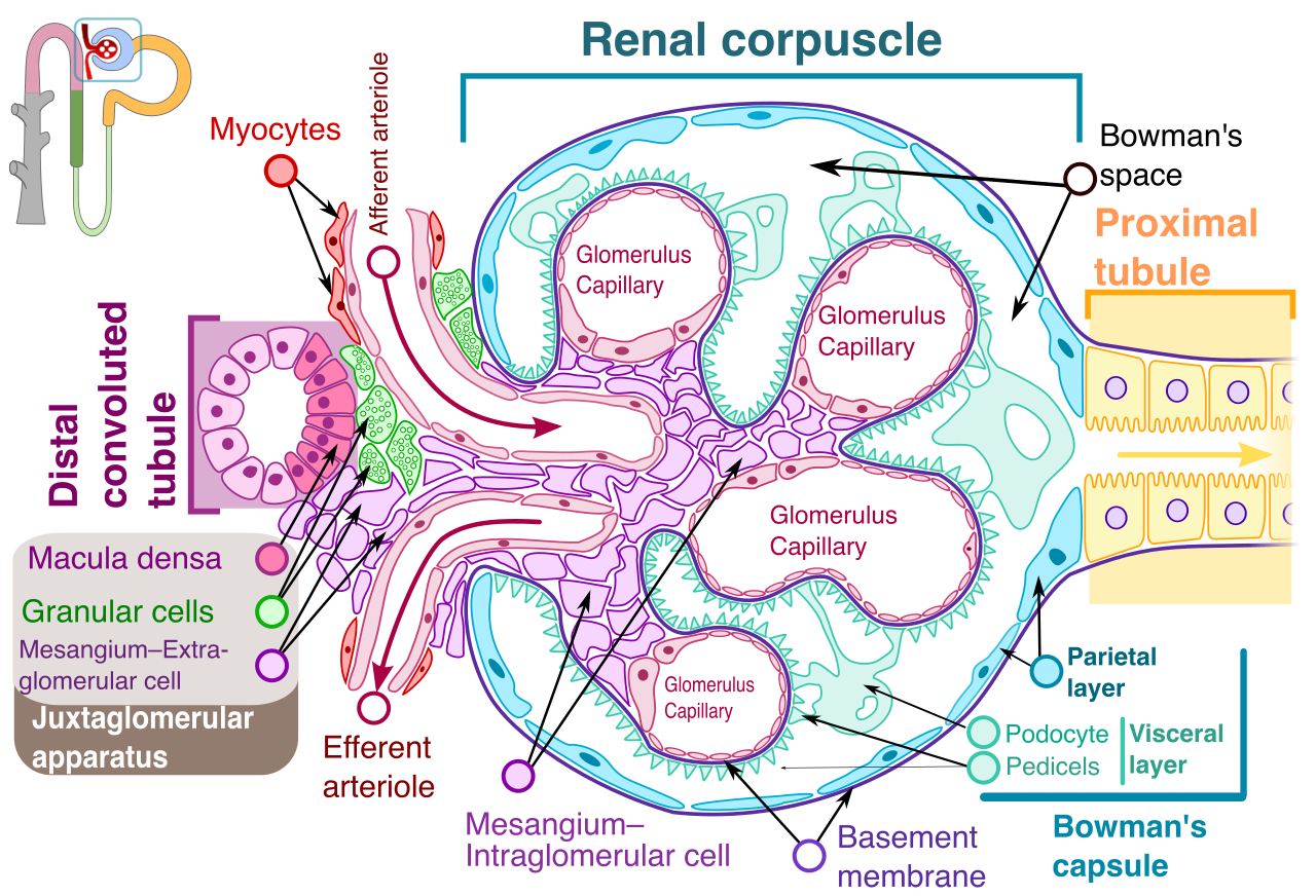
Why This Matters: If you see red blood cells (hematuria) or proteins (proteinuria) in a patient's urine, it immediately tells you that the structural integrity of the glomerulus has failed.
2. Tubular Reabsorption (Keeping the Good Stuff)
Once the "drawer" is dumped into the tubule, the body must quickly grab back the vital nutrients before they are flushed away. Tubular reabsorption selectively returns essential substances from the nephron filtrate back into the systemic bloodstream.
The proximal convoluted tubule is the primary site of tubular reabsorption in the nephron. The efficiency here is staggering:
- Virtually all filtered glucose is reabsorbed back into the blood within the proximal convoluted tubule.
- Virtually all filtered amino acids are reabsorbed back into the blood within the proximal convoluted tubule.
Next, the fluid enters the Loop of Henle, a masterclass in concentration gradients:
- The descending limb of the Loop of Henle is highly permeable to water, allowing water to leave the tubule and return to the blood.
- The ascending limb of the Loop of Henle is highly impermeable to water. Instead, the ascending limb of the Loop of Henle actively transports sodium and chloride ions out of the tubular fluid.
3. Tubular Secretion (Taking Out the Trash)
Filtration isn't perfect. Some wastes must be manually and actively pumped from the blood into the urine. Tubular secretion involves the active transfer of specific materials from peritubular capillaries into the renal tubular lumen.
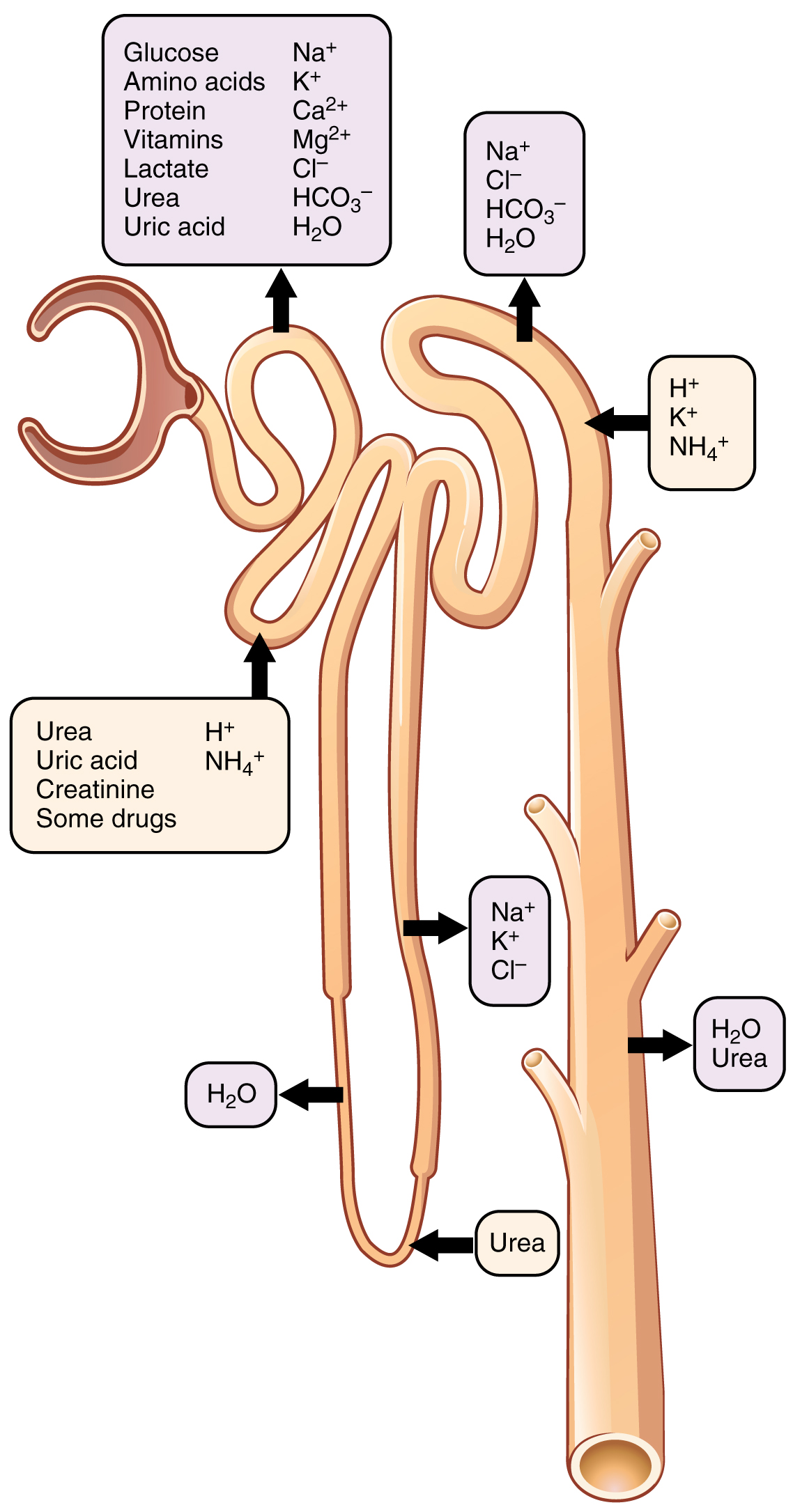
Tubular secretion primarily occurs in the distal convoluted tubule and the collecting duct. Two critical ions are managed here to keep a patient alive:
- Hydrogen ions are actively secreted into the renal tubule to help regulate systemic blood pH.
- Potassium ions are actively secreted into the renal tubule to prevent dangerous hyperkalemia. (Hyperkalemia—high blood potassium—can trigger fatal cardiac arrhythmias).
Hormonal Regulation: Controlling Volume and Pressure
The nephron's function is intimately controlled by hormones to manage overall blood pressure and hydration status.
Renin: When you are dehydrated or bleeding, blood pressure drops. The kidneys secrete the regulatory enzyme renin in direct response to a decrease in systemic blood pressure. This kicks off a chemical cascade (the RAAS system) that ultimately summons aldosterone.
Aldosterone: Released by the adrenal glands, aldosterone specifically stimulates the active reabsorption of sodium ions in the distal convoluted tubule. Because nature dictates that water passively follows reabsorbed sodium ions out of the renal tubule via osmosis, keeping sodium means keeping water. At the same time, aldosterone specifically stimulates the active secretion of potassium ions into the distal convoluted tubule. By pulling sodium and water back into the blood, aldosterone effectively increases overall blood volume and blood pressure by increasing renal sodium and water retention.
Antidiuretic Hormone (ADH): Produced in the brain, antidiuretic hormone increases the water permeability of the late distal tubules and collecting ducts. Without ADH, the collecting duct is like a waterproof pipe. With ADH, pores open up, allowing water to escape the urine and return to the blood. Thus, increased antidiuretic hormone secretion leads to increased water reabsorption and the production of highly concentrated urine.