Immune, Integumentary, and Reproductive Systems
Not sure you’re ready?
Take the ~3-minute readiness diagnostic and see where you stand.
The human body operates as an impossibly intricate, besieged fortress. Every second of every day, it encounters microscopic invaders intent on pillaging its resources, fluctuating external temperatures threatening its delicate internal chemistry, and the biological imperative to pass its genetic legacy to the next generation. To survive this relentless barrage, the body relies on three specialized operational networks: the integumentary system, acting as the impenetrable outer wall; the immune system, deploying distinct tiers of defensive forces; and the reproductive system, engineering the astonishing feat of human propagation. For a clinician, mastering these systems is not an exercise in memorizing isolated anatomy; it is learning the fundamental rules of engagement that govern a patient’s survival, resilience, and vitality.
When you assess a patient's skin—noting its temperature, color, or the presence of a pressure ulcer—you are examining their primary interface with the dangerous outside world. The integumentary system consists of the skin, hair, nails, and associated exocrine glands. It is not just a passive wrapper; it is an active, synthetic, and defensive organ.
The Three Layers of the Skin
The skin is architecturally divided into three distinct layers, each engineered with a highly specific clinical purpose.
| Layer | Clinical & Structural Significance | Key Components |
|---|---|---|
| Epidermis | The shield. It blocks pathogens and prevents water loss. | Keratin, Melanocytes |
| Dermis | The industrial zone. Contains blood supply, nerves, and glands. | Collagen, Elastin, Blood Vessels, Glands |
| Hypodermis | The foundation. Provides insulation and energy reserves. | Adipose (fat) tissue, Loose Connective Tissue |
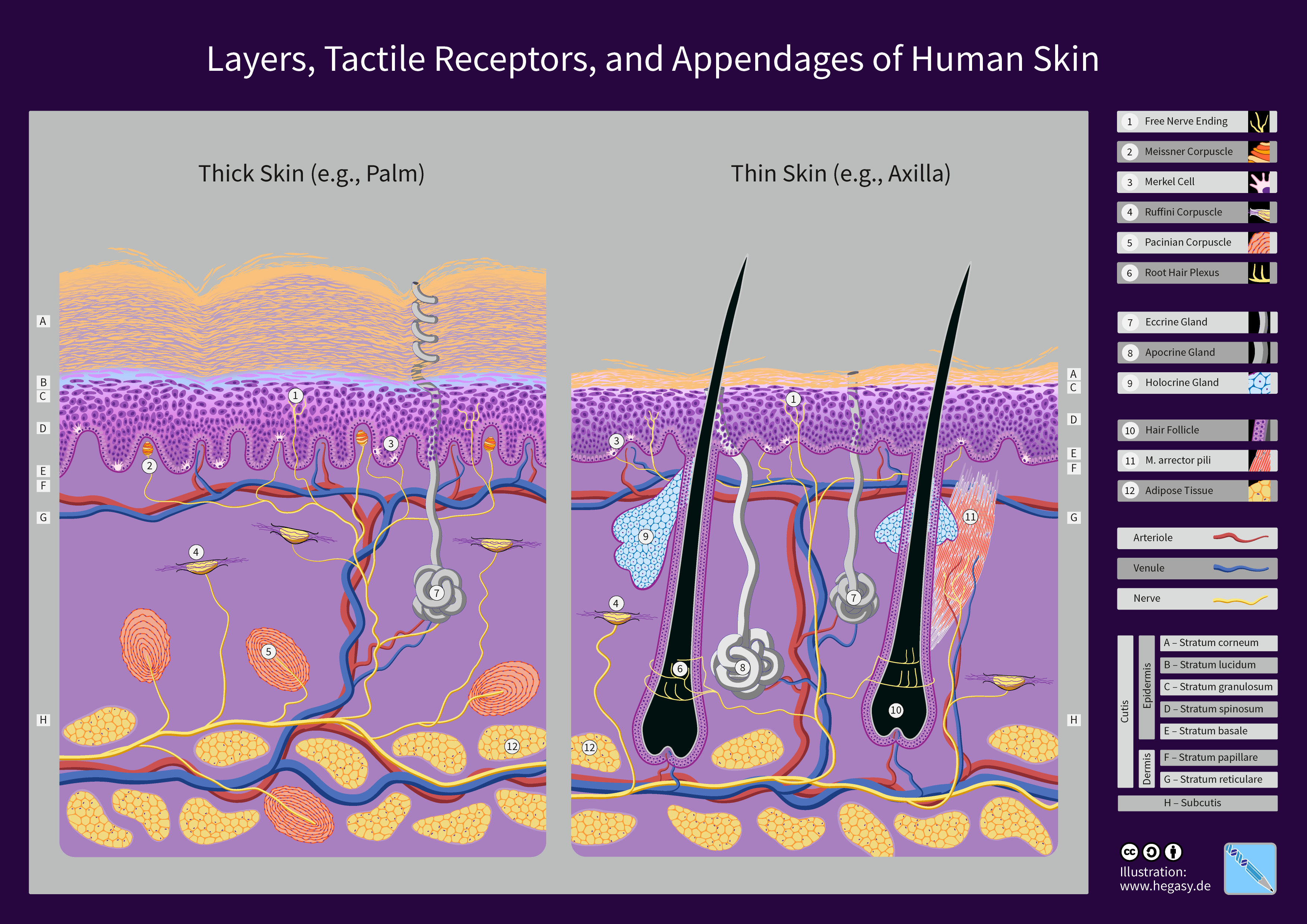
1. The Epidermis The epidermis is the most superficial and outermost layer of the human skin. Notably, the epidermis is completely avascular, meaning the epidermis lacks a direct blood supply. Because it is avascular, superficial scratches do not bleed; the outermost cells are entirely dead, sacrificed to form a protective crust.
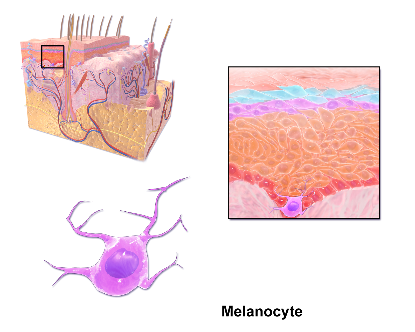
These cells are packed with keratin, a tough, fibrous protein found abundantly in the epidermis, hair, and nails. Functionally, keratin provides structural strength and water resistance to the outer surface of the body, preventing you from absorbing water like a sponge in the rain or drying out like a husk in the sun. Furthermore, melanocytes are specialized cells located in the epidermis that produce a pigment called melanin. Why does skin darken in the sun? Melanin absorbs ultraviolet radiation to protect cellular DNA in the skin from damage, preventing catastrophic mutations like melanoma.
2. The Dermis Beneath the dead, keratinized armor lies the living machinery. The dermis is the middle layer of the skin located directly beneath the epidermis. This layer is rich in infrastructure: the dermis contains blood vessels, sensory nerves, hair follicles, and sweat glands.
When you assess an older patient and notice their skin tears easily, you are witnessing a breakdown of the dermal matrix. Collagen and elastin fibers within the dermis provide the skin with structural strength and elasticity.
3. The Hypodermis (Subcutaneous Layer) Below the dermis is the foundation. The hypodermis, also known as the subcutaneous layer, is the deepest layer associated with the skin. Think of this layer when administering a "sub-Q" injection, like insulin. The hypodermis is primarily composed of fat-storing adipose tissue and loose connective tissue. This fat is not merely passive weight; adipose tissue in the hypodermis provides thermal insulation and serves as an energy storage site for the body.
Thermoregulation and Glandular Function
Beyond acting as a wall, the integumentary system plays a major role in regulating internal body temperature. It acts as the body's radiator system.
- Heat Loss: If the body overheats, vasodilation of blood vessels in the dermis brings warm blood to the surface to allow excess heat to escape the body (which is why a patient with a fever appears flushed). Concurrently, the evaporation of sweat from the skin surface rapidly removes excess heat from the human body.
- Heat Conservation: In freezing environments, vasoconstriction of blood vessels in the dermis restricts blood flow to the skin surface to conserve core body heat, leading to pale, cold extremities.
To manage fluids and temperature, the skin utilizes specialized exocrine glands:
- Sebaceous Glands: These glands secrete an oily and lipid-rich substance called sebum. Think of it as biological lotion; sebum naturally lubricates the skin and hair while preventing excessive water loss from the body surface.
- Sudoriferous (Sweat) Glands: These are specialized exocrine glands in the skin that produce and secrete sweat.
- Eccrine sweat glands are widely distributed across almost the entire surface of the human body. They primarily function in thermoregulation by releasing a clear, watery sweat.
- Apocrine sweat glands are heavily concentrated in the axillary and genital regions. These glands activate during puberty and release a thicker, protein-rich secretion that produces body odor when broken down by skin bacteria.
Clinical Bonus: The skin is also a metabolic organ! The skin chemically synthesizes vitamin D when dermal blood vessels are exposed to ultraviolet sunlight. This vitamin is absolutely critical for the body to absorb calcium.
When the integumentary shield is breached—perhaps by a surgical incision or a rusty nail—the immune system takes command. It operates in three escalating tiers of defense.
The First Line of Defense: Physical and Chemical Barriers
Before microscopic invaders can cause an infection, they must enter the body. The immune system's first line of defense consists of physical and chemical barriers.
- Intact skin acts as a physical barrier to prevent the entry of external pathogens.
- If a pathogen tries to enter through an airway, mucous membranes trap pathogens in sticky mucus before the pathogens can establish an infection.
- If a pathogen is swallowed in contaminated food, it faces a chemical vat: stomach acid destroys many swallowed pathogens due to its highly acidic low pH.
The Second Line of Defense: Nonspecific Innate Immunity
If a pathogen successfully breaches the skin or mucosa, the local alarms trigger. The second line of defense is a nonspecific innate immune response.
Innate immunity is a non-specific defense mechanism present in the human body from birth. It does not care what species of bacteria has entered; it reacts to all damage the exact same way. If you have ever watched a localized wound react to a splinter, you have witnessed this. Inflammation is a localized tissue response to injury or infection characterized by redness, heat, swelling, and pain.
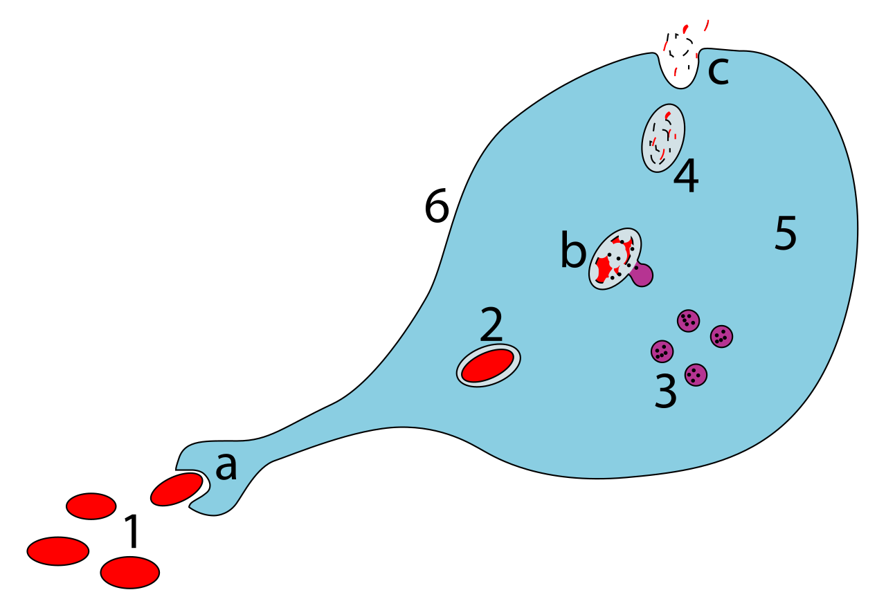
The heat and swelling occur because blood vessels leak fluid to rush biological "infantry" to the site. These infantry are phagocytes, which are white blood cells that engulf and destroy foreign particles. The heavy artillery of this group are the macrophages. Macrophages are a type of large phagocyte that consume dead host cells and invading pathogens—essentially operating as the battlefield clean-up crew.
The Third Line of Defense: Specific Adaptive Immunity
When the innate response is overwhelmed, the body deploys its "special forces." The third line of defense is the adaptive immune response. Unlike the innate system, adaptive immunity is a highly specific defense mechanism developed over time after exposure to specific antigens.
This system relies on two elite classes of lymphocytes: B cells and T cells.
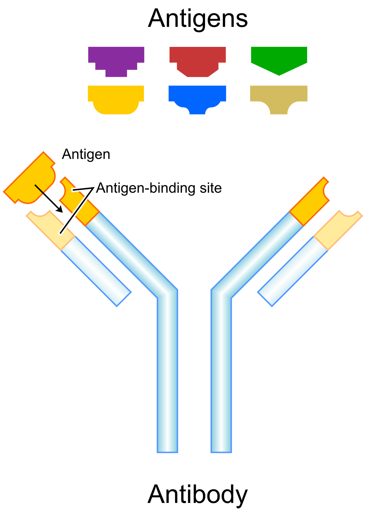
1. Humoral Immunity (B Cells) B cells are specialized lymphocytes responsible for humoral immunity (immunity occurring in the bodily fluids, or "humors"). Rather than fighting in hand-to-hand combat, B cells produce and secrete specialized protective proteins called antibodies. Antibodies bind to specific antigens to neutralize pathogens or mark the pathogens for destruction by other immune cells. Think of antibodies as targeted homing beacons painted onto the enemy.
2. Cell-Mediated Immunity (T Cells) T cells are specialized lymphocytes responsible for cell-mediated immunity. They engage in direct cellular warfare.
- Cytotoxic T cells directly attach to and destroy infected host cells or cancer cells. When a virus hijacks your own cell, the Cytotoxic T cell recognizes the compromised cell and forces it to self-destruct.
- Helper T cells coordinate the overall immune response by releasing chemical messengers called cytokines. They are the generals of the immune system. (This is why HIV, which specifically targets Helper T cells, is so devastating—it decapitates the immune system's chain of command).
Once the pathogen is defeated, the adaptive system creates an archive of the threat. Memory cells provide long-lasting immunity by recognizing and rapidly responding to a previously encountered pathogen.
Active vs. Passive Immunity:
- Active immunity occurs when the body produces its own antibodies in response to an infection or a vaccine. Your immune system did the heavy lifting.
- Passive immunity occurs when a person receives pre-formed antibodies produced by another organism. Examples include anti-venom or a nursing infant receiving antibodies through breast milk. The protection is immediate, but temporary.
While the skin protects the individual and the immune system defends it, the reproductive system looks toward the future of the species.
The Male Reproductive System: Production and Delivery
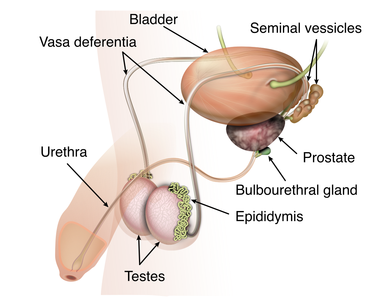
The male reproductive system is designed to produce, store, and deliver male gametes known as sperm.
Primary Organs and Storage: The testes are the primary male reproductive organs. Operating as microscopic factories, the testes generate sperm cells and secrete the primary male sex hormone called testosterone.
Temperature control is vital here. The scrotum is a sac of skin that houses the testes outside the main abdominal cavity. Why place such critical organs externally? Because the external location of the scrotum keeps the testes at a slightly lower temperature necessary for viable sperm production.
Once manufactured, sperm need to mature. The epididymis is a tightly coiled tube situated on top of the testes where sperm mature and are stored.
Transport and Semen Production: During ejaculation, sperm begin their journey. The vas deferens is a muscular tube that transports mature sperm from the epididymis up toward the ejaculatory duct.
Along the way, three key accessory glands secrete fluids to create semen, providing the sperm with energy and armor:
- The seminal vesicles secrete a thick, fructose-rich fluid that provides a cellular energy source for swimming sperm. They need fuel for the long swim.
- The prostate gland secretes a slightly alkaline fluid into the passing semen during ejaculation. This is a critical protective measure; alkaline prostatic fluid helps neutralize the highly acidic environment of the female reproductive tract to protect sperm viability.
- The bulbourethral glands secrete a clear, lubricating mucus into the urethra prior to ejaculation.
Finally, the semen exits the body. The urethra in males serves as a common anatomical passageway for the excretion of both urine and semen.
The Female Reproductive System: Cultivation and Support
The female reproductive system is designed to produce ova and physically support a developing fetus until birth.
Primary Organs and Transport: The ovaries are the primary female reproductive organs. They operate on a monthly cycle where the ovaries generate egg cells and secrete the female sex hormones estrogen and progesterone.
Upon ovulation, the egg is caught by the plumbing network. The fallopian tubes are narrow ducts that transport released eggs from the ovaries toward the uterus. A massive clinical pearl for nursing exams: fertilization of an egg by a sperm cell most commonly occurs within the fallopian tubes, not in the uterus.
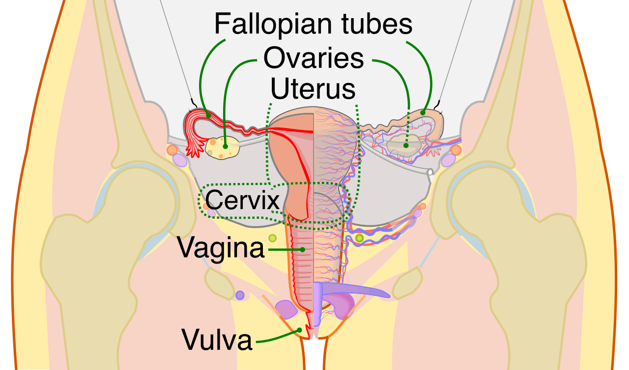
Gestation and Birth: Once fertilized, the resulting embryo travels down to its new home. The uterus is a hollow, muscular organ where a fertilized egg implants and continuously develops into a fetus.
The internal environment of the uterus is carefully maintained. The endometrium is the highly vascularized inner mucosal lining of the uterus. Anticipating a fertilized egg every month, the endometrium naturally thickens during the menstrual cycle to prepare for a potential embryonic implantation. If no implantation occurs, this lining sheds (menstruation).
Guarding the exit of the uterus is the cervix, which is the lower, narrow neck portion of the uterus that connects directly to the vaginal canal. Finally, the vagina is an elastic, muscular canal that serves as the birth canal and the receptacle for the penis during sexual intercourse.
Professor's Closing Advice for the HESI A2
To conquer this section of the exam, do not view these systems in isolation. Remember why they function the way they do. The skin isn't just tissue; it's a water-proof, UV-absorbing, temperature-regulating barrier. The immune system isn't just a list of cells; it's a chronological battlefield (Barriers → Innate Inflammation → Adaptive T/B Cells). The reproductive systems aren't just plumbing; they are precision-engineered environments designed to protect delicate gametes and sustain new life. Keep these functional goals in mind, and the physiological details will effortlessly fall into place.