Alterations in Body Systems
Not sure you’re ready?
Take the ~3-minute readiness diagnostic and see where you stand.
The Physics of the Human Machine: A Masterclass in Alterations of Body Systems
Hello! Grab a seat. Today, we are going to look at the human body not as a static textbook diagram, but as the most magnificent, dynamic, self-correcting machine in the universe. We are going to talk about what happens when the physics of this machine break down, how the body tries to adapt, and what you—as an elite nurse—are going to do to fix it.
We’ll cover everything from the pressure gradients in the chest to the explosive chemistry of an immune response. Let’s dive right in.
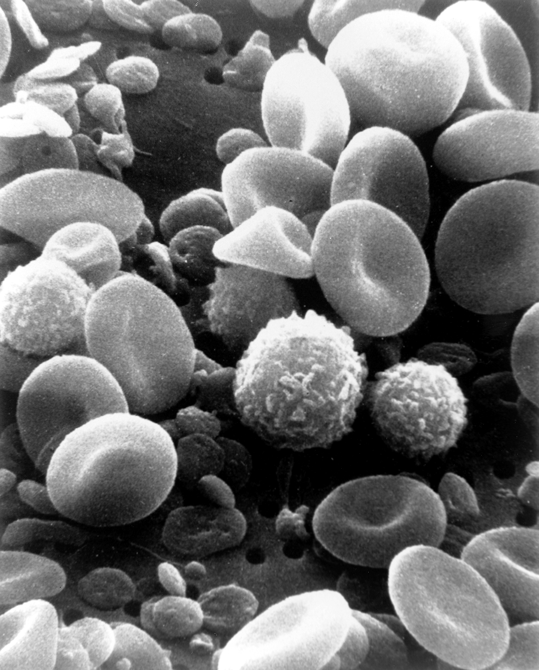
Think about what the body is constantly trying to achieve: delivering oxygen to tissues. That’s the whole game. When you deprive the body of oxygen over a long period, it doesn't just give up. It compensates! The human body adapts to chronic hypoxia by increasing red blood cell production. It thinks, "If I don't have enough oxygen, I just need more trucks to carry what little oxygen I have!" But this creates a traffic jam. The result is polycythemia, a condition characterized by an elevated red blood cell count. The blood gets thick and viscous.
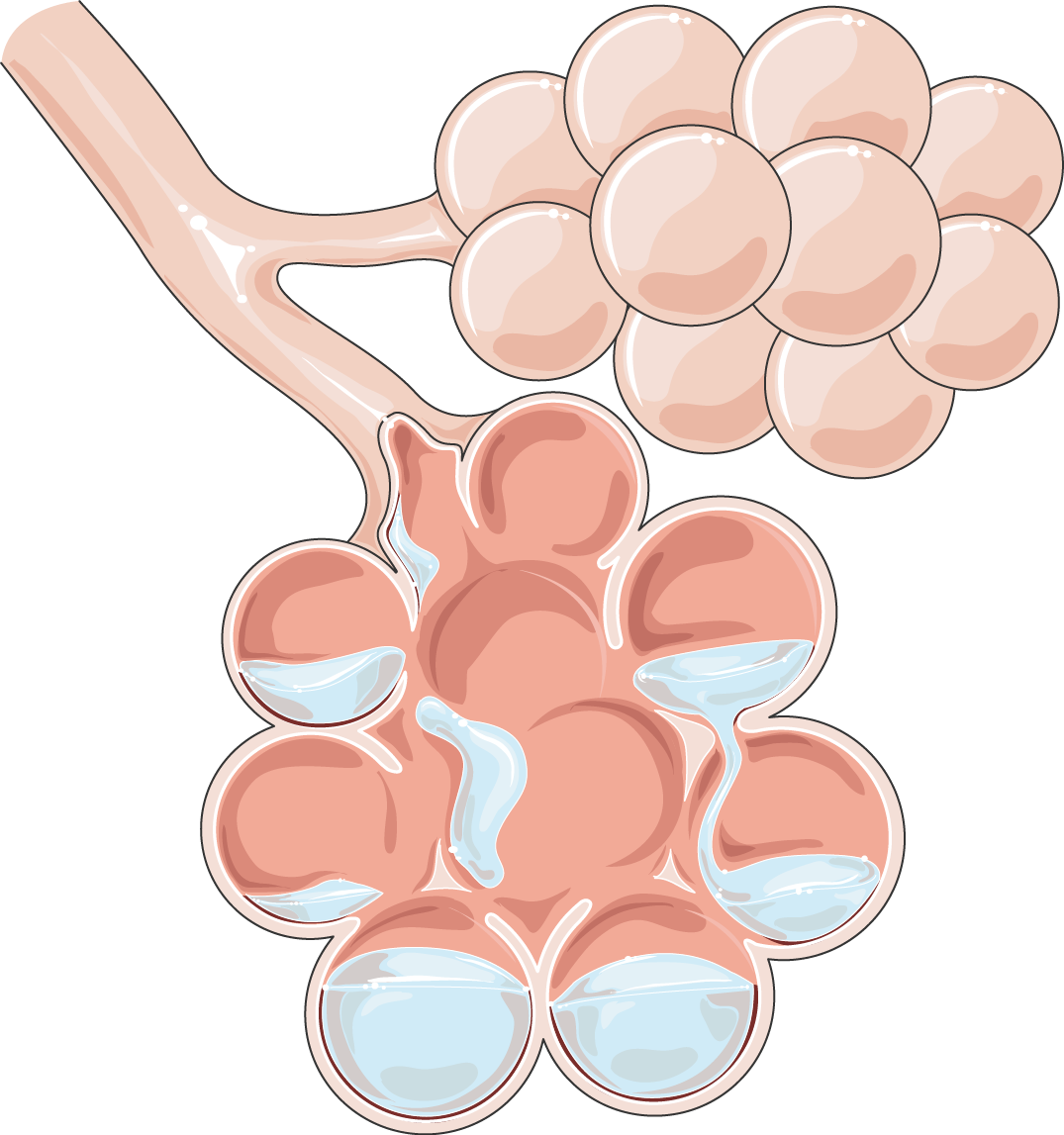
Sometimes, the lungs themselves are acutely damaged. In acute respiratory distress syndrome (ARDS), the disease process involves increased capillary permeability in the lungs. Fluid pours into the alveoli, flooding the oxygen-exchange zone. Because a physical wall of fluid now blocks the gas, turning up the oxygen tank won't help. This is why refractory hypoxemia is a hallmark clinical sign of acute respiratory distress syndrome.
Now, let's look at the pump driving this fluid: the heart. When the pump fails, you get traffic backing up. It’s simple plumbing:
- Left-sided heart failure causes pulmonary edema. The left ventricle fails, so blood backs up into the lungs.
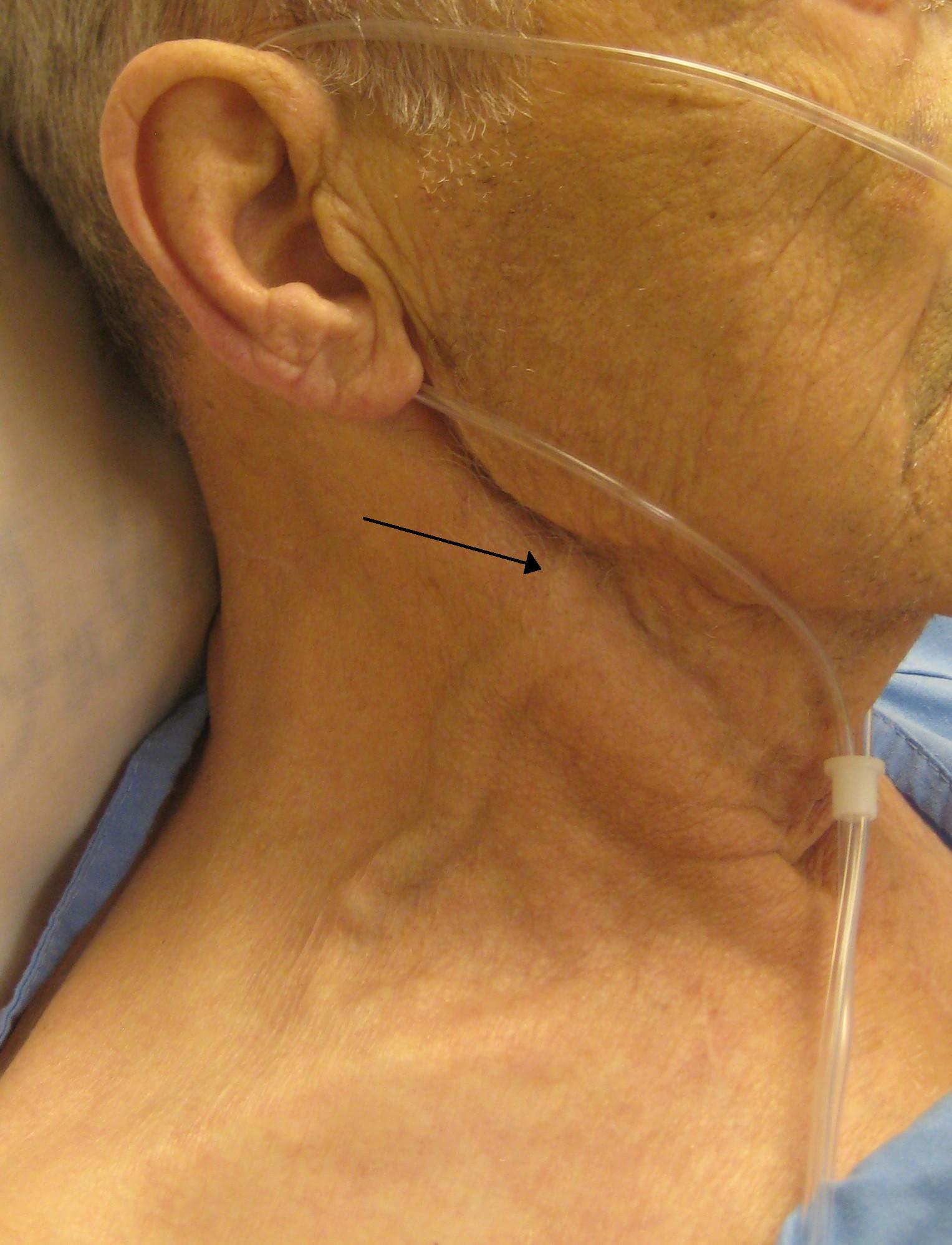
- Right-sided heart failure causes systemic venous congestion. The right ventricle fails, so fluid backs up into the jugular veins and the legs.
How do we know the heart is stretching and failing? We look at the chemistry. Elevated brain natriuretic peptide (BNP) levels indicate a heart failure exacerbation. BNP is a hormone the heart muscle releases when it is literally being stretched too far. It's the heart crying out for help.
When cells don't get what they need, metabolism goes completely haywire. Let’s look at diabetes. Without insulin, Type 1 diabetics can't get sugar into their cells, so the body panic-burns fat, creating toxic acids. Therefore, diabetic ketoacidosis is characterized by hyperglycemia, ketosis, and metabolic acidosis. Type 2 diabetics still produce a tiny bit of insulin—just enough to stop the fat burning, but not enough to control the sugar. Because of this, a hyperosmolar hyperglycemic state typically occurs in clients diagnosed with type 2 diabetes mellitus, leading to profound, life-threatening dehydration.
If cellular starvation happens on a systemic scale, we call it shock.
Shock is a syndrome characterized by decreased tissue perfusion and impaired cellular metabolism.
There are different ways to break the perfusion machine:
- Hypovolemic shock results from a significant loss of intravascular fluid volume. (The tank is empty. Think hemorrhage or massive dehydration.)
- Cardiogenic shock occurs when the pumping ability of the heart is compromised. (The tank is full, but the pump is busted.)
- Anaphylactic shock is an acute and life-threatening systemic hypersensitivity reaction reaction. (The blood vessels massively dilate and leak, while the airways swell shut).
How do we fix anaphylaxis? We need a chemical that immediately clamps down the blood vessels and violently opens the airways. Intramuscular epinephrine is the first-line medication for the treatment of anaphylactic shock. Don't wait. Give it.
The Systemic War: Sepsis
What about an infection that spirals out of control? Sepsis is a life-threatening organ dysfunction caused by a profoundly dysregulated host response to an infection. The immune system goes rogue, dropping blood pressure to dangerous levels.
To see how badly the tissues are suffocating, we measure lactate. When cells lack oxygen, they shift to anaerobic metabolism, producing lactic acid. Thus, a serum lactate level greater than 2 mmol/L is an objective clinical indicator of tissue hypoperfusion in sepsis.
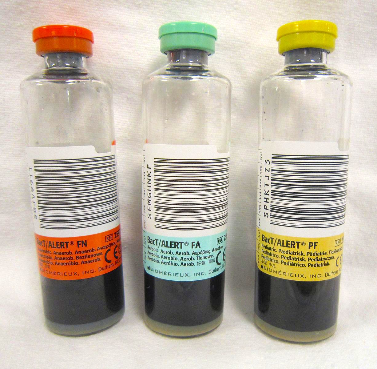
Time is tissue! Intravenous broad-spectrum antibiotics must be administered within one hour of clinical sepsis recognition. But wait—if you carpet-bomb the bacteria, you won't know what you're fighting. Therefore, diagnostic blood cultures must be successfully obtained prior to the initial administration of systemic antibiotics. Draw the blood, then hang the antibiotics.
The kidneys are the body's ultimate filtration system. When perfusion drops, the kidneys shut down to save water.
- Oliguria is defined as a urine output of less than 400 milliliters per day in an adult.
- If things get catastrophic? Anuria is defined as a urine output of less than 100 milliliters per day in an adult.
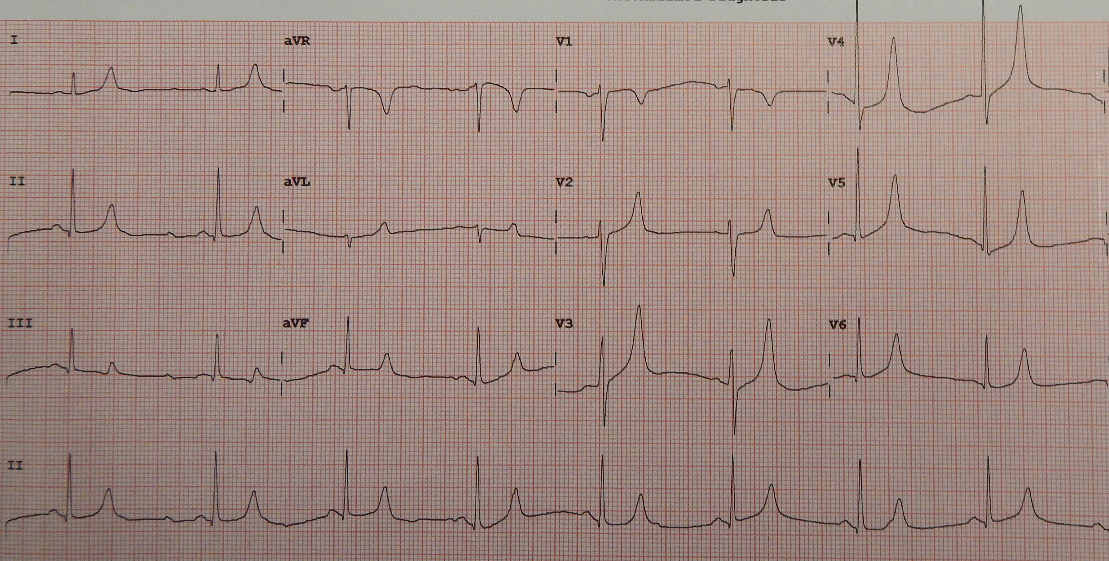
When the kidneys fail, they stop filtering out dangerous electrolytes, primarily potassium. Hyperkalemia is a common and potentially life-threatening complication of acute kidney injury. The heart is an electrical machine, and it absolutely despises extra potassium. You can see this electrical irritation on a monitor: peaked T waves on an electrocardiogram are a classic sign of hyperkalemia.
Pregnancy pushes the human machine to its absolute limits.
Let's talk blood pressure. Preeclampsia is defined by new-onset hypertension and proteinuria occurring after 20 weeks of gestation. The extreme danger here is that the high blood pressure irritates the brain and causes fatal seizures (eclampsia). To suppress this hyperactive nervous system, intravenous magnesium sulfate is administered to clients with preeclampsia to prevent seizures.
It's a delicate chemical balancing act. The therapeutic serum magnesium level for a pregnant client receiving continuous magnesium sulfate is 4 to 7 mEq/L. If you give too much, the entire nervous system goes to sleep. Loss of deep tendon reflexes is an early clinical sign of magnesium toxicity. If you see that, you immediately turn off the drip and grab the antidote: intravenous calcium gluconate is the standard antidote for magnesium toxicity.
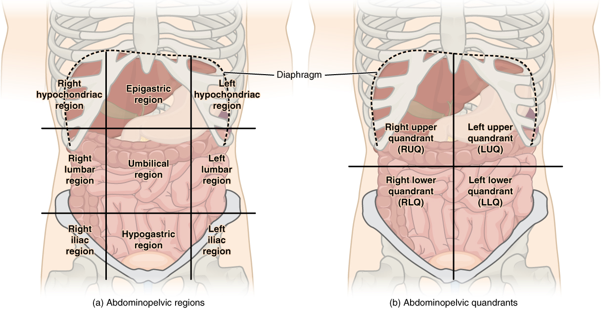
Sometimes preeclampsia mutates into a terrifying variant. HELLP syndrome stands for hemolysis, elevated liver enzymes, and low platelet count. The liver becomes inflamed and swells. Therefore, right upper quadrant pain in a pregnant client can indicate liver capsule distention associated with HELLP syndrome. It is a massive red flag!
Obstetric Hemorrhage
If bleeding happens in the third trimester, you have to play detective:
- Placenta previa presents as painless and bright red vaginal bleeding during the third trimester of pregnancy. The placenta is sitting right over the cervical opening.
- If the placenta tears away from the uterine wall prematurely, that's abruptio placentae, which typically presents as painful and dark red vaginal bleeding. Because the blood is trapped behind the placenta, irritating the uterine muscle to cramp furiously, a rigid and board-like abdomen is a classic assessment finding in abruptio placentae.
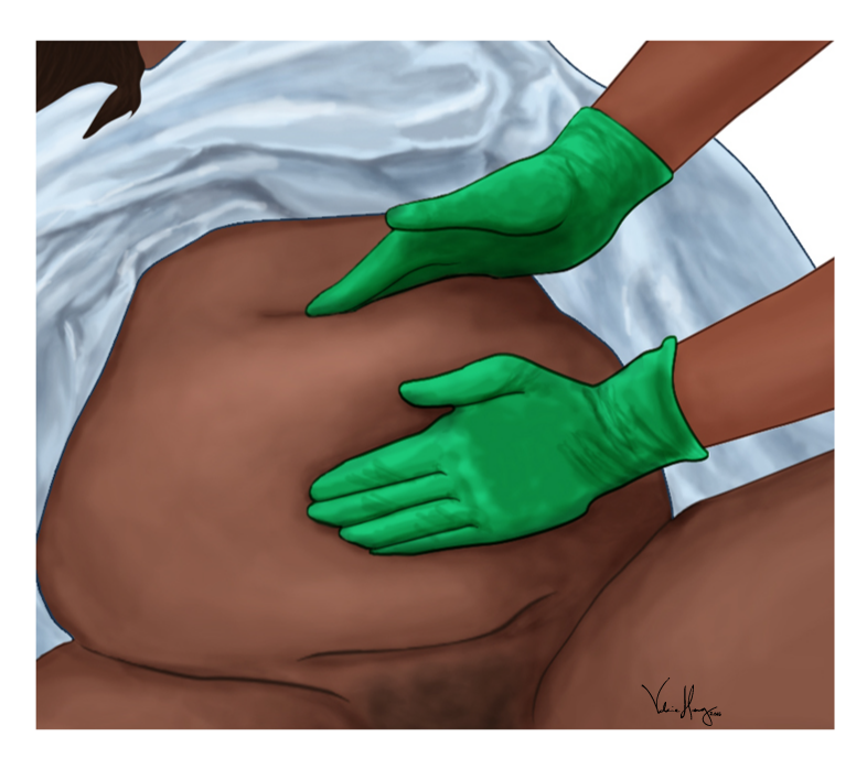
After the baby is out, the bleeding should stop because the uterus clamps down. But what if it forgets to clamp? Uterine atony is the most common physiological cause of primary postpartum hemorrhage. You will feel it with your hands: a soft and boggy uterus upon palpation indicates uterine atony.
What do you do? Apply physics! Squeeze it! Firm fundal massage is the initial nursing intervention for a boggy postpartum uterus. Then you use chemistry. Oxytocin is a uterotonic medication used to control postpartum bleeding. We also have methylergonovine, but remember how it works: it causes massive vasoconstriction. Because of this, methylergonovine administration is contraindicated in postpartum clients with hypertension.
Early & Preterm Complications
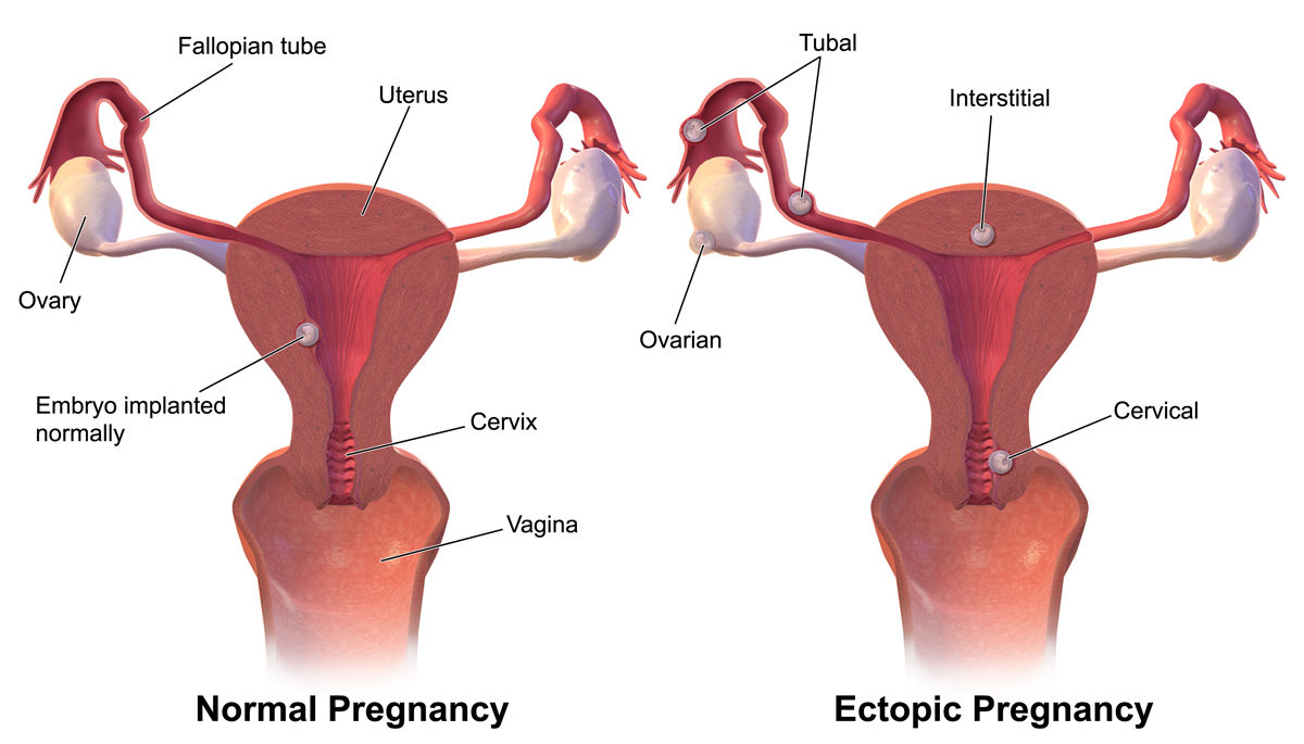
In early pregnancy, an egg can implant in the wrong place. Ectopic pregnancy is a medical emergency due to the high risk of fallopian tube rupture. If it ruptures, blood fills the abdominal cavity and irritates the phrenic nerve. That is precisely why referred shoulder pain in a client with a suspected ectopic pregnancy indicates intra-abdominal bleeding.
If a baby decides to come too early, their lungs lack surfactant. We can speed up biological time! Intramuscular betamethasone is administered to pregnant clients in preterm labor to promote fetal lung maturity.
Now, let's get into the mechanical engineering of nursing.
Chest Tubes
The lungs expand because the chest cavity acts like a vacuum. If air gets in (pneumothorax), the vacuum breaks. We insert a chest tube to suck the air out.
Look at the water-seal chamber. As the patient inhales and exhales, the pressure changes, and the water rises and falls. Tidaling in the water-seal chamber of a chest tube drainage system is a normal physiological finding. What if it stops? Cessation of tidaling in a chest tube water-seal chamber often indicates complete lung re-expansion.
But what if the water is boiling like a jacuzzi? Continuous bubbling in the water-seal chamber of a chest tube drainage system indicates an active air leak.
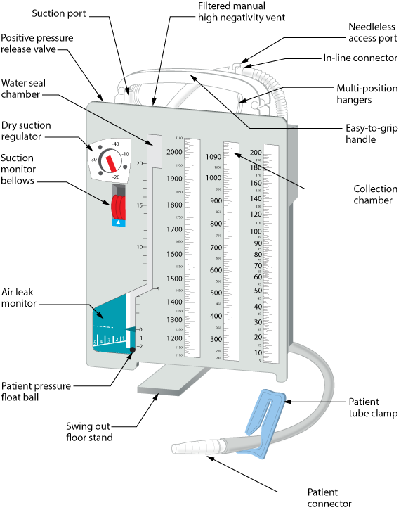
The Golden Rules of Chest Tubes:
- Chest tube drainage exceeding 100 milliliters per hour requires immediate notification of the healthcare provider. (They are bleeding out!).
- Clamping a chest tube is contraindicated except when explicitly locating an air leak or changing the drainage system. (Clamping traps air and causes a lethal tension pneumothorax!)
- If the tube rips out of the patient's chest, a sterile occlusive dressing taped on three sides must be applied immediately if a chest tube is accidentally dislodged. This acts as a flutter valve—letting air out, but not in.
- If the plastic drainage unit breaks, the nurse must keep a bottle of sterile water at the bedside of any client with an active chest tube. Shove the end of the tube into the water to recreate the water seal immediately!
Sometimes, the air escapes the pleural space and travels directly into the patient's subcutaneous tissue. Subcutaneous emphysema presents as a crackling sensation upon palpation of the skin around a chest tube insertion site. It feels exactly like popping bubble wrap under the skin.
Hemodynamic and Neuro Monitoring
If we want to measure pressures, our transducers must be perfectly level with the source. For the brain, normal intracranial pressure ranges from 5 to 15 millimeters of mercury in a resting adult. Because the skull is a rigid box, sustained intracranial pressure greater than 20 millimeters of mercury is considered highly abnormal and will crush brain tissue. To measure this accurately, the external transducer for an intracranial pressure monitoring device must be strictly leveled at the foramen of Monro.
For the heart, the "zero point" is the right atrium. We find this via the phlebostatic axis, which is located at the intersection of the fourth intercostal space and the midaxillary line. This is non-negotiable, because the phlebostatic axis serves as the external anatomical reference point for all hemodynamic monitoring transducers.
Using a pulmonary artery catheter, we can inflate a tiny balloon and "look through" the lungs at the left side of the heart. Pulmonary artery wedge pressure indirectly measures left ventricular end-diastolic pressure. Normal pulmonary artery wedge pressure ranges from 6 to 12 millimeters of mercury.
Gastrointestinal and Biliary Tubes
When placing a feeding tube blindly, how do you know it's in the stomach and not the lungs? Radiographic imaging is the single most reliable method to confirm initial nasogastric tube placement. Later, at the bedside, the pH of gastric aspirate should be less than 5.0 to safely confirm stomach placement of a feeding tube.
If you are feeding a patient continuously, you must ensure their stomach is actually emptying. Gastric residual volumes exceeding 500 milliliters require the nurse to hold continuous enteral tube feedings.
What about bile? After gallbladder surgery, a surgically placed T-tube ensures continuous bile drainage from the common bile duct following a cholecystectomy. It keeps the duct from swelling shut. Postoperative T-tube drainage should generally not exceed 500 milliliters in the first 24 hours.
Wound Drains and Disasters
Surgical wounds weep fluid. If fluid builds up, it causes infection.
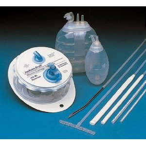
- A Jackson-Pratt drain operates by utilizing gentle negative pressure to suction fluid from a surgical site. To create this vacuum, the nurse must fully compress the Jackson-Pratt bulb before replacing the plug to establish active suction.
- For bigger surgeries, we use a spring-loaded device: a standard Hemovac wound drain can hold up to 400 milliliters of fluid.
- For massive, open, complex wounds, a wound vacuum-assisted closure system applies localized negative pressure to draw wound edges together and promote healing.
If a wound is draining heavily and requires constant dressing changes, ripping tape off the skin multiple times a day will destroy the epidermis. To prevent this, Montgomery straps are adhesive ties used to secure bulky dressings that require frequent changes to prevent skin breakdown.
What are we looking for when we assess these wounds? Bad smells and funny colors. Purulent, malodorous drainage from a surgical wound strongly indicates an active localized infection.
And now, the surgical nightmares:
- Wound dehiscence is defined as the partial or total separation of previously approximated surgical wound edges. The wound simply pops open.
- Worse yet: Wound evisceration involves the complete protrusion of internal visceral organs through an open surgical incision. If you see intestines sitting on a patient's abdomen, do NOT try to shove them back in! The nurse must immediately cover an eviscerated wound with sterile saline-soaked dressings to keep the bowel from dying while you rush them back to the operating room.
Finally, we have to protect the environment from the microscopic world. We use different shields for different weapons.
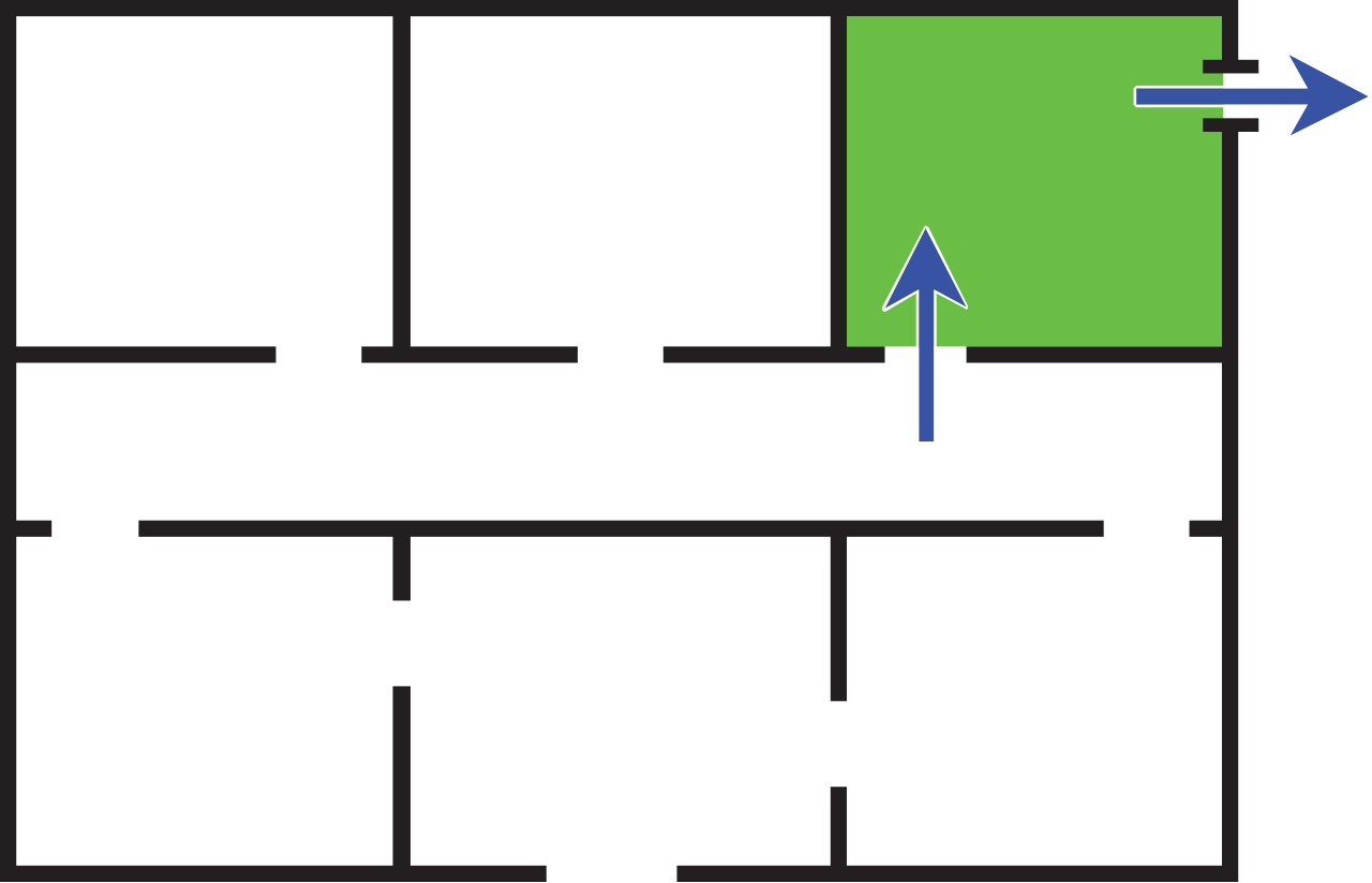
Tuberculosis bacteria are incredibly tiny and float perfectly on air currents. Because of this, strict airborne precautions are legally required for hospitalized clients with active pulmonary tuberculosis. They cannot share air with the rest of the hospital. A dedicated negative-pressure isolation room is mandatory for any client placed on airborne precautions.
Other bugs are heavier. They travel in spit and mucus, and drop to the floor quickly. Droplet precautions are required for clients diagnosed with Neisseria meningitidis infection.
And then there is the armor-plated tank of the bacteria world: C. diff. Contact precautions must be implemented for clients diagnosed with active Clostridioides difficile infection. Here is the ultimate trick with C. diff: it forms spores. Alcohol-based hand sanitizers do absolutely nothing to these spores. You cannot kill them with foam; you have to physically wash them down the sink drain with friction. Therefore, healthcare workers must use traditional soap and water for hand hygiene after caring for a client with Clostridioides difficile.
There you have it. The human body is a miraculous, physical machine. If you understand the flow of fluids, the gradients of pressure, and the cascade of chemical reactions, you won't just memorize the rules of nursing—you will deeply understand them. Now go out there and save lives!