Therapeutic Procedures
Not sure you’re ready?
Take the ~3-minute readiness diagnostic and see where you stand.
Welcome to the fascinating world of therapeutic procedures! If you want to master nursing, you have to stop memorizing disconnected facts and start thinking about the mechanics of the human body. Think of the body like an intricate, perfectly balanced machine of physics, chemistry, and fluid dynamics.
When we do a procedure—whether we are chemically altering a patient's consciousness, adjusting the negative pressure vacuum of their lungs, or flushing out their plumbing—we are stepping into that machinery. Our job is to control the variables, anticipate the reactions, and safely hand the controls back to the patient.
Let’s break down the mechanics of anesthesia, therapeutic devices, and post-procedural care. Put on your thinking caps. We’re going to figure out why these things work.
The nervous system is an electrical highway. When we need to do surgery, we have to block the traffic of pain signals. Depending on the procedure, we might shut down a single driveway, a regional interstate, or the entire electrical grid.
1. General Anesthesia: Shutting Down the Grid
General anesthesia depresses the central nervous system to induce unconsciousness and amnesia. You are taking the brain completely offline. But when you mess with the central thermostat, things can sometimes go catastrophically wrong.
The nightmare scenario you must always watch for is malignant hyperthermia, a life-threatening complication of general anesthesia. What happens? A genetic anomaly causes the skeletal muscles to dump calcium, leading to massive, uncontrolled muscle contractions.
- Early signs: Because the muscles are hypermetabolic, they demand massive amounts of oxygen and produce a ton of CO2. The body tries to compensate. Therefore, the early signs of malignant hyperthermia include tachycardia, tachypnea, and muscle rigidity.
- Late sign: All that metabolic friction creates heat. Surprisingly, an elevated body temperature is a late sign of malignant hyperthermia. By the time they are burning up, you are already behind schedule!
- The Antidote: You must immediately grab dantrolene sodium, the specific pharmacological antidote for malignant hyperthermia, which acts as a muscle relaxant by stopping that calcium release.
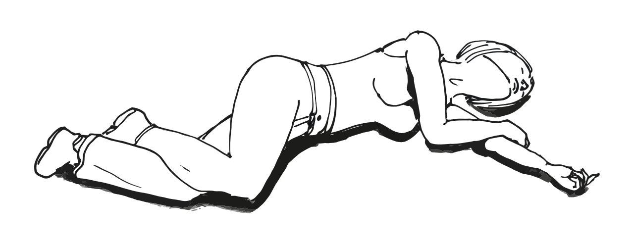
Recovery Physics: Once the surgery is over, gravity and reflexes are your main concerns. Clients recovering from general anesthesia are placed in a lateral recovery position to prevent aspiration of vomit. If they vomit, gravity pulls it out of the mouth, not down the trachea. Furthermore, watch their hemodynamics! A rapid heart rate and decreasing blood pressure following a surgical procedure indicate potential hemorrhage. The heart pumps faster to compensate for the dropping pressure in the pipes.
2. Regional Anesthesia: Closing the Interstate
Regional anesthesia blocks pain sensations in a specific area of the body without causing loss of consciousness.
- Epidural anesthesia is a type of regional anesthesia injected into the epidural space (just outside the sac of fluid around the spinal cord).
- Spinal anesthesia goes deeper, injecting local anesthetic directly into the subarachnoid space (into the cerebrospinal fluid).
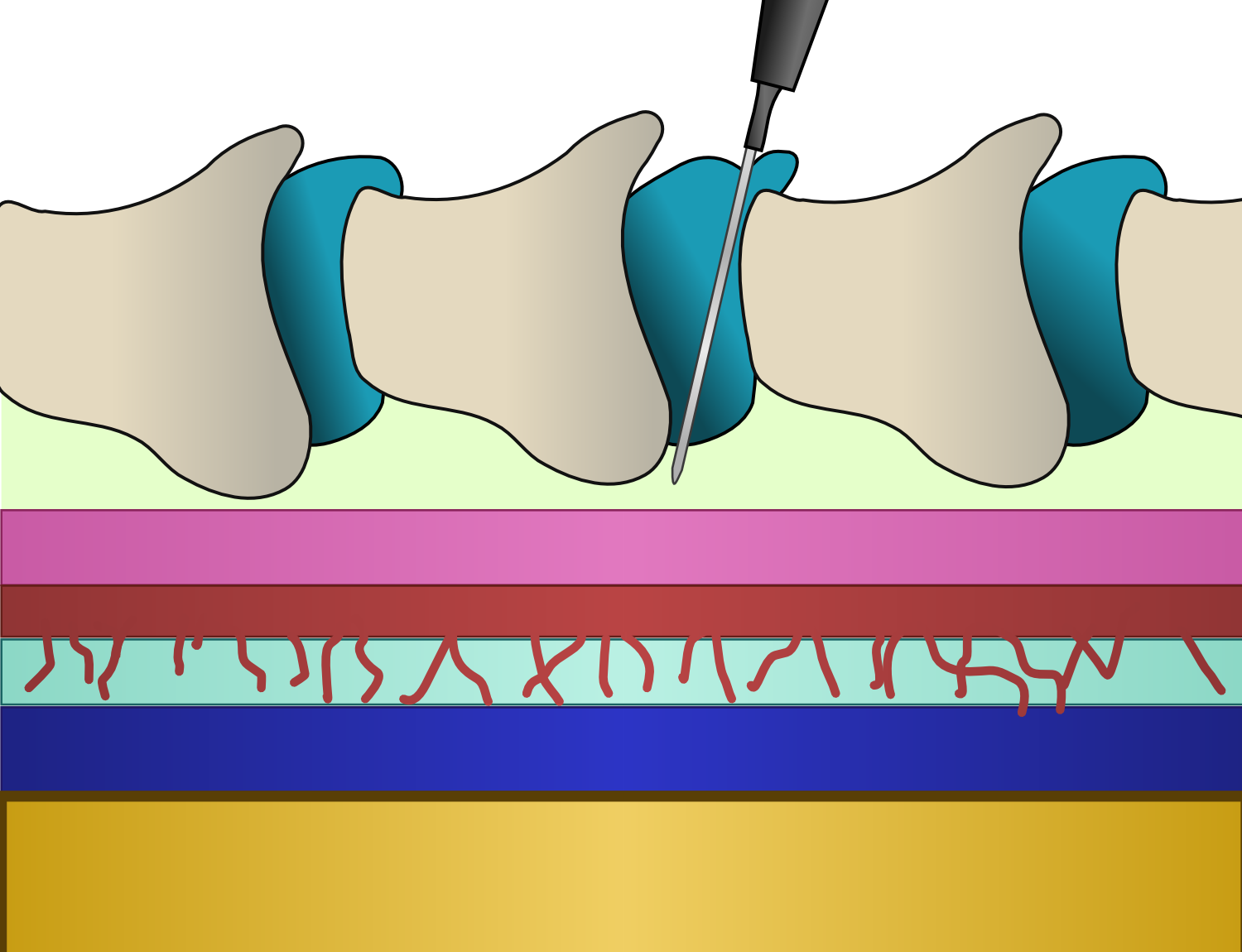
Because these blocks vasodilate the lower half of the body, hypotension is a common complication of epidural and spinal anesthesia. Blood pools in the legs, and pressure drops. Furthermore, because spinal anesthesia punctures the dura mater, a spinal headache can occur after spinal anesthesia due to cerebrospinal fluid leakage. The brain loses its hydrostatic cushion!
The golden rule of recovery here? Assessing the return of sensation and motor function is a critical nursing assessment after regional anesthesia. You must prove the electrical grid is coming back online top-to-bottom.
3. Local Anesthesia: Blocking the Driveway
Local anesthesia produces a reversible loss of sensation in a small, targeted body area. How does it work? It's elegant electrochemistry. Local anesthetics ending in -caine block sodium channels to inhibit nerve impulse transmission. If sodium can't rush into the nerve cell, there is no electrical spark. The nerve is effectively silenced!
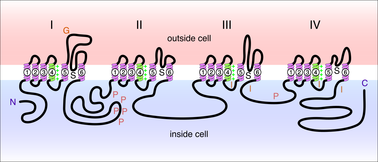
But the body absorbs these drugs quickly. How do we keep the drug at the site longer? Epinephrine is added to local anesthetics to prolong the anesthetic effect through localized vasoconstriction. It shrinks the local blood vessels so the anesthetic can't wash away.
4. Moderate Sedation: The "Autopilot" Trance
Unlike general anesthesia, moderate sedation depresses client consciousness while allowing the client to maintain a patent airway independently. They are relaxed, slightly amnesiac, but the autopilot is on. In fact, clients undergoing moderate sedation retain the physical ability to respond appropriately to verbal commands. If you tell them to take a deep breath, they will.
Because the patient relies on their own reflexes, a pre-procedure fasting period is required before moderate sedation to reduce the risk of pulmonary aspiration.
The Nurse’s Role During Moderate Sedation:
- Absolute Focus: Things can go from "moderate" to "deep" in a fraction of a second. Therefore, the nurse monitoring a client during moderate sedation must have no other concurrent clinical duties.
- Monitoring Data: You need objective data. Continuous pulse oximetry and capnography are required monitoring parameters during these procedures. You must measure both oxygenation and ventilation. For example, the administration of propofol for moderate sedation requires continuous monitoring of respiratory rate and depth, as it is a potent respiratory depressant.
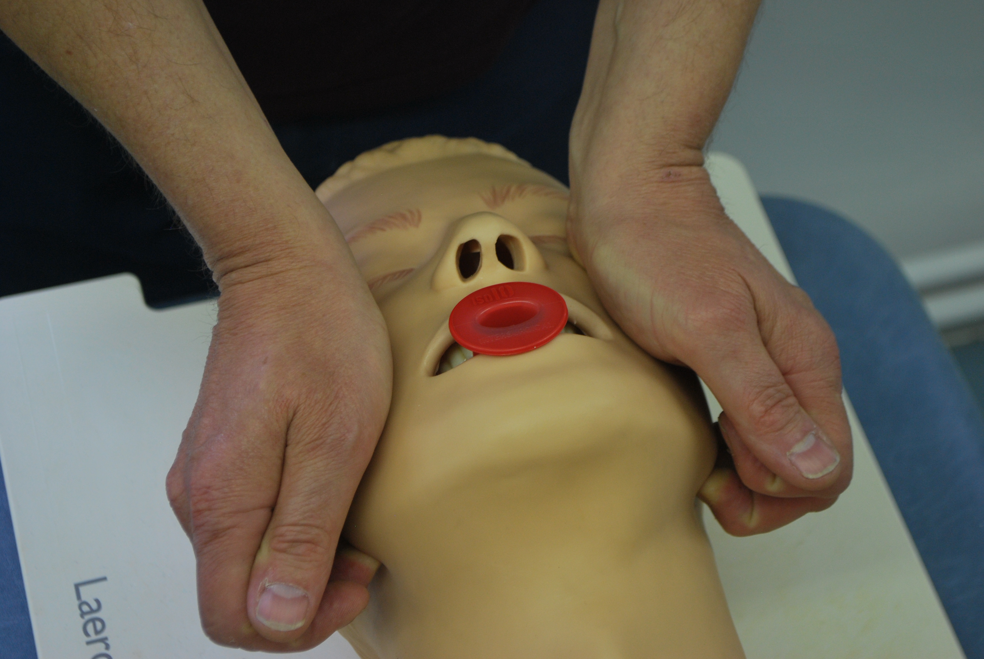
- Immediate Interventions: If you see oxygen desaturation during moderate sedation, it requires immediate airway stimulation or repositioning (like a jaw-thrust maneuver) to open the pipes.
- Safety Nets: Always be prepared for the worst. The nurse must ensure emergency resuscitation equipment is immediately available in the room.
The Reversal Agents (Antidotes): If you overshoot the runway, you need to knock the drugs off their receptors immediately.
| Drug Class | Reversal Agent | Mechanism / Notes |
|---|---|---|
| Benzodiazepines | Flumazenil | Flumazenil is the pharmacological reversal agent for benzodiazepines used during moderate sedation. |
| Opioids | Naloxone | Naloxone is the pharmacological reversal agent for opioids used during moderate sedation. |
Discharge Rules: You can't just let them walk out the door. Clients must meet specific discharge criteria including stable vital signs before leaving a facility after moderate sedation. Because their judgment and reflexes are still cloudy, post-procedure discharge instructions after moderate sedation must require the client to be accompanied by a responsible adult.
Now let's talk about the plumbing. When we put tubes into the human body, we are relying on strict laws of pressure, gravity, and fluid dynamics.
1. Chest Tubes: Restoring the Vacuum
The pleural space surrounding the lungs is a brilliant biological vacuum (negative pressure). If air or fluid gets in, the vacuum is broken, and the lung collapses. A chest tube is inserted into the pleural space to remove air, fluid, or blood and restore that negative pressure.
To make this work, we use a closed drainage system. And remember your gravity: Chest tube drainage systems must be continuously kept below the level of the client's chest. If you lift it up, the fluid drains right back into the patient!
The Magic of the Water Seal: The heart of this device is the water seal chamber. The water seal chamber of a chest tube drainage system prevents ambient air from re-entering the pleural space. It acts as a one-way valve. Air can bubble out through the water, but it can't swim backward up the tube. Crucially, keeping the chest tube collection chamber upright ensures the water seal functions correctly. If you knock it over, the seal is broken!
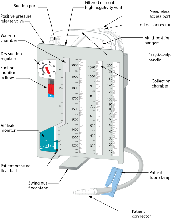
Reading the Water Seal Chamber (The Physics of Bubbles and Waves):
- Tidaling is the normal vertical fluctuation of fluid in the water seal chamber during respiration. When the patient breathes in, pressure changes, and the water level rises and falls. It tells you the tube is patent and the physics are working!
- The cessation of tidaling in a chest tube system indicates lung re-expansion or an obstruction in the tube. (Either the lung is fully healed and filling the space, or the tube is kinked).
- Intermittent bubbling in the water seal chamber corresponds to respirations and is an expected finding when treating a pneumothorax. As the lung expands on exhalation, it pushes the trapped air out into the chamber.
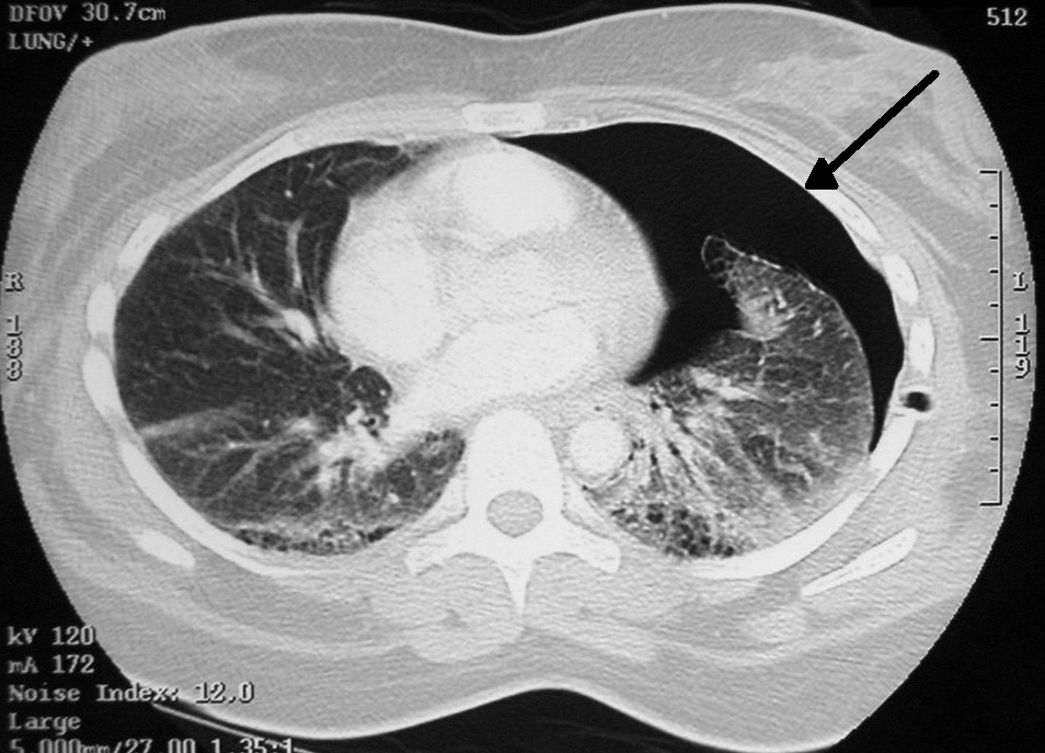
- Continuous bubbling in the water seal chamber indicates an air leak in the chest tube system. This is bad. It means ambient air is sneaking into the system somewhere.
Chest Tube Emergencies & Rules:
- Never clamp unnecessarily! Clamping a chest tube is only performed briefly to check for air leaks or to change the drainage system. Clamping traps air inside the chest and causes a lethal tension pneumothorax.
- The Dislodgement Crisis: What if the tube gets accidentally ripped out of the patient's chest? You now have a hole sucking air into the chest cavity! An un-clamped chest tube accidentally dislodged from the client requires immediate application of a sterile occlusive dressing taped on three sides.
- Why exactly three sides? It is brilliant, improvised physics. Taping a chest tube dressing on three sides allows air to escape during exhalation and prevents air entry during inhalation. You've just created a one-way flutter valve!
2. Continuous Bladder Irrigation (CBI): The River of Dilution
After prostate surgery, the bladder bleeds. If blood sits in the warm bladder, it clots. If it clots, the urethra blocks, urine backs up to the kidneys, and you have a massive, painful emergency.
Our solution? A constant river of fluid. Continuous bladder irrigation prevents blood clot formation in the bladder after prostate surgery.
The Mechanics of CBI:
- The Hardware: To put fluid in while letting urine and fluid out simultaneously, a three-way Foley catheter is required for the administration of continuous bladder irrigation. (One port for the balloon, one for irrigation in, one for drainage out).
- The Flow Rate: How fast do you run it? It’s completely dynamic! The irrigation fluid flow rate in continuous bladder irrigation is adjusted to keep the urine drainage light pink or clear.
- The Adjustments: If you look at the drainage bag and see bright red urine with large clots during continuous bladder irrigation, [it] indicates a need to increase the irrigation rate. Turn up the dial! Flush it faster!
Calculating True Output: If you put 3,000 mL of irrigation fluid into a patient, and empty 4,000 mL from their Foley bag, did they make 4,000 mL of urine? No!
The CBI Formula True urine output during continuous bladder irrigation is calculated by subtracting the total irrigation volume infused from the total catheter drainage volume. (4,000 mL total drainage - 3,000 mL irrigation = 1,000 mL True Urine)
CBI Complications: You must be extremely vigilant. A client with a continuous bladder irrigation system must be assessed frequently for bladder distention. If the outflow gets blocked by a clot while the inflow is still running, the bladder will rapidly stretch to the point of rupture. Sudden severe abdominal pain during continuous bladder irrigation indicates potential catheter obstruction or bladder spasm. If this happens, stop the inflow immediately and assess the patency of the tube!
The procedure is over. The patient is stable. Now, we have to send them home. If we don't teach them how to monitor their own biological machinery, they will end up right back in our emergency department.
Infection Control
A surgical incision is a breach in the body's armor. Post-procedure home management education includes specific signs of surgical site infection to report to a healthcare provider. You can't just tell them to "watch for infection"—you must be objective. Teach them that increasing redness, warmth, or purulent drainage at a procedural site are objective signs of infection.
Protecting the Work
Tissues take time to knit back together. Home management education must provide specific instructions on post-procedural activity restrictions. Whether it is avoiding heavy lifting to prevent a hernia, or abstaining from driving while taking narcotic pain medications, give them absolute, concrete boundaries.
Managing Devices at Home
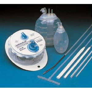
Sometimes patients go home with plumbing attached—like a Jackson-Pratt drain or a T-tube. They cannot manage what they do not understand. Clients must be taught how to correctly measure and empty therapeutic drainage devices at home before discharge. Have them do a "teach-back" demonstration. Watch them squeeze the bulb to re-establish negative pressure. Watch them record the output.
Final Thoughts
When you assess a post-procedure patient, you are evaluating how well they have regained control of their own biology. Is the electrical system (anesthesia) resetting normally? Is the fluid dynamics system (bleeding, urine, chest tubes) maintaining equilibrium? Master these principles of physics and physiology, and you won't just memorize the NCLEX answers—you will understand exactly why they are correct.
Happy studying, and keep asking "why"!