Changes and Abnormalities in Vital Signs
Not sure you’re ready?
Take the ~3-minute readiness diagnostic and see where you stand.
Welcome to the fascinating world of human telemetry. When we take a patient’s vital signs, we aren't just writing numbers on a chart; we are lifting the hood of a highly complex, beautifully engineered machine to see how the engine is running. Every temperature shift, every pulse wave, every breath, and every millimeter of mercury tells a story of adaptation, compensation, or failure.
To master vital signs for the NCLEX-RN, you can't just memorize isolated numbers. You have to understand why the body is doing what it's doing. Let's break down the mechanics of life.
Before we touch a thermometer or a blood pressure cuff, we must understand the overarching principles of physiological monitoring.
First, context is everything. Changes in vital signs must be evaluated in the context of the client's individual baseline measurements. A blood pressure of 90/60 might be a catastrophic drop for a hypertensive patient, but perfectly normal for a 20-year-old marathon runner. If you ever notice a sudden, unexplained change in a client's vital signs, do not wait for the next scheduled rounds. A sudden, unexplained change requires immediate nursing reassessment.
We also have to trust our clinical skills over our electronics. Machines get confused. Therefore, the nurse must verify abnormal automated vital sign readings with manual measurements.
Finally, we have to account for the environment and the patient's recent actions:
- Acute pain stimulates the sympathetic nervous system (your "fight or flight" response) to increase the heart rate and increase blood pressure.
- Smoking causes transient increases in blood pressure and heart rate due to systemic vasoconstriction. Because of this, the nurse should wait 15 to 30 minutes to measure vital signs if the client has recently smoked.
Your body is a furnace. It generates heat through metabolism and loses heat to the environment. The normal adult oral temperature range is 36.5 to 37.5 degrees Celsius. But where we measure this heat matters immensely.
If we want accuracy, we have to respect the anatomy. If a client has recently consumed hot or cold liquids, the oral cavity is temporarily altered. The nurse should wait 15 to 30 minutes to measure an oral temperature in this case.
Routes of Measurement
We often compare other routes to the oral baseline:
- Rectal temperatures are generally 0.5 degrees Celsius higher than oral temperatures because they are closer to the heavily insulated core.
- Axillary temperatures are generally 0.5 degrees Celsius lower than oral temperatures because the armpit is exposed to the periphery.
- Tympanic thermometers assess core body temperature via the tympanic membrane (the eardrum), which shares an incredibly rich blood supply with the hypothalamus—the body's thermostat.
The Pathophysiology of Temperature Changes
What happens when the engine runs too hot or too cold?
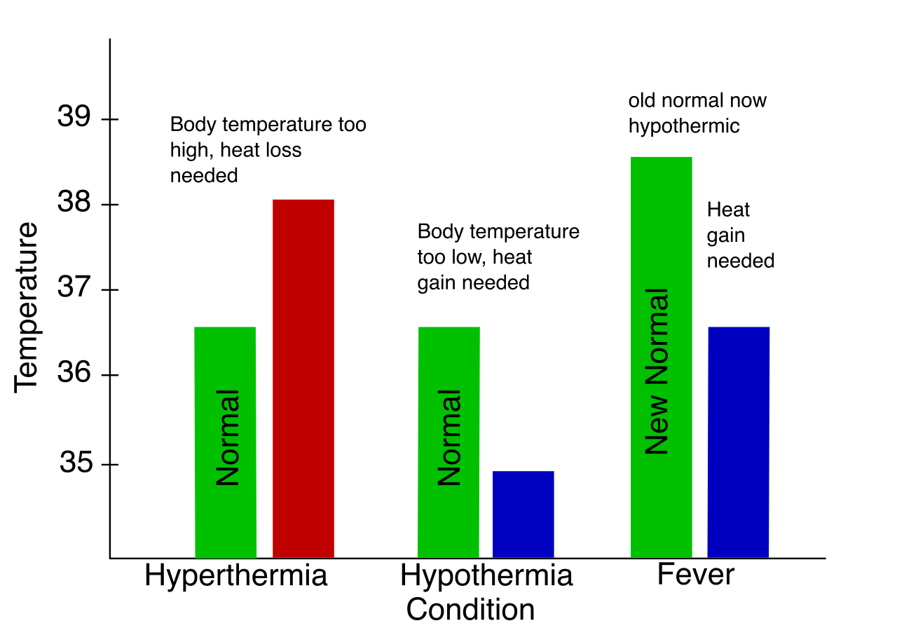
Hyperthermia is an elevation in body temperature above the normal physiological range.
When body temperature is elevated, the body's basal metabolic rate increases. Think of it as a fire burning faster; it needs more oxygen and expels more waste. Therefore, an increased basal metabolic rate leads to a compensatory increase in the heart rate (to deliver the blood) and a compensatory increase in the respiratory rate (to bring in more oxygen).
Hypothermia is defined as a core body temperature below 35 degrees Celsius.
Conversely, freezing the engine slows everything down. Severe hypothermia depresses the myocardium and decreases the heart rate, leading to dangerous bradycardia.
The heart is a pump pushing fluid through elastic pipes. The normal resting heart rate for an adult is 60 to 100 beats per minute.
- Tachycardia is defined as a resting adult heart rate greater than 100 beats per minute.
- Bradycardia is defined as a resting adult heart rate less than 60 beats per minute.
Assessing the Rhythm
We assess the heart centrally (the apical pulse) and peripherally (like the radial pulse). The apical pulse is assessed at the fifth intercostal space at the midclavicular line—right over the apex of the heart. If the client has an irregular rhythm, a quick 15-second check won't cut it. An apical pulse should be counted for one full minute to ensure accuracy in clients with irregular rhythms.
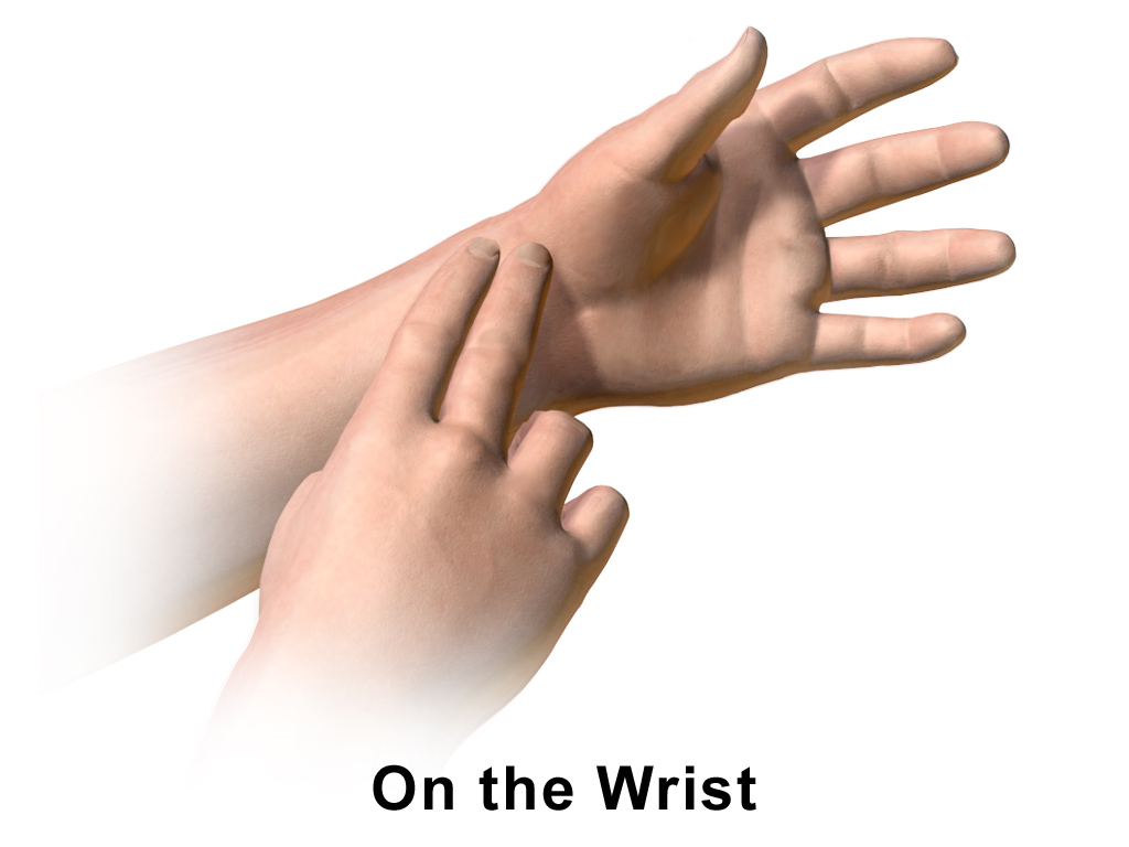
Sometimes, the pump is struggling. A pulse deficit occurs when the apical pulse rate exceeds the radial pulse rate. Why does this happen? A pulse deficit indicates that some cardiac contractions are too weak to produce a palpable peripheral pulse. The heart is beating, but the shockwave never reaches the wrist!
If you cannot feel a peripheral pulse at all, do not immediately panic—use technology. A Doppler ultrasound device is used to assess non-palpable peripheral pulses by bouncing sound waves off moving red blood cells.
Pulse Volume and Compensation
The feel of the pulse tells a hemodynamic story:
- A bounding peripheral pulse feels full and powerful. It indicates increased stroke volume or increased pulse pressure.
- A thready peripheral pulse feels weak and easily obliterated. It indicates decreased stroke volume.
Why does the heart speed up or slow down?
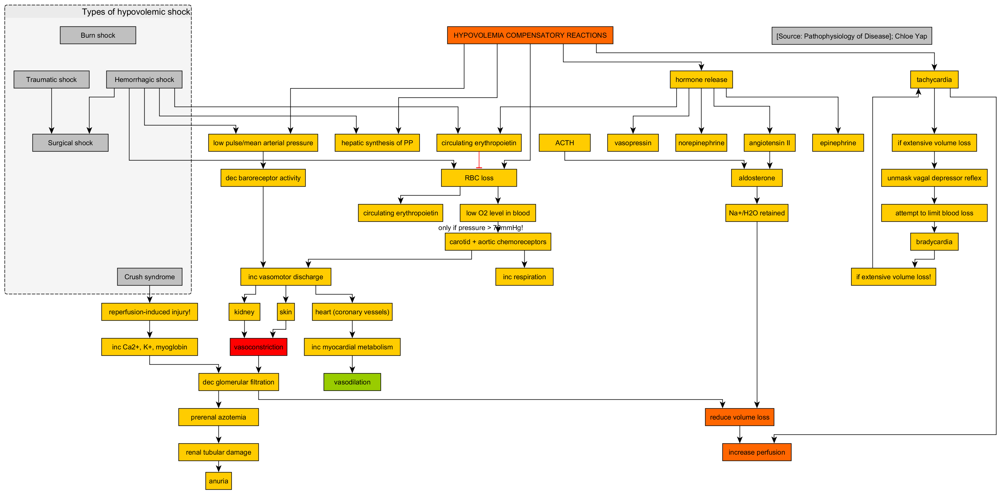
- If the tank is leaking (loss of blood or fluids), hypovolemia causes a compensatory increase in the heart rate to maintain cardiac output.
- On the flip side, bearing down or certain throat stimulations trigger the vagus nerve. Vagal nerve stimulation causes a decrease in the heart rate.
The Shock Profiles
When a patient is crashing, their vital signs tell you how they are crashing:
- New onset tachycardia accompanied by hypotension suggests acute hemorrhage or distributive shock. The tank is empty or inappropriately dilated, so the heart pumps furiously to compensate.
- Neurogenic shock presents uniquely with bradycardia and hypotension. Why? Because the sympathetic nervous system has been severed (usually via spinal cord injury), leaving the parasympathetic nervous system entirely unopposed.
The normal resting respiratory rate for an adult is 12 to 20 breaths per minute.
- Tachypnea is a respiratory rate greater than 20.
- Bradypnea is a respiratory rate less than 12.
Psychomotor tip: If someone knows you are watching them breathe, they will consciously change their breathing pattern. Therefore, respiratory assessment should occur without the client realizing their breathing is being monitored (often done while pretending to still take their radial pulse).
What Drives the Breath?
- Hypoxia (low oxygen) stimulates the peripheral chemoreceptors to increase the respiratory rate. The body is gasping for air.
- Conversely, opioid medications depress the central nervous system and cause a decrease in the respiratory rate. This is why we carefully monitor respiratory rates in post-operative patients on morphine.
Abnormal Breathing Patterns
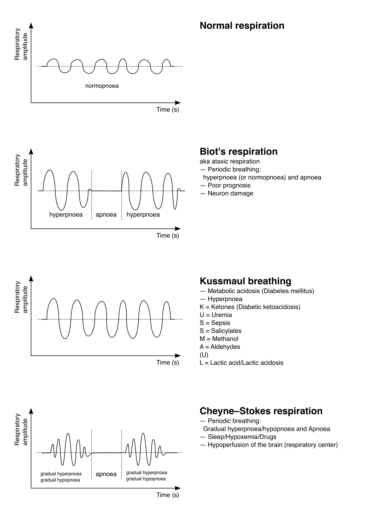
Nature provides brilliant, yet alarming, visual cues when acid-base or neurological balances fail.
| Pattern | Description | Pathophysiological Meaning |
|---|---|---|
| Cheyne-Stokes | Alternating periods of deep breathing and apnea. | Often seen in dying patients or severe brain injury (the brainstem's respiratory center is failing). |
| Kussmaul | Abnormally deep and rapid respirations. | A compensatory mechanism for metabolic acidosis. (e.g., Diabetic Ketoacidosis). The body is desperately trying to blow off acidic carbon dioxide (CO2). |
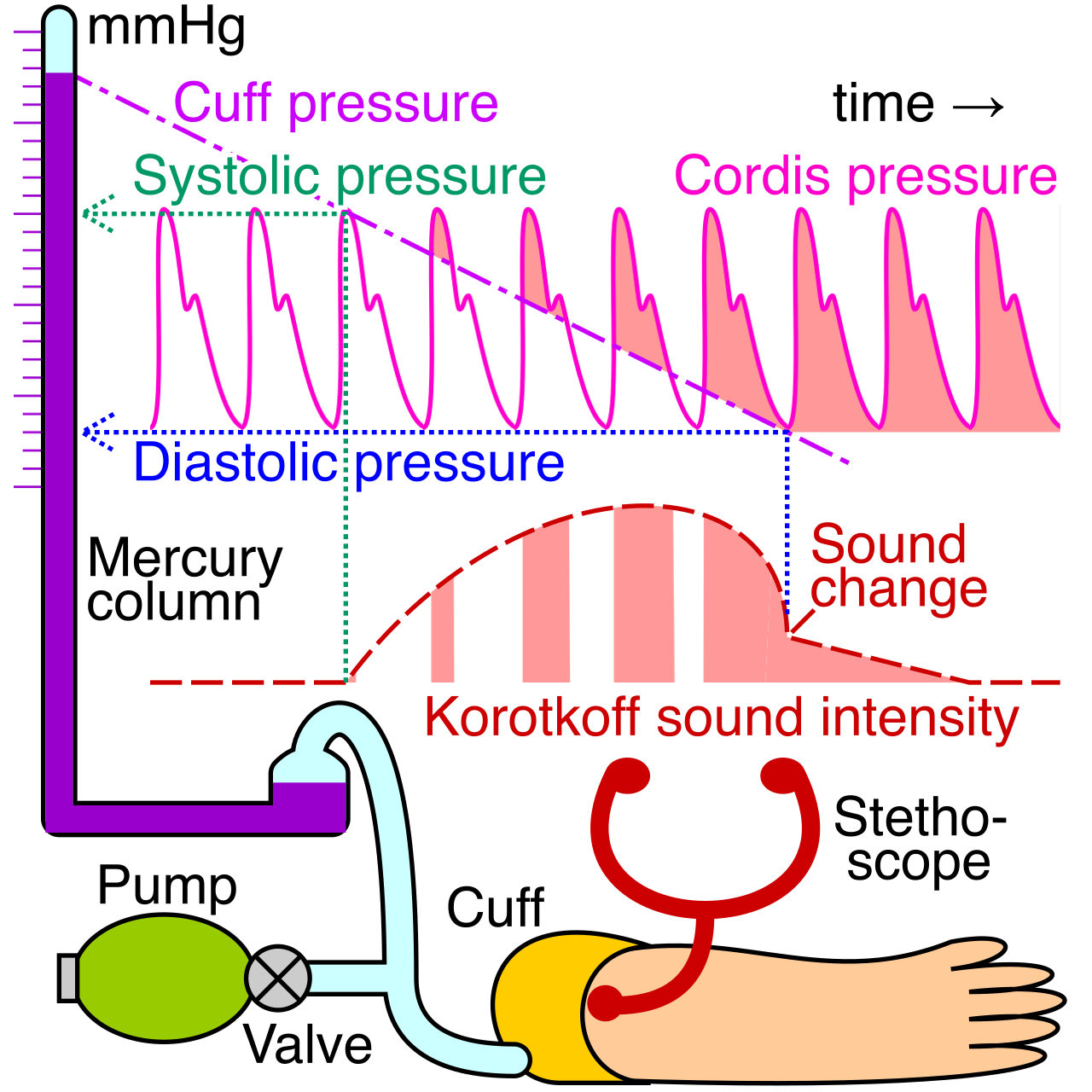
Blood pressure is the tension of the fluid against the pipes. Normal adult systolic blood pressure is less than 120 millimeter of mercury, and normal adult diastolic blood pressure is less than 80 millimeter of mercury.
The difference between these two numbers is vital. Pulse pressure is the numerical difference between the systolic and diastolic blood pressure values. A wide pulse pressure can indicate increased intracranial pressure, while a narrow one can indicate heart failure or hypovolemia.
The Mechanics of the Hemodynamics
The pipes and the blood volume strictly control the pressure:
- Decreased blood volume causes a decrease in blood pressure.
- Systemic vasodilation (opening the pipes) causes a decrease in blood pressure.
- Systemic vasoconstriction (clamping the pipes) causes an increase in blood pressure.
The Psychomotor Physics of the BP Cuff
If you measure BP incorrectly, you are treating a phantom problem. The cuff must fit the arm perfectly based on pure physics:
- The width of the blood pressure cuff bladder should cover forty percent (40%) of the client's arm circumference.
- The length of the blood pressure cuff bladder should encircle eighty percent (80%) of the client's arm circumference.
What happens if you use the wrong size?
- A blood pressure cuff that is too narrow will result in an artificially HIGH blood pressure reading. (It takes too much pressure to force a tiny cuff to occlude the artery).
- A blood pressure cuff that is too wide will result in an artificially LOW blood pressure reading. (The wide cuff occludes the artery too easily).
Positioning and deflation speed are equally critical:
- The client's arm should be positioned at the level of the heart.
- Positioning the arm ABOVE the heart level causes a falsely LOW reading (fighting gravity).
- Positioning the arm BELOW the heart level causes a falsely HIGH reading (gravity adds pressure).
- Crossing the legs during measurement causes a falsely elevated systolic reading due to compressed venous return in the lower body translocating blood upward.
- Deflating the cuff too slowly causes venous congestion in the arm, skewing the numbers.
- Deflating the cuff too quickly results in a falsely LOW systolic reading and a falsely HIGH diastolic reading. (You miss the first sound, and you mistake the muffled sounds for the complete cessation of sound).
Pathological Blood Pressure Phenomena
Orthostatic Hypotension
When you stand up, gravity pulls your blood into your legs. A healthy body instantly vasoconstricts to push it back up. If this reflex fails:
Orthostatic hypotension involves a drop in systolic blood pressure of at least 20 mmHg OR a drop in diastolic blood pressure of at least 10 mmHg when moving from supine to standing.
To prove this, measuring orthostatic blood pressure requires assessing blood pressure and heart rate in the supine, sitting, and standing positions.
Paradoxical Blood Pressure (Pulsus Paradoxus)
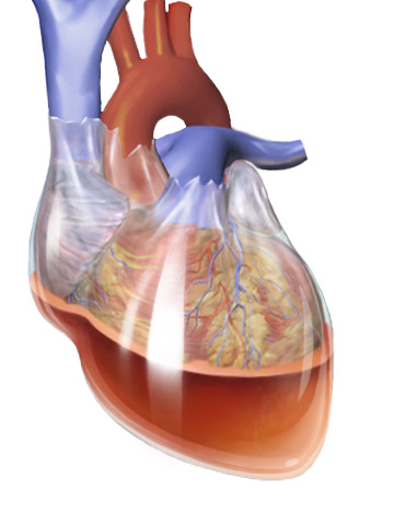
Paradoxical blood pressure is a decrease in systolic blood pressure of more than 10 millimeter of mercury during inspiration.
Why? When you breathe in, negative pressure pulls blood into the right side of the heart. If fluid is trapped in the sac around the heart (cardiac tamponade), the bulging right ventricle crushes the left ventricle, causing a massive drop in systemic output. This makes paradoxical blood pressure a classic clinical sign of cardiac tamponade.
Cushing's Triad
If the pressure inside the skull (intracranial pressure) becomes massive, the brain desperately tries to keep blood flowing in against the squeeze. Severely increased intracranial pressure causes an increase in systolic blood pressure.
If you see the following three signs, the brainstem is herniating. Cushing's triad is a late sign of severely increased intracranial pressure:
- Systolic hypertension with a widening pulse pressure
- Pathological bradycardia
- Irregular respirations
Pulse oximetry measures the red blood cells, which act as boxcars carrying oxygen. Specifically, pulse oximetry measures the percentage of hemoglobin binding sites occupied by oxygen.
A normal pulse oximetry reading in a healthy adult is 95 percent to 100 percent.
Pitfalls of the Pulse Ox
The pulse oximeter relies on light absorption. Therefore, things that block or alter light reception will trick the machine:
- Dark nail polish can interfere with accurate pulse oximetry readings on the fingers. (Remove it!).
- Poor peripheral perfusion causes inaccurate pulse oximetry readings. If the fingers are freezing cold or the patient is in shock, blood isn't reaching the sensor.
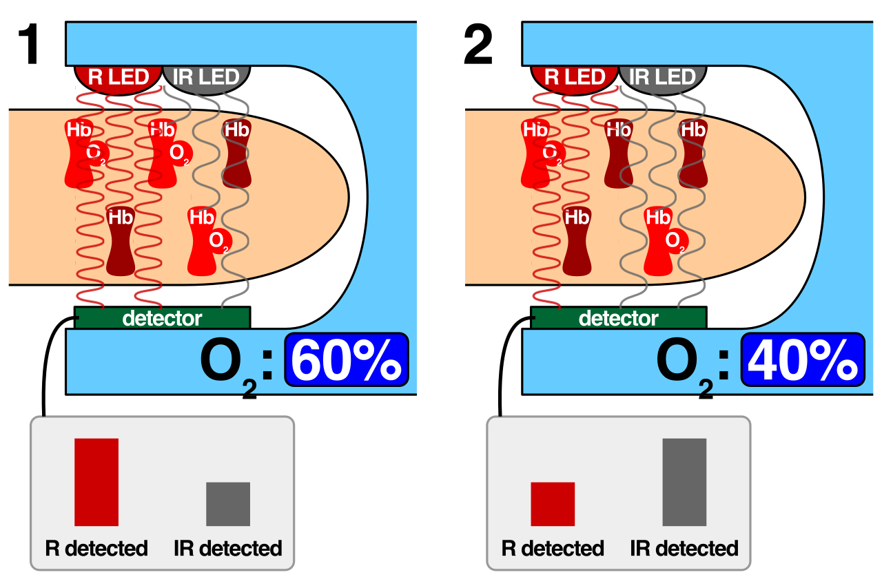
The Carbon Monoxide Trap: Carbon monoxide (CO) has an affinity for hemoglobin that is 200 times stronger than oxygen. It will bully oxygen off the binding sites. The pulse oximeter only sees that the "boxcars" are full, but it cannot tell they are full of poison. Therefore, carbon monoxide poisoning produces falsely elevated pulse oximetry readings.
Finally, if the machine isn't lying, you must act fast. An acute decrease in oxygen saturation requires immediately assessing the client's airway patency. Always look at your patient first. Is their airway open? Are they choking? Secure the plumbing before you stare at the monitor.
Conclusion
Vital signs are the ultimate physiological report card. When you see a high heart rate, ask yourself: Is this pain? Is it hypovolemia? Is it compensation for a fever? When you see a low blood pressure, ask yourself: Is my cuff too wide, or is this patient going into shock?
Apply the physics, trust your manual assessments, and always, always trace the vital sign back to the underlying physiology. That is how you think like an elite nurse, and that is how you conquer the NCLEX-RN.