Diagnostic Tests
Not sure you’re ready?
Take the ~3-minute readiness diagnostic and see where you stand.
The Physiology of Clues: A Bedside Scientist’s Guide to Diagnostic Testing
Let’s get right to the heart of the matter. When you step onto the nursing unit, you are not merely a technician pushing buttons on a machine or drawing fluid into a tube. You are a scientist at the bedside. The human body is an endlessly fascinating, exquisitely tuned biological engine. When it breaks down, it doesn’t usually send us an email; it gives us clues. Drops in pressure, erratic electrical rhythms, strange proteins spilling into the urine.
Diagnostic testing is how we listen to those clues. Our job is to apply nursing procedures, perform the tests, compare the findings, and act on what the physiology is screaming at us. Let’s explore exactly how you intercept these signals and save lives.
Think of the heart as a brilliant, rhythmic electrical storm. Before the muscle can pump blood, an electrical signal has to tell it to do so. An electrocardiogram (ECG) records the electrical activity of the heart to identify arrhythmias and myocardial ischemia. It shows us if the wiring is short-circuiting or if a part of the heart is starving for oxygen.
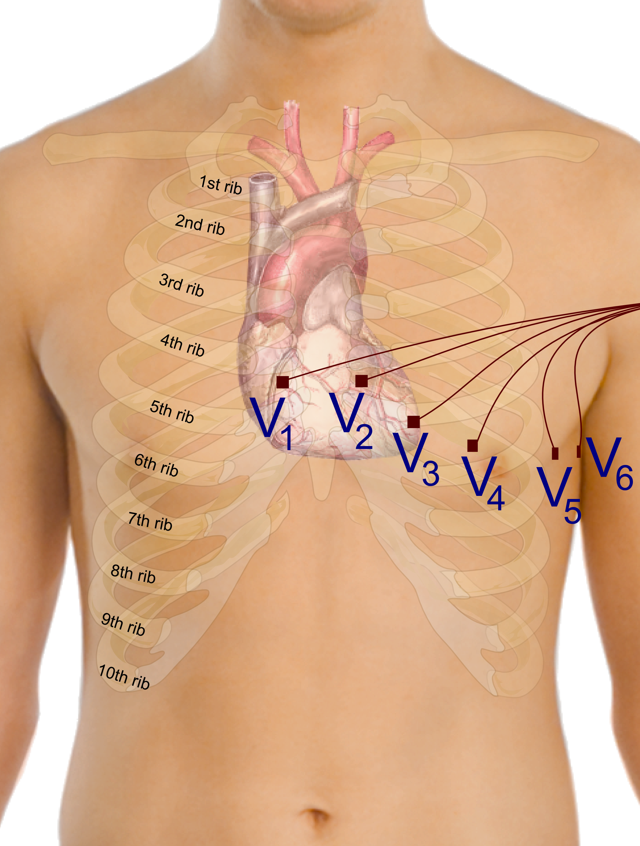
And who is the chief electrician? You are. Registered nurses are authorized to perform 12-lead electrocardiograms. Placing the leads isn't random; it’s like setting up microphones around a stage to capture the sound from every possible angle. To get an accurate reading, you must be structurally precise:
- The V1 electrode of a 12-lead electrocardiogram is placed at the fourth intercostal space on the right sternal border.
- The V2 electrode of a 12-lead electrocardiogram is placed at the fourth intercostal space on the left sternal border.
- The V4 electrode of a 12-lead electrocardiogram is placed at the fifth intercostal space at the midclavicular line. (We skip right to V4 because V3 sits halfway between V2 and V4).
- The V6 electrode of a 12-lead electrocardiogram is placed at the fifth intercostal space at the midaxillary line.
The Potassium Connection
Because this system runs on electricity, it is incredibly sensitive to the ions in our blood—most notably, potassium. Potassium is responsible for repolarization, the "reset" phase of the heart cell's electrical cycle.
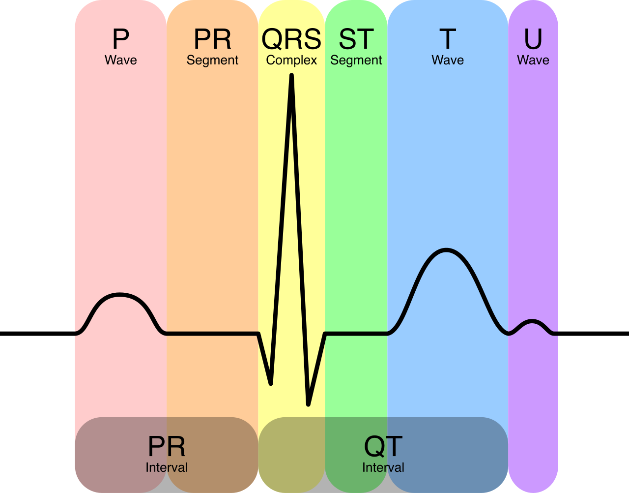
Feynman Rule of Thumb: Changes in potassium change the "T wave" (the electrical reset).
- A serum potassium level greater than 5.0 mEq/L requires electrocardiogram monitoring for tall peaked T waves. If there's too much potassium, the reset is violently fast, causing the T wave to shoot up like a tent pole.
- A serum potassium level less than 3.5 mEq/L requires electrocardiogram monitoring for U waves. If there's too little potassium, the reset is sluggish and delayed, creating an entirely extra hump—the U wave.
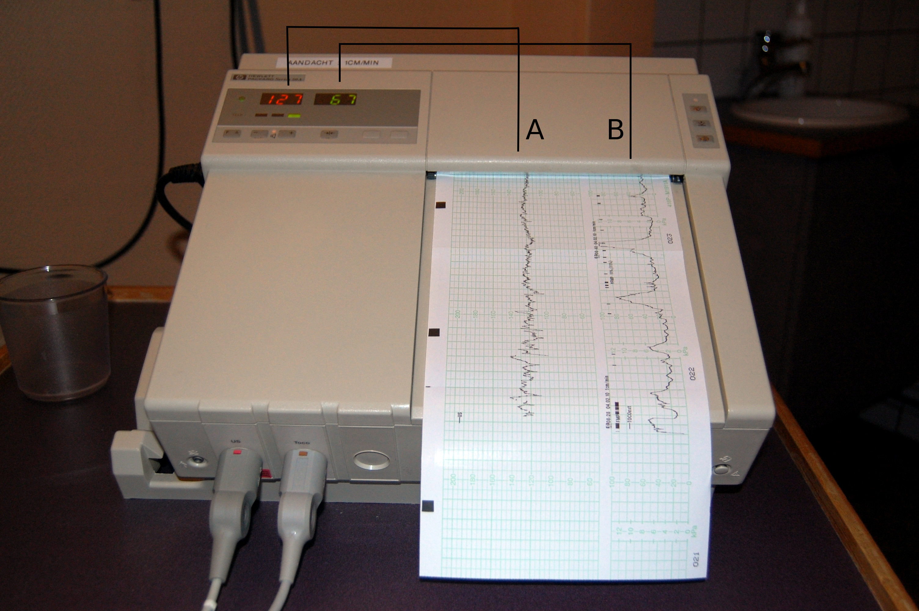
When we monitor a pregnant patient, we are essentially trying to eavesdrop on a life we cannot see. We do this by measuring stress and oxygenation.
A continuous fetal monitor evaluates the relationship between fetal heart rate patterns and uterine contractions. Every time the uterus contracts, it momentarily squeezes the blood vessels, acting as a natural stress test on the baby. A normal fetal heart rate baseline is between 110 and 160 beats per minute.
When the heart rate drops during a contraction, we call it a deceleration. The timing of that drop tells you exactly what the anatomy is doing.
Decoding Decelerations
| Deceleration Type | What it Indicates | The Physics/Anatomy | Nursing Intervention |
|---|---|---|---|
| Early | Fetal head compression | The baby's head is squeezed by the birth canal, causing a vagal response. It matches the contraction perfectly. | Nature at work! Early decelerations in fetal heart rate do not require acute nursing interventions. |
| Variable | Umbilical cord compression | The baby or uterus is randomly squeezing the "hose" (the umbilical cord). The drops look sharp, like V's or W's. | Reposition the mother to get the weight off the cord. |
| Late | Uteroplacental insufficiency | The placenta is failing. The baby runs out of oxygen after the peak of the contraction, causing a delayed, late drop. | CRITICAL ACTION REQUIRED. |
Late decelerations in fetal heart rate indicate uteroplacental insufficiency. The baby is drowning in the physiological equivalent of thin air. You must step in to restore oxygen delivery immediately!
- Nursing interventions for late fetal heart rate decelerations include repositioning the pregnant patient onto her left side. This removes the heavy uterus from the maternal vena cava, instantly boosting blood return to the heart and placenta.
- Nursing interventions for late fetal heart rate decelerations include administering supplemental oxygen to the pregnant patient.
- Nursing interventions for late fetal heart rate decelerations include discontinuing intravenous oxytocin infusions. Oxytocin causes contractions. If contractions are starving the baby of oxygen, you must stop the drug causing them!
Fetal Diagnostic Tests
Sometimes, we want to check on the baby before labor begins. We use two main tests to evaluate fetal well-being:
- The Nonstress Test (NST): Think of this as checking if a resting baby is healthy. A nonstress test evaluates fetal well-being by monitoring fetal heart rate in response to fetal movement. Just as your heart rate jumps when you run up the stairs, a healthy fetus's heart rate jumps when it kicks. A reactive nonstress test indicates adequate fetal oxygenation. (Reactive = Good!)
- The Contraction Stress Test (CST): This is a deliberate challenge. A contraction stress test evaluates the ability of the fetus to tolerate the physiological stress of uterine contractions. We induce mild contractions to see how the placenta holds up.
- A negative contraction stress test indicates the absence of late fetal heart rate decelerations during uterine contractions. (Negative = Good! The baby passed the test).
- A positive contraction stress test is an abnormal finding indicating late decelerations with at least half of the monitored uterine contractions. (Positive = Bad! The placenta is failing).
In nursing, static numbers are rarely as important as trends. A single snapshot only tells you where the patient is, but a trend tells you where they are going. Nurses must compare current laboratory values to baseline results to identify clinical deterioration.
- A progressive elevation in serum creatinine levels indicates worsening renal function. The kidneys are the body's filtration system. If creatinine (waste) is building up in the blood, the filter is clogged or dying.
- An upward trend in cardiac troponin levels indicates progressive myocardial tissue injury. Troponin belongs inside heart muscle cells. If you find it rising in the blood, it means heart cells are bursting open and dying.
- A downward trend in hemoglobin levels suggests active internal or external hemorrhage. If hemoglobin is progressively vanishing, the blood is leaking out somewhere—either onto the floor or into a body cavity. You must find the leak.
Whenever these numbers hit emergency thresholds, you are the crucial link in the chain of survival. Nurses must notify the primary healthcare provider of any critical laboratory value immediately.
Coagulation Dynamics
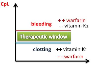
We routinely manipulate the blood's ability to clot using medications like Heparin and Warfarin. But if we overshoot, the patient could bleed to death. You must watch the dials:
- An activated partial thromboplastin time (aPTT) exceeding the therapeutic range in a patient receiving heparin requires a decrease in the heparin infusion rate.
- An international normalized ratio (INR) greater than the prescribed therapeutic target requires an assessment for active bleeding. Always check the patient first—look for bleeding gums, bruising, or blood in the urine.
Let's talk about the radiology department. When a patient needs a CT scan, we often inject intravenous (IV) contrast dye to make the blood vessels glow on the scan. But there is a biological cost: these contrast molecules are incredibly heavy and hard on the kidneys. Intravenous contrast dye can cause acute kidney injury (AKI).
Because of this risk, nurses must verify serum creatinine and blood urea nitrogen (BUN) levels before a patient receives intravenous contrast dye. If the kidneys are already struggling, the contrast dye could tip them into failure.
Here is a brilliant piece of pharmacology you must never forget: the diabetes drug Metformin. If a patient develops an acute kidney injury from contrast dye while taking Metformin, their kidneys will stop excreting the Metformin. The drug will build up to toxic levels. Metformin combined with intravenous contrast dye increases the risk of lactic acidosis, a deadly metabolic state. Therefore, Metformin must be withheld for 48 hours after the administration of intravenous contrast dye until we prove the kidneys survived the insult.
After invasive diagnostic procedures, nursing care becomes an exercise in applying basic physics—gravity, pressure, and fluid dynamics—to prevent catastrophic complications.
Liver Biopsy
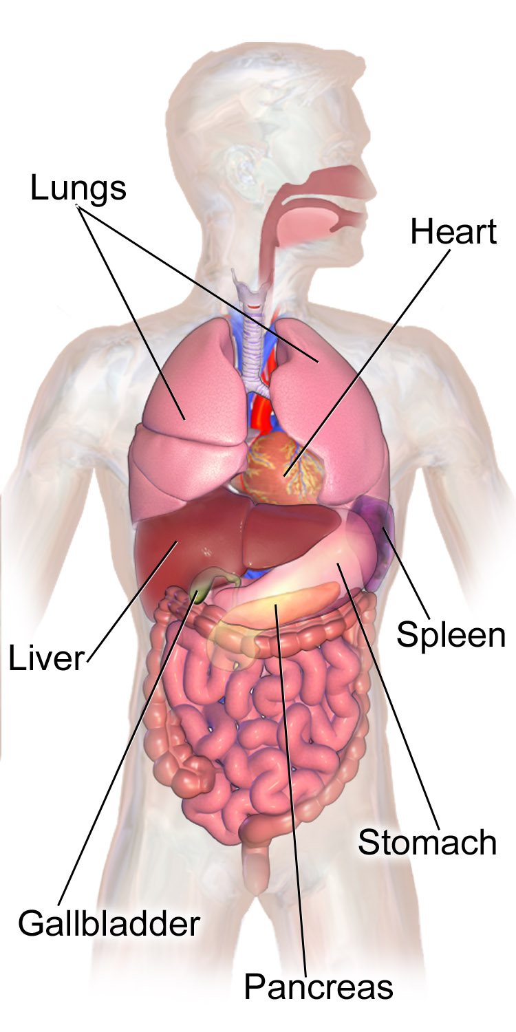
When a doctor punches a needle into the liver to take a sample, they are poking an organ heavily engorged with blood. Patients must lie on their right side after a liver biopsy. Why? Because the liver sits on the right side of the abdomen. Lying on the right side after a liver biopsy provides anatomical pressure to the biopsy site to prevent hemorrhage. You are literally using the weight of the patient's own body to hold pressure on the internal wound!
Lumbar Puncture
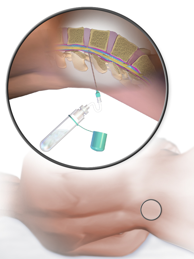
We tap the spine to test the cerebrospinal fluid (CSF). But when we pull the needle out, we leave a tiny hole in the dural sac. If the patient sits up, gravity will pull the CSF out of that hole, dropping the fluid pressure around the brain. Patients must lie supine for several hours after a lumbar puncture. The physics are simple: Lying supine after a lumbar puncture reduces the risk of a post-dural puncture headache caused by cerebrospinal fluid leakage.
Thoracentesis
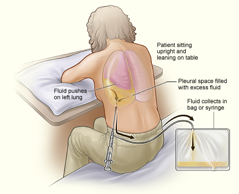
When we insert a needle into the pleural space to drain fluid around the lungs, there is always a risk we might accidentally nick the lung itself, allowing air to escape into the chest cavity. Because of this, nurses must monitor the patient for a pneumothorax after a thoracentesis. How do we know for sure that the lung is safe? A chest radiograph is required to confirm the absence of a pneumothorax after a thoracentesis.
Bronchoscopy
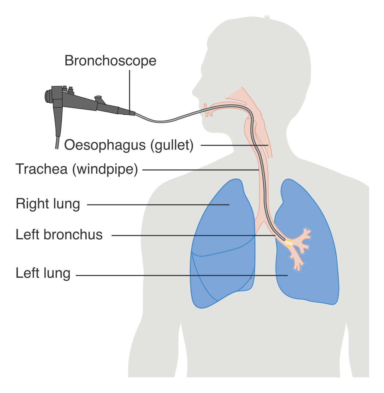
During a bronchoscopy, we numb the back of the patient's throat to slide a camera down into the lungs. If you give that patient a glass of water immediately afterward, the water will glide straight past the numb airway and into the lungs, causing aspiration pneumonia. Nurses must assess the gag reflex before allowing oral intake after a bronchoscopy. No gag, no water. Period.
Finally, never forget how much diagnostic power you hold right at the bedside. You do not always need to wait for the central laboratory or the radiology department. You have tools at your immediate disposal.
- Registered nurses are authorized to perform bedside blood glucose monitoring. In seconds, you can identify if a comatose patient is simply profoundly hypoglycemic.
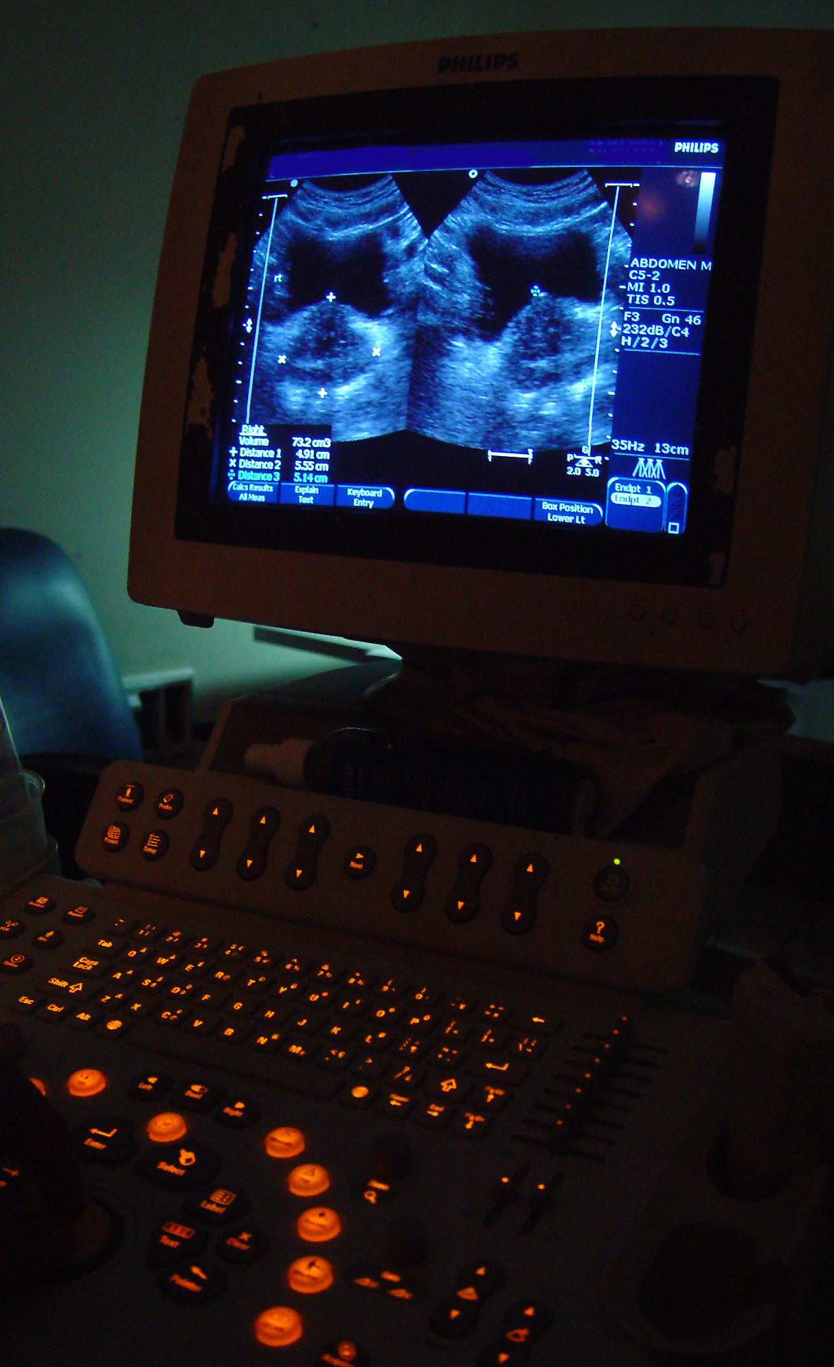
- Registered nurses are authorized to perform bedside bladder ultrasound scans. If a patient hasn't voided in 8 hours, is their kidney failing, or is their bladder just full? A bedside bladder ultrasound measures the volume of urine retained in the bladder, instantly solving the mystery.
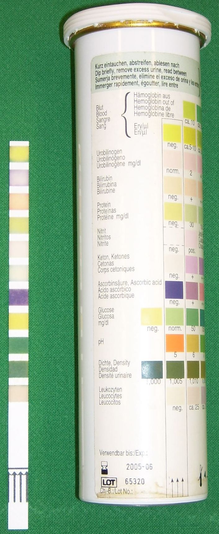
- We also command the urinalysis. Urine dipstick testing provides immediate identification of protein in the urine, which could warn us of preeclampsia in a pregnant patient or kidney damage. Furthermore, urine dipstick testing provides immediate identification of ketones in the urine, alerting us to starvation states or diabetic ketoacidosis.
The Takeaway
Diagnostic testing isn't just paperwork; it is the fascinating intersection of human physiology, physics, and critical thinking. By performing your scope of practice, watching the trends, and understanding the why behind the procedures, you become the ultimate protector of your patient. You catch the clues before the catastrophe. That is what it means to be an exceptional nurse.