Mobility and Immobility
Not sure you’re ready?
Take the ~3-minute readiness diagnostic and see where you stand.
The Machinery of Motion: A Masterclass on Mobility and Immobility
Welcome! Imagine the human body as a brilliantly designed, high-performance engine. It didn’t evolve to lie flat on a mattress; it evolved to run, hunt, and constantly fight gravity. When you take a patient and put them on prolonged bed rest, you are essentially turning off the engine while leaving the battery running. Physics and chemistry start to turn against the body.
As nurses, we are the mechanics. We have to simulate the effects of gravity and motion, or the machine breaks down. Today, we are going to look under the hood at the complications of immobility. We will uncover exactly why these breakdowns happen and, more importantly, how we fix them. Let’s dive in.
Think of your patient’s circulatory system like a sprawling network of rivers. The heart pumps the blood out, but getting the venous blood back up the legs against gravity? That requires help.
Normally, walking does this. The skeletal muscles in your legs act as a secondary heart. But immobility reduces skeletal muscle contractions, which directly decreases venous return to the heart.
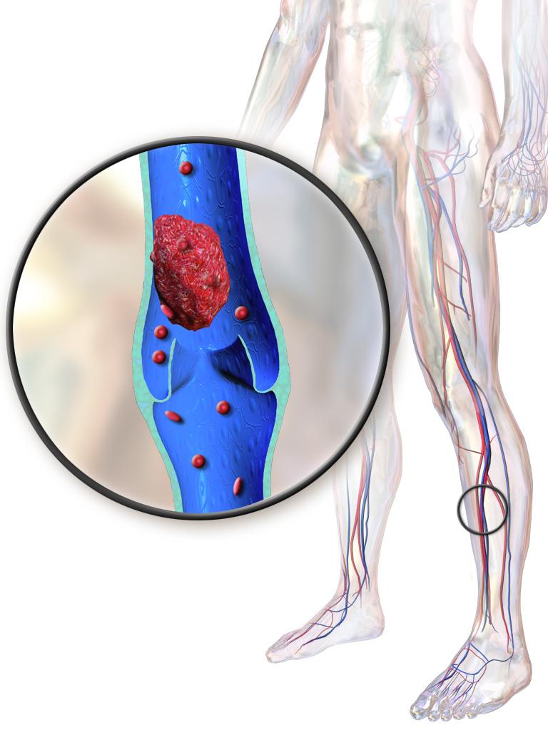
When the river slows down, the water gets stagnant. Decreased venous return causes blood pooling in the lower extremities, and stagnant blood loves to clot. This blood pooling in the lower extremities increases the risk of deep vein thrombosis (DVT). Here is the terrifying part: a deep vein thrombosis can dislodge and travel to the lungs, and a dislodged deep vein thrombosis traveling to the lungs causes a pulmonary embolism. That is a life-threatening crisis.
Engineering the Fix: Promoting Circulation
How do we replace the action of walking? We use gravity, mechanics, and targeted exercise.
- Gravity: Elevating the lower extremities above the level of the heart uses gravity to increase venous return. Water flows downhill.
- Active Mechanics: Have the patient perform ankle pump exercises. These engage the calf muscles to push venous blood upward toward the heart.
- External Compression:
- Sequential compression devices (SCDs) pump air into sleeves to promote venous return from the lower extremities. By constantly squeezing and releasing, sequential compression devices help prevent deep vein thrombosis. But beware: sequential compression devices must be removed periodically to assess the underlying skin for breakdown.
- Anti-embolic stockings apply external mechanical pressure to the legs to promote venous return. To work correctly, anti-embolic stockings must be measured accurately using the circumference and length of the leg.
-
⚠️ The Tourniquet Trap: Never fold these stockings! Rolling the top band of anti-embolic stockings downward creates a localized tourniquet effect. This is a disaster because the tourniquet effect from rolled anti-embolic stockings impedes venous blood flow back to the heart—the exact opposite of what we want!
The Golden Rule of DVTs: If you suspect a clot, hands off! Massaging the calves of an immobile patient can dislodge an existing deep vein thrombosis.
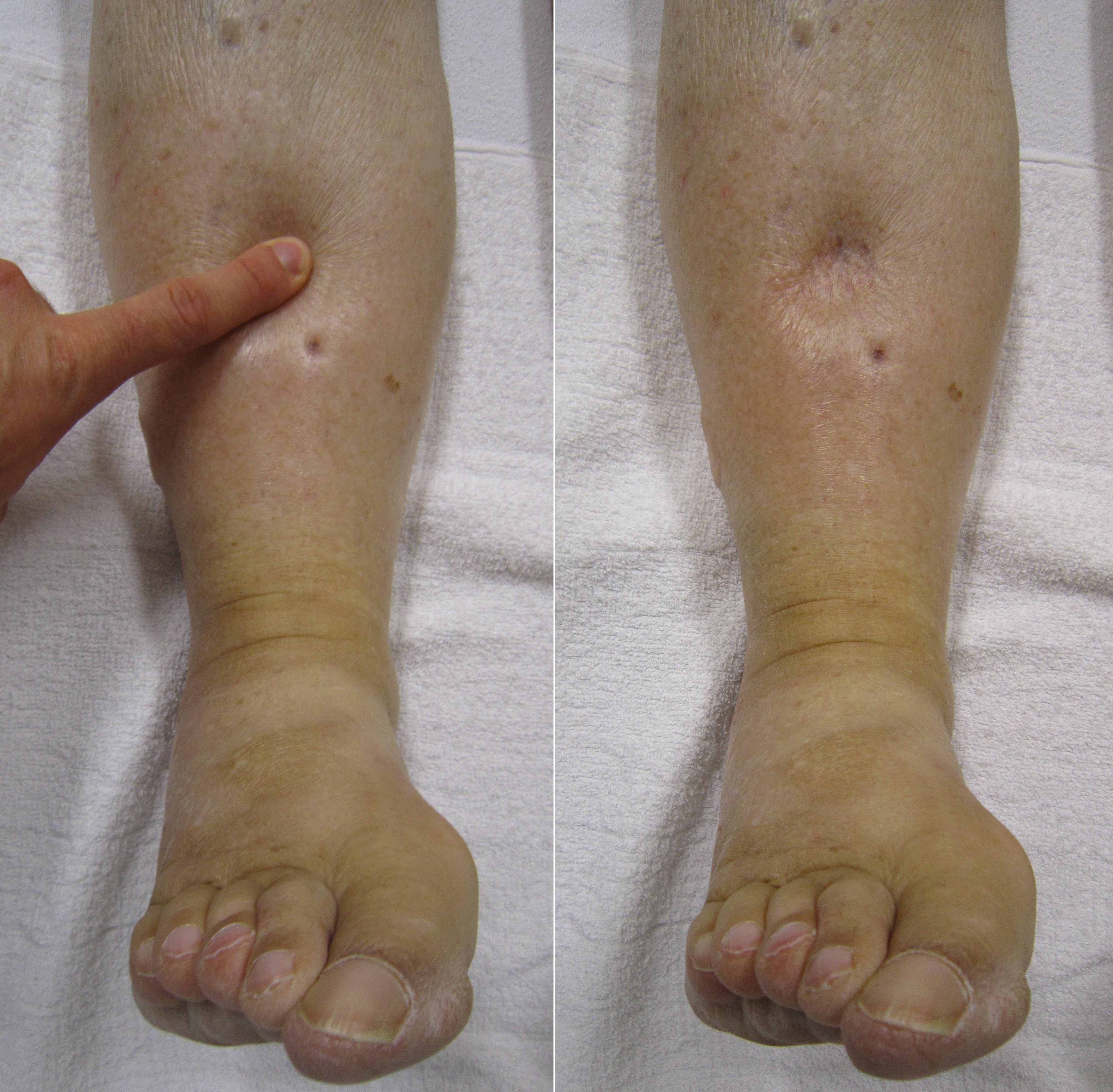
Did it work? How do you know if your plumbing interventions are successful? The nurse evaluates the response to circulation interventions by checking for the absence of peripheral pitting edema. No swelling means the fluid is making it back to the heart.
Let’s talk about blood pressure. In your neck and aorta, you have specialized sensors called baroreceptors. When you stand up, gravity tries to pull all your blood into your boots. The baroreceptors instantly sense the drop in pressure and constrict your vessels to keep blood in your brain.
But prolonged bed rest reduces the baroreceptor reflex response. They basically fall asleep on the job. Because of this, a reduced baroreceptor reflex response causes orthostatic hypotension upon standing.
Definition: Orthostatic hypotension is a drop in systolic blood pressure of at least 20 millimeters of mercury when moving from a lying to a standing position. When you stand your patient up, do it slowly!
Your lungs are a pair of bellows. To pull air into the deep tissues, your chest cavity has to expand. But lying flat makes the chest heavy and restricted. Immobility decreases respiratory chest expansion.
This mechanical failure causes two massive problems:
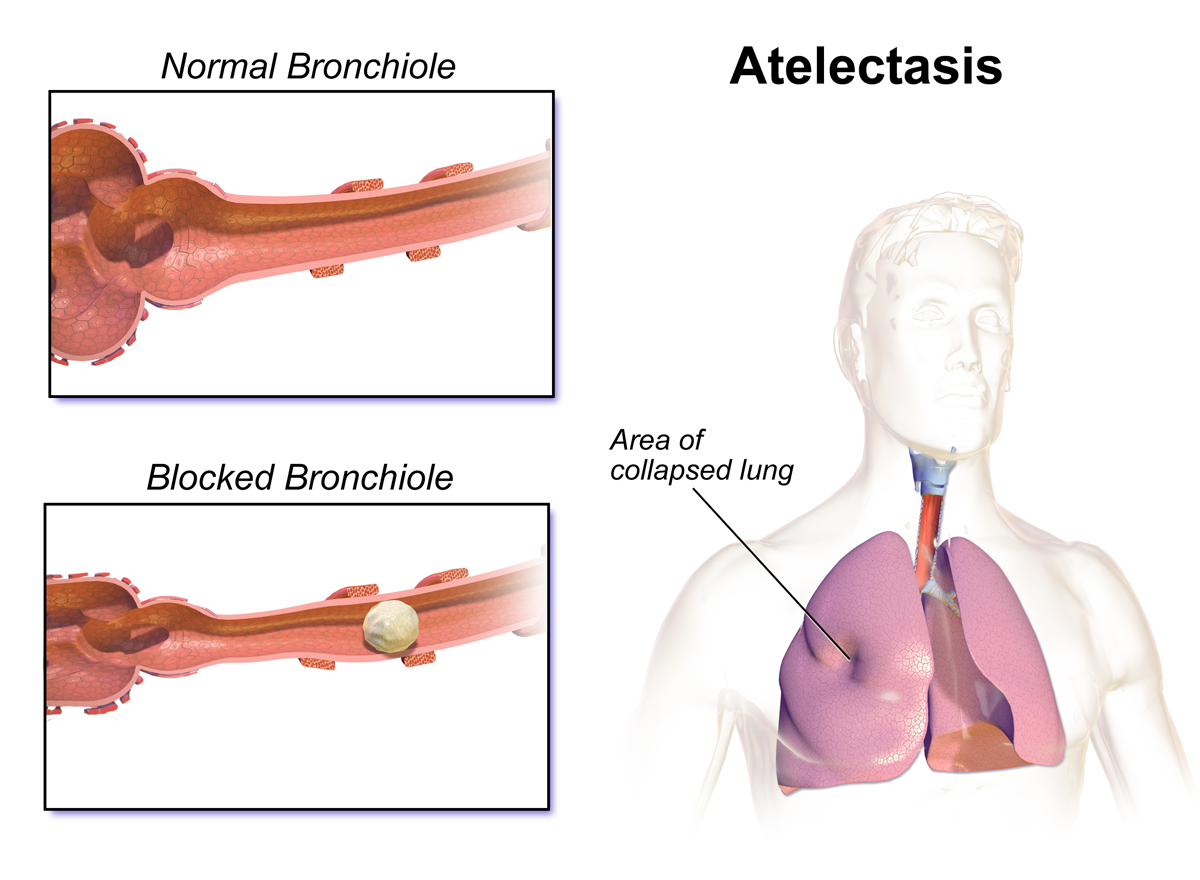
- The Collapse: Decreased respiratory chest expansion increases the risk of atelectasis. What is that? Atelectasis is the collapse of lung alveoli. Imagine millions of tiny balloons sticking together and deflating. No alveoli = no gas exchange.
- The Swamp: Without deep breaths, decreased respiratory chest expansion causes pooling of respiratory secretions in the lungs. Bacteria love warm, stagnant swamps. Pooled respiratory secretions increase the risk of hypostatic pneumonia.
Inflating the Bellows
- Positioning: Positioning an immobile patient in a semi-Fowler position facilitates maximum chest cavity expansion. Let the diaphragm drop!
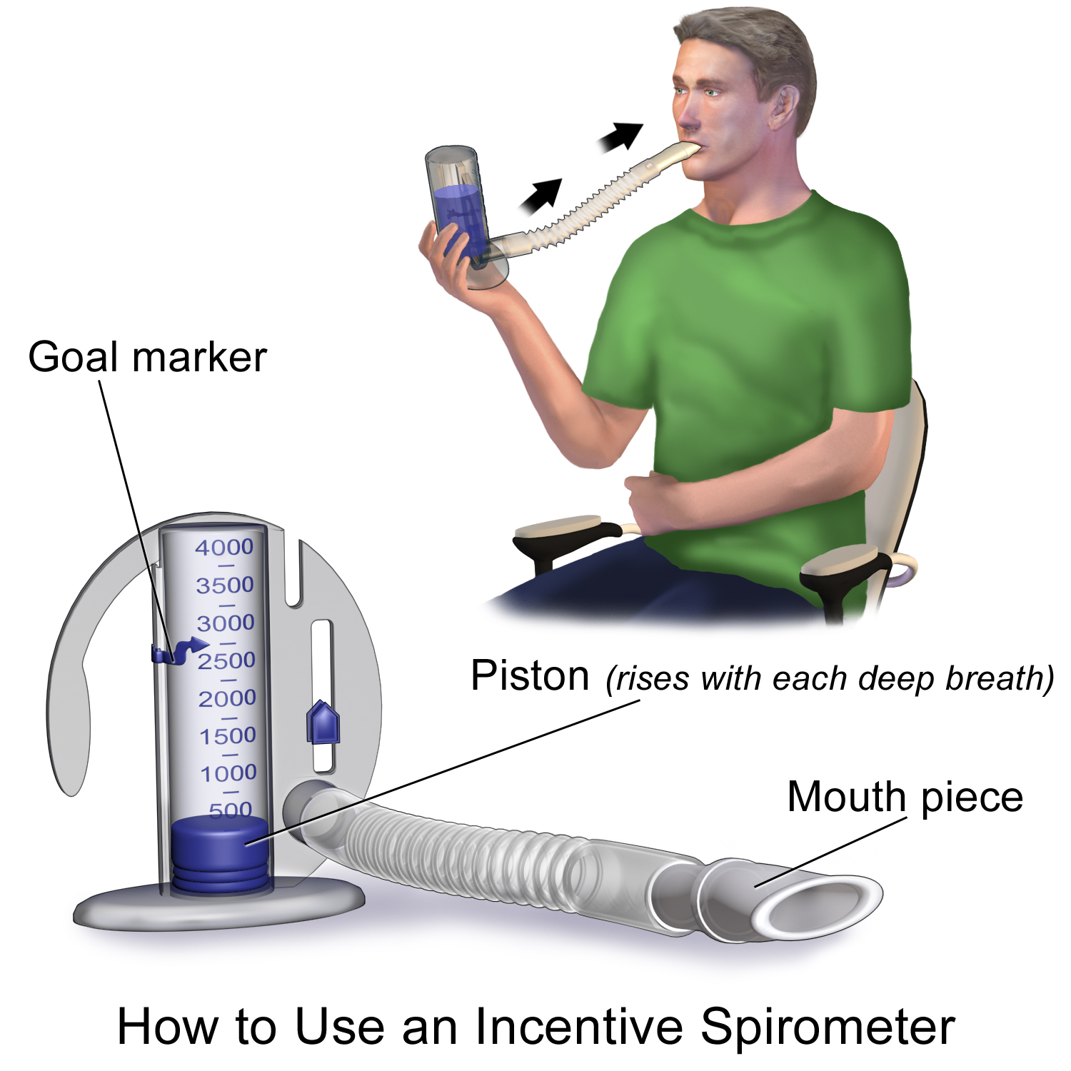
- Mechanics: Incentive spirometry promotes sustained deep breathing. This isn't just about breathing deeply; the negative pressure from deep breathing via incentive spirometry inflates the alveoli to prevent atelectasis in immobile patients.
- Chemistry: You must give them fluids! Adequate oral fluid intake thins respiratory secretions in immobile patients. Why? Because thinner respiratory secretions are easier for an immobile patient to expectorate through coughing.
A body at rest shuts down its internal factories. Actually, early ambulation accelerates the return of normal gastrointestinal and respiratory function. But if they can't walk, here is what happens:
- The Gut: Immobility decreases gastrointestinal peristalsis, and decreased gastrointestinal peristalsis increases the risk of constipation.
- The Kidneys: Urine doesn't drain well when lying flat. Immobility causes urinary stasis in the renal pelvis. This stagnant pool means that urinary stasis increases the risk of urinary tract infections.
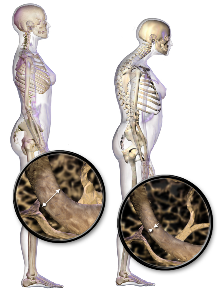
- The Bones: Nature is a ruthlessly efficient accountant. If you don't use your bones to bear weight, the body stops investing in them. Immobility alters bone metabolism, and altered bone metabolism from immobility causes calcium to leach out of the bones. This loss of structural integrity means calcium leaving the bones leads to disuse osteoporosis. Furthermore, all that calcium dumps into the bloodstream. Increased serum calcium from bone breakdown increases the risk of renal calculi (kidney stones).
- Nutrition: Without movement, muscle atrophies. Nitrogen balance becomes negative during immobility due to extensive muscle tissue breakdown. A negative nitrogen balance delays physiological wound healing. To fix this metabolic deficit, increased dietary protein intake is required to correct a negative nitrogen balance associated with immobility.
Your skin is a living organ that requires a constant supply of blood. But prolonged physical pressure on tissues reduces local blood flow. When blood flow stops, oxygen stops. Reduced local blood flow leads to tissue ischemia, and unrelieved tissue ischemia causes pressure injuries.
We use data to fight this. The Braden Scale assesses the risk of a patient developing a pressure injury. Remember the six domains it checks: The Braden Scale evaluates sensory perception, moisture, activity, mobility, nutrition, and friction.
Pro Tip: On the Braden Scale, a lower numerical score indicates a higher risk for pressure injuries. A score of 9 is a red alert; a 23 means they are perfectly fine.
The Assessment and the Fix
Skin assessments must include thorough inspection of bony prominences, specifically where the skeleton presses hardest against the mattress. Common bony prominences susceptible to pressure injuries include the sacrum, heels, elbows, and occiput (back of the head). If you push on a red spot and it stays red, you have found trouble. Non-blanchable erythema of intact skin indicates a Stage 1 pressure injury.
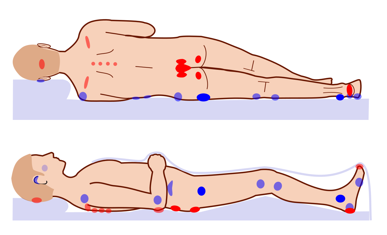
But pressure isn't our only enemy. We also fight physics in the form of shear. Shearing forces occur when the epidermis remains stationary against a surface while the underlying tissues shift. Imagine the top layer of skin gripping the sheets while the heavy skeleton slides downward. Shearing forces physically tear blood vessels in the underlying subcutaneous tissue.
- Warning: Because of gravity, elevating the head of the bed greater than 30 degrees increases the risk of a shearing injury on the sacrum. Keep them positioned carefully!
Repositioning an immobile patient relieves mechanical pressure on bony prominences.
- Immobile patients lying in a bed must be repositioned at least every two hours.
- Immobile patients sitting in a chair must shift body weight at least every fifteen minutes.
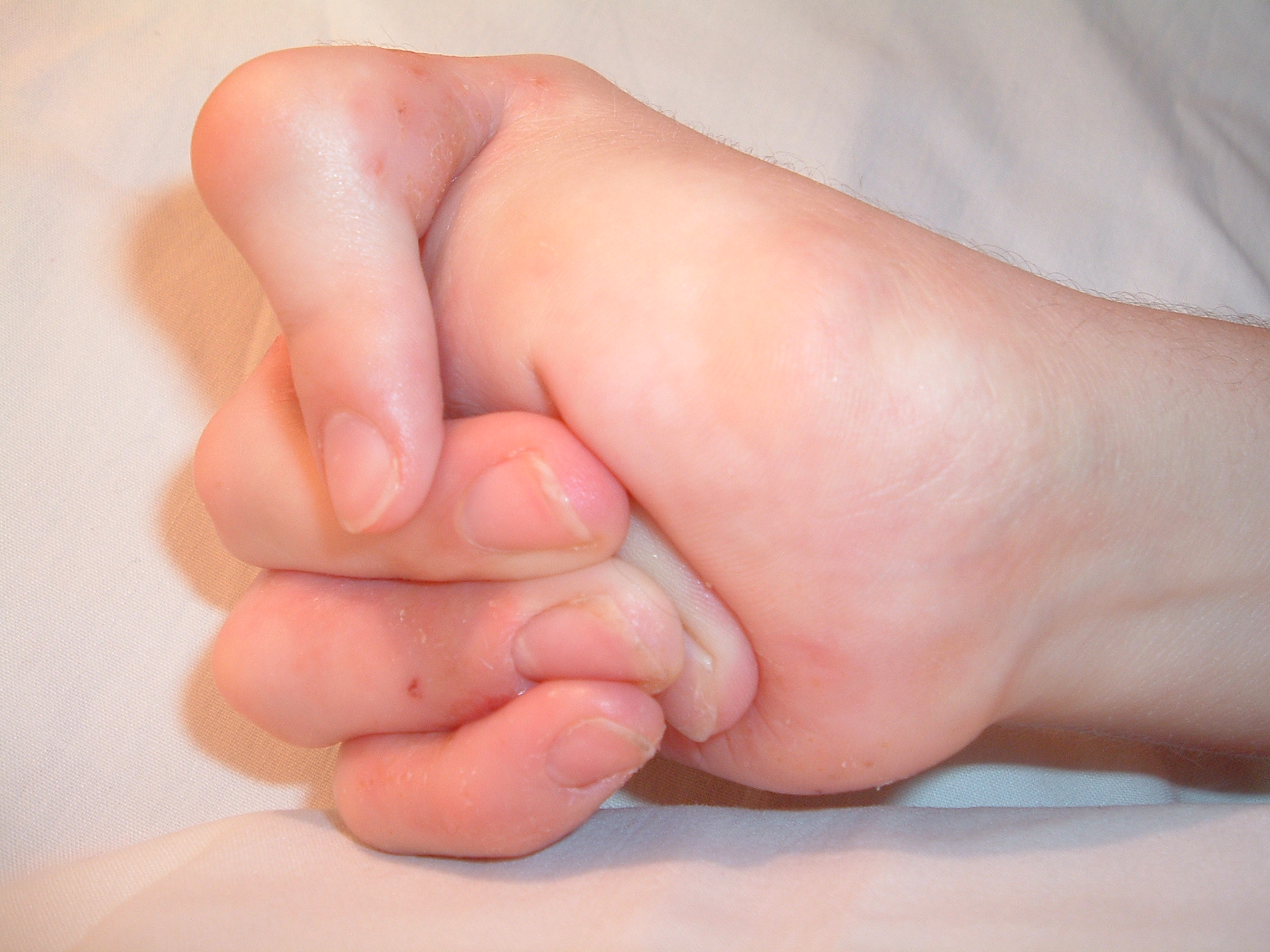
Joints are made to move. If left bent or unused, they permanently freeze. Joint contractures result from the permanent shortening of muscle fibers and connective tissue. The body literally sews the joint shut.
How do we stop it? Motion! Frequent range of motion exercises prevent joint contractures.
- Active range of motion exercises require the patient to independently move a joint through the full extent of movement. Because the patient does the work, active range of motion exercises maintain existing muscle strength.
- Passive range of motion exercises require the nurse to manually move the joints of the patient. This prevents contractures without maintaining muscle strength.
Positioning to Prevent Specific Contractures
| The Device | The Function | The "Why" |
|---|---|---|
| Footboard | Prevents foot drop by maintaining the foot in continuous dorsiflexion. | Foot drop is a permanent joint contracture of the ankle that fixes the foot in a state of plantar flexion. |
| Trochanter Rolls | Prevent external rotation of the hips in a supine patient. | Keeps the femurs aligned straight, mimicking a natural standing posture. |
| Hand Rolls | Maintain the thumb in slight adduction and in opposition to the fingers. | Hand rolls prevent flexion contractures of the fingers, stopping the hand from curling into a permanent, useless fist. |
| Chair Alert | Avoid sitting them up for too long! | Prolonged hip flexion in a seated position increases the risk of hip flexion contractures. |
Sometimes we have to enforce immobility to heal a trauma.
Casts and Compartment Syndrome
Orthopedic casts immobilize a specific body part to facilitate bone healing. But encasing a limb in fiberglass creates a rigid, unforgiving tunnel. If the tissue swells, the pressure has nowhere to go.
An excessively tight orthopedic cast impairs arterial circulation to the distal extremity and compresses peripheral nerves in the affected extremity. If the pressure gets high enough, you get a catastrophic event: compartment syndrome is a surgical emergency caused by increased pressure within a confined anatomical muscle space. Tight orthopedic casts and restrictive dressings can cause compartment syndrome.
_(annotated).jpg)
🚨 The Ultimate Red Flag: Severe pain unrelieved by opioid medication is an early clinical sign of compartment syndrome.
You must continuously perform neurovascular checks distal to (below) the device:
- Nerve function: The nurse evaluates nerve function distal to an orthopedic cast by assessing tactile sensation and motor movement. Paresthesia (numbness/tingling) is an early clinical sign of peripheral nerve compression from an orthopedic device.
- Circulation (Blood Flow): The nurse evaluates circulation distal to an orthopedic device by assessing the strength of peripheral pulses. We also check color and refill! Pallor (paleness) distal to an orthopedic device indicates decreased arterial blood flow, while cyanosis (blue) distal to an orthopedic device indicates severe localized tissue hypoxia. Finally, the nurse evaluates circulation distal to an orthopedic cast by assessing capillary refill time. A capillary refill time greater than three seconds indicates impaired tissue perfusion.
The Physics of Traction
Traction uses weights and pulleys to overcome muscle spasms and align bones.
- Skin traction applies a pulling force directly to the skin using tape or a specialized boot.
- Skeletal traction applies a pulling force directly to the bone using surgically implanted pins or wires.
Rules of Traction:
- The physical weights in a therapeutic traction system must hang freely without touching the floor.
- If they touch down, physics fails: weights resting on the floor in a traction system completely eliminate the intended pulling force on the patient.
- A nurse must never remove the weights from a skeletal traction system. Only a physician can do this.
Pin Care: Because skeletal traction pierces the body armor (skin), the nurse must assess the pin insertion sites of skeletal traction for signs of localized infection.
- Normal: Clear serous fluid drainage from a skeletal traction pin site is a normal expected finding in the first 72 hours.
- Infection: Purulent yellow drainage from a skeletal traction pin site indicates an active infection.
Specialty Orthopedic Devices
- Continuous Passive Motion (CPM): A continuous passive motion machine repeatedly moves a target joint through a prescribed range of motion to prevent joint stiffness after joint replacement surgery. But remember the mechanics: the nurse must ensure the anatomical joints of the patient align correctly with the mechanical joints of the continuous passive motion machine.
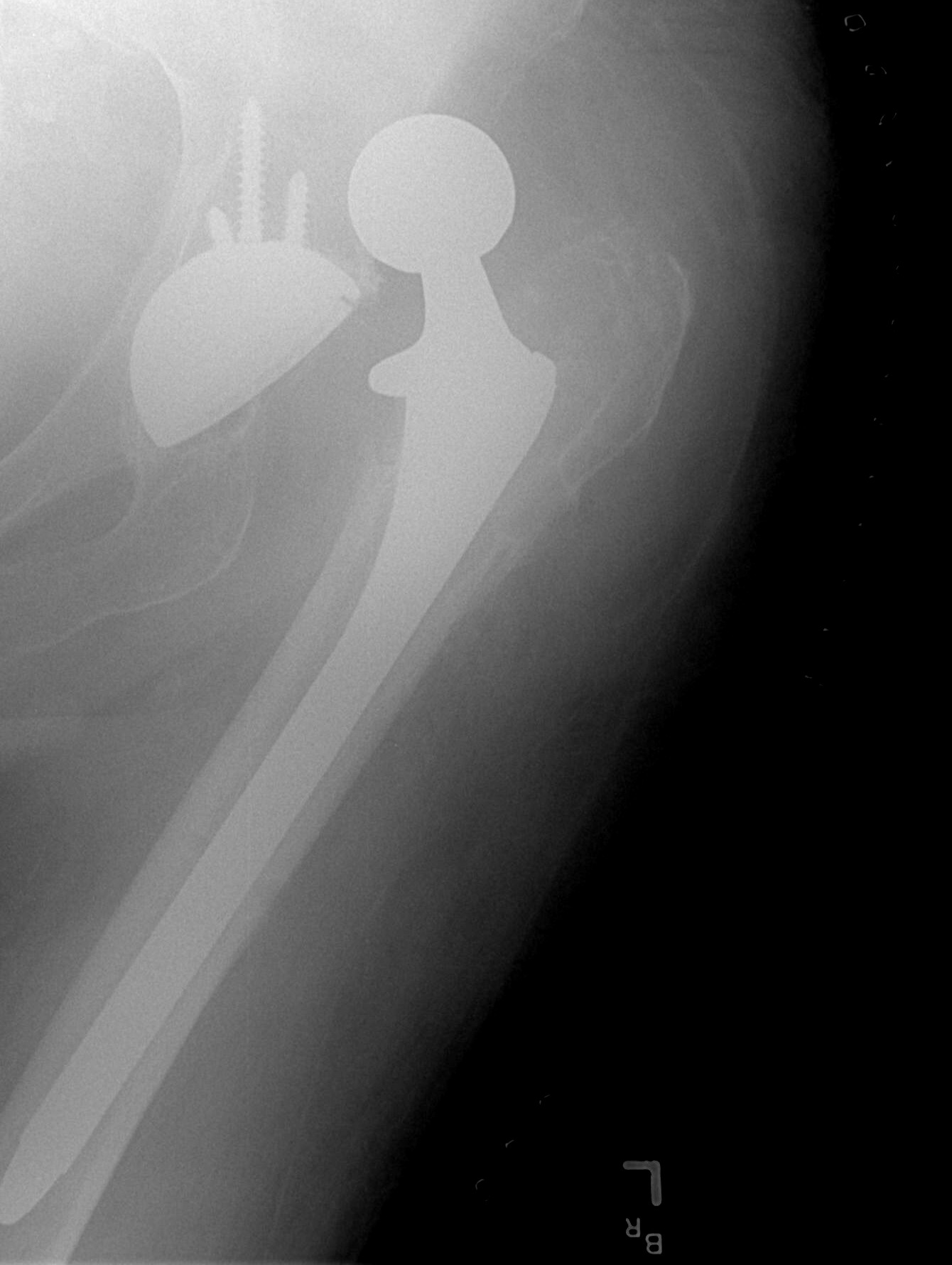
- Total Hip Arthroplasty: An abduction pillow placed between the legs prevents hip adduction. Keeping the legs spread wide apart is necessary to prevent joint dislocation after a total hip arthroplasty.
- Cervical Spine Trauma: A cervical collar must maintain the neck in a neutral anatomical position to prevent further spinal cord injury. However, strapping hard plastic to a neck poses a skin risk. The nurse must regularly assess the skin directly under a cervical collar for signs of pressure injuries.
Final Thoughts for the NCLEX
Nature breaks down what it does not use. Your job as a nurse is to be the ultimate antagonist to immobility. You inflate the lungs, you move the joints, you feed the muscles, and you inspect the skin. Understand why the machinery breaks, and you will always know exactly how to protect the patient. Keep this framework in mind, and you are going to master these concepts on the NCLEX!