Unexpected Response to Therapies
Not sure you’re ready?
Take the ~3-minute readiness diagnostic and see where you stand.
Here’s a fundamental truth about medicine that they don't always print in the glossy brochures: every single therapy we introduce into the human body is an experiment.
You administer a drug, perform a procedure, or hook up a line, and you expect a specific result. But the human body is a spectacularly complex, dynamic, and sometimes rebellious system. When things go sideways, you are no longer just a nurse—you are a detective, a physicist, and a first responder rolled into one.
Whenever a client exhibits an unexpected adverse therapy response, your foundational, non-negotiable first move is simple: a client exhibiting an unexpected adverse therapy response requires immediate vital sign assessment. You need data. You need to know what the machine is doing right now.
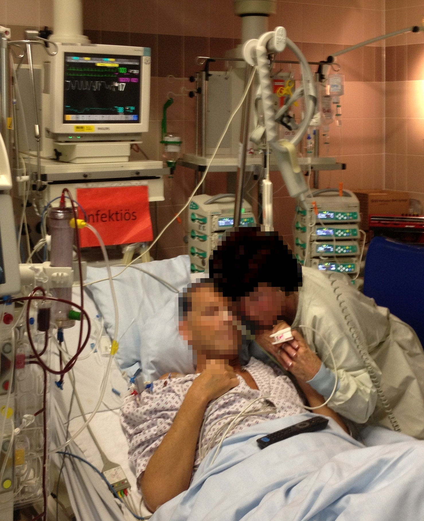
Let’s take a walk through the beautiful, chaotic physics and chemistry of the body when therapies go wrong, and explore exactly how we catch the fall.
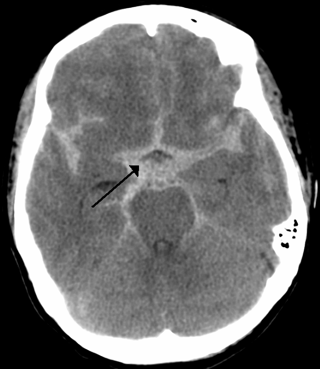
Think of the skull as a sturdy, inflexible vault. Inside, you have brain tissue, cerebrospinal fluid, and blood. Normal intracranial pressure ranges from 5 to 15 millimeters of mercury. If anything takes up extra space—like an unexpected bleed from a procedure—that pressure goes up, and the brain has nowhere to go.
How do we know the pressure is rising? The brain is a sensitive electrical network. Long before the vital signs crash, the "lights flicker." Therefore, an early sign of increased intracranial pressure is a change in the level of consciousness.
If you miss that early sign, the body enters a state of absolute physiological panic, resulting in Cushing's triad, which is a late clinical sign of increased intracranial pressure. The brain, desperate for oxygen, tells the heart to pump harder, while the brainstem starts getting crushed.
Cushing's Triad Includes:
- A widening pulse pressure (systolic goes up to force blood into the tight skull, diastolic stays low).
- Bradycardia (a reflex to the extreme hypertension).
- Irregular respirations (the brainstem respiratory centers are getting squished).
The Plumbing Solution: Gravity and Flow
If the pressure is high, we use physics to our advantage. Elevating the head of the bed to 30 degrees promotes venous drainage from the brain. By simply utilizing gravity, promoting venous drainage from the head decreases intracranial pressure.
But watch the pipes! Flexion of the neck impedes venous return from the brain. If you kink the jugular veins by letting the patient's chin drop to their chest, impeded venous return from the brain increases intracranial pressure. Keep that neck straight!
When a client bleeds out—say, a surgical complication—they lose the crucial fluid that gives the heart something to pump.
The body reacts to blood loss logically. The heart says, "I have less volume to pump, so I’m going to pump faster to keep the delivery rate up!" Because of this, tachycardia is an early clinical sign of hemorrhagic shock. But eventually, the tank runs dry. The heart's compensation fails, and the pressure plummets. Hypotension is a late clinical sign of hemorrhagic shock.
Immediate intervention for external hemorrhage requires applying direct manual pressure to the bleeding site. Stop the leak! But what if you can't see the leak? An expanding hematoma at a surgical site indicates active internal bleeding.
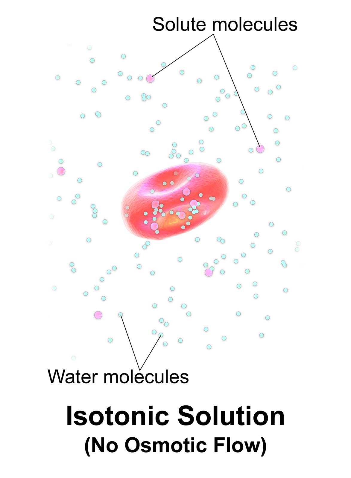
To fix the shock, we must refill the pipes. Fluid resuscitation with isotonic crystalloids is the initial intravenous treatment for hemorrhagic shock. We use isotonic fluids because they stay in the vascular space where we desperately need them.
When we introduce foreign substances—blood, drugs, or contrast—the immune system or metabolic pathways can completely overreact.
Blood Transfusion Reactions
If you suspect the patient is reacting to donor blood, the first nursing action for a suspected blood transfusion reaction is to immediately stop the transfusion.
How do we identify the reaction type?
- Hemolytic reactions: The body is actively destroying the donor red blood cells. The cellular debris clogs the kidneys. Thus, hemolytic blood transfusion reactions manifest with severe lower back pain (kidney pain) and hematuria (blood in the urine).
- Febrile non-hemolytic reactions: The client reacts to white blood cell remnants in the donor blood, causing an acute temperature increase during a transfusion.
Once you stop the blood, suspected blood transfusion reactions require maintaining intravenous access with a normal saline infusion. But here is a critical mechanical detail: normal saline administration during a blood transfusion reaction requires completely new intravenous tubing. If you flush the old tubing, you are literally pushing the remaining offending blood right into the patient!
The Ultimate Overreaction: Anaphylaxis
Anaphylaxis is an immune system explosion. It causes two immediate, life-threatening physical changes: sudden airway bronchoconstriction (closing the breathing tubes) and sudden systemic vasodilation (dropping blood pressure to zero).
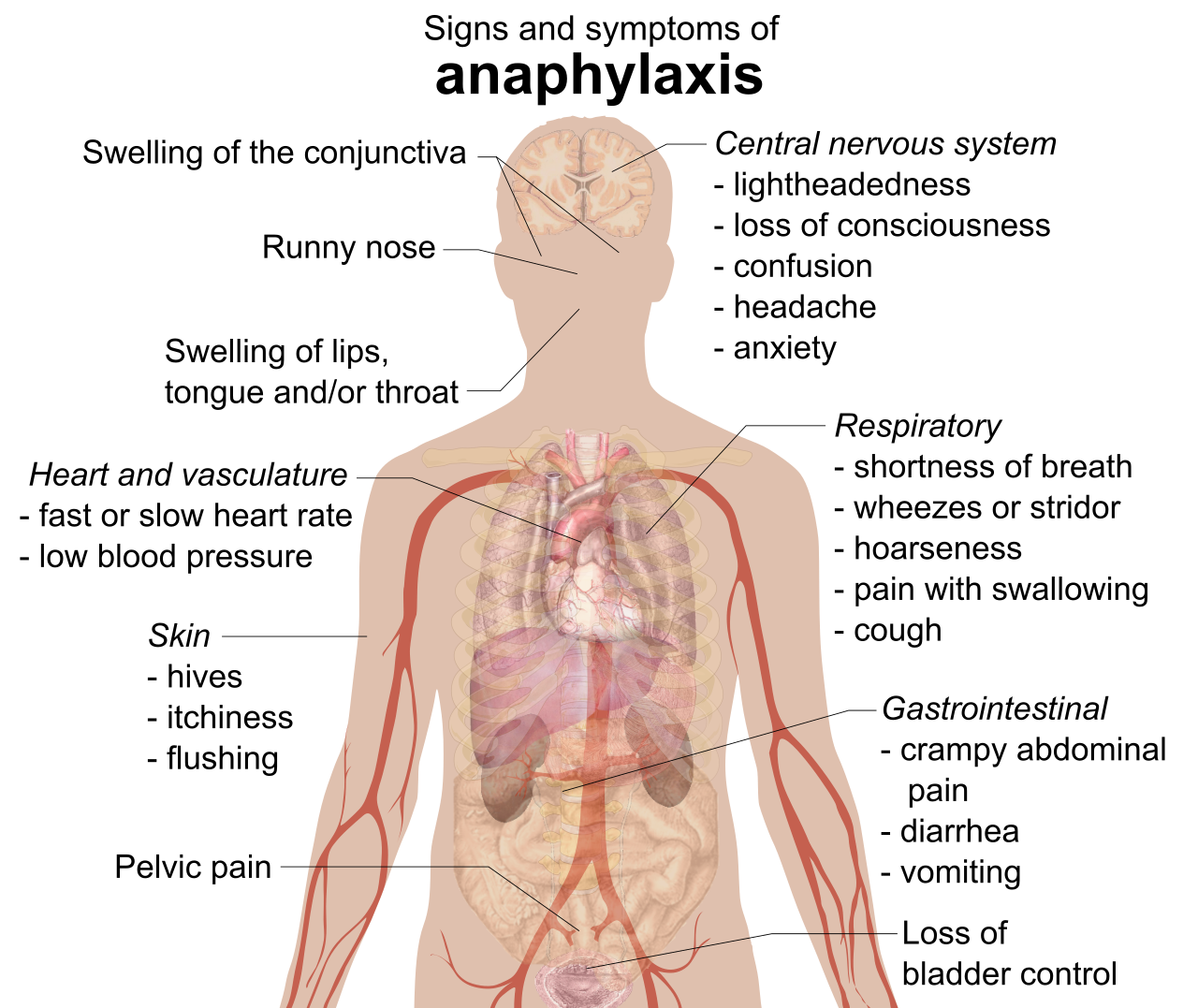
Epinephrine is the first-line medication for anaphylactic reactions to client therapies. It fixes both problems: it opens the airways and constricts the blood vessels. Where do we give it? Administering epinephrine intramuscularly in the anterolateral thigh provides rapid medication absorption during anaphylaxis because that muscle is massive and highly vascular.
Other Surprising Reactions
- Red Man Syndrome: This is an unexpected infusion-related reaction to intravenous vancomycin. It is not an allergy; it's a rate-dependent histamine release that presents with intense flushing of the upper body. The fix? Simple physics. The initial intervention for red man syndrome involves slowing the vancomycin infusion rate.
- Stevens-Johnson Syndrome: This is a nightmare scenario—a severe adverse cutaneous reaction to specific medications. It presents with painful blistering of the skin and mucous membranes, essentially burning the patient from the inside out. Immediate discontinuation of the offending medication is required.
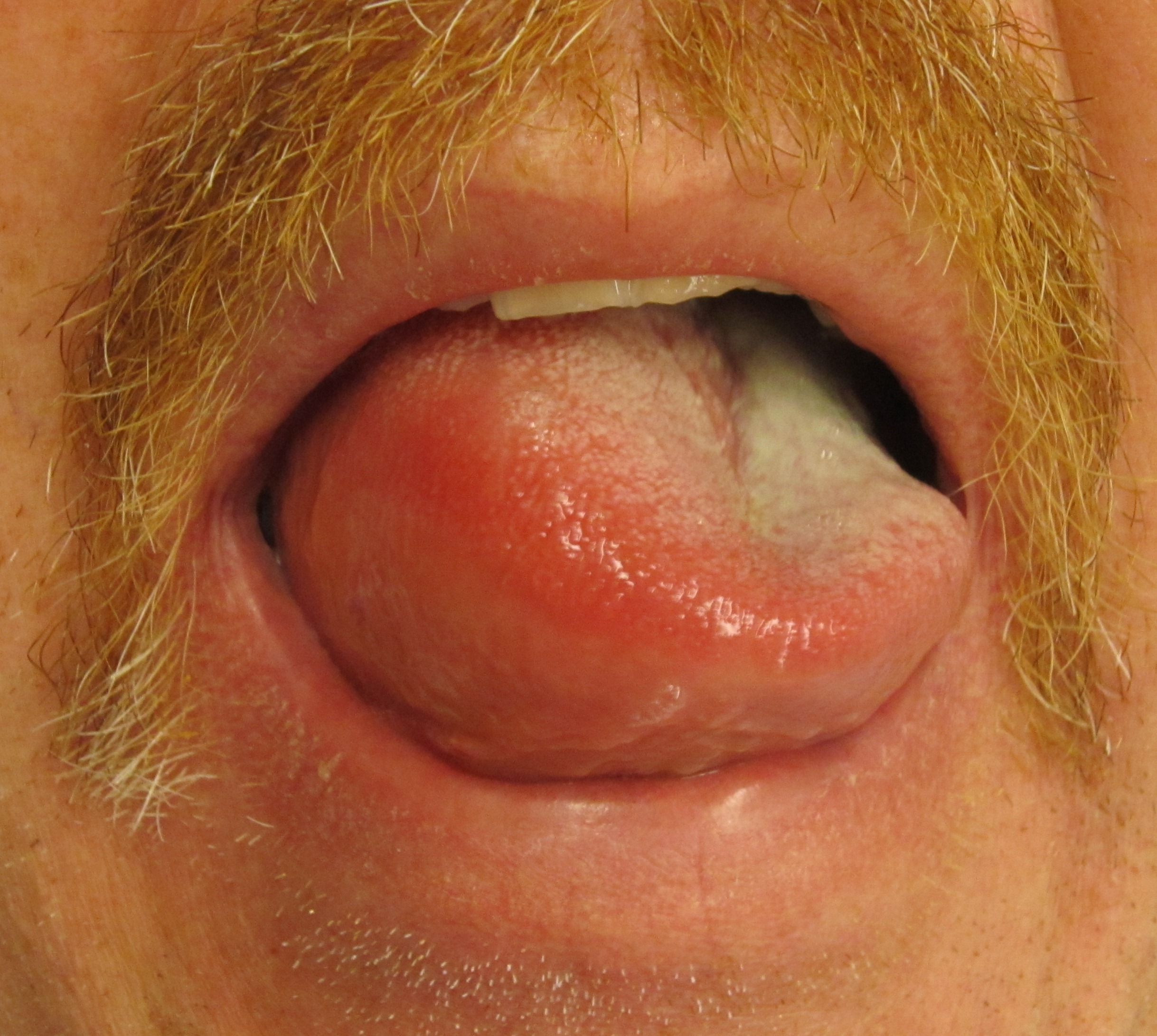
- Angioedema: This is a life-threatening adverse reaction to angiotensin-converting enzyme (ACE) inhibitors. It involves rapid swelling of the lips and tongue. Because the swelling directly obstructs the trachea, airway protection is the priority nursing intervention for medication-induced angioedema.
- Contrast-Induced Nephropathy: Radiocontrast media is heavy, thick, and tough on the kidneys, sometimes causing an acute decline in renal function after exposure. How do we protect them? Dilution! Administering intravenous hydration before a radiocontrast procedure reduces the risk of contrast-induced nephropathy.
Sometimes, the drugs that manipulate neurotransmitters tip the scales too far. Let's look at the brain's chemical storms.
Serotonin Syndrome vs. Neuroleptic Malignant Syndrome (NMS)
These two look similar to the untrained eye, but chemically, they are distinct.
| Feature | Serotonin Syndrome | Neuroleptic Malignant Syndrome (NMS) |
|---|---|---|
| Trigger | Occurs after initiating or increasing the dose of a serotonergic medication. | A severe adverse reaction to antipsychotic medications. |
| Muscle Tone | Manifests with spontaneous muscle clonus (rhythmic jerking) and hyperreflexia (jumpy reflexes). | Presents with severe diffuse muscle rigidity (lead-pipe stiffness). |
| Temperature | Elevated, but usually less severe. | Presents with extreme hyperthermia. |
Narrow Therapeutic Margins: Lithium and Digoxin
Some drugs have a razor-thin line between "therapeutic" and "toxic."
- Lithium Toxicity: Lithium acts a lot like sodium in the body. If a patient gets dehydrated, the kidneys hold onto lithium like it's water. Therefore, dehydration significantly increases the risk of developing lithium toxicity. Lithium toxicity manifests with severe neurological symptoms, and one of the most reliable early alarms is coarse hand tremors (not to be confused with expected fine tremors).
- Digoxin Toxicity: Digoxin helps the heart pump, but it binds to the exact same receptor sites on the heart cells as potassium. If potassium is low, digoxin takes over all the empty seats, causing toxicity. Thus, hypokalemia increases the risk of digoxin toxicity. Digoxin toxicity presents with visual disturbances. What kind? A very famous one: seeing yellow or green halos around lights is a classic symptom of digoxin toxicity.
Reversing the Damage (Antidotes)
When we intentionally depress the nervous system but go too far, we need the "undo" button.
- Administration of flumazenil reverses the adverse central nervous system depression caused by benzodiazepines.
- Administration of naloxone reverses the adverse respiratory depression caused by opioid analgesics.
The operating room and intensive care units are places of high-stakes interventions. Things happen fast.
Malignant Hyperthermia: The Metabolic Furnace
Imagine a genetic switch that gets accidentally flipped, causing the body's metabolism to go into overdrive. Malignant hyperthermia is a severe adverse reaction to certain inhaled anesthetic gases and depolarizing muscle relaxants (like succinylcholine).
Because the body is burning through energy, it produces massive amounts of waste gas. An early sign of malignant hyperthermia is an unexplained rapid increase in end-tidal carbon dioxide. The muscles are firing non-stop, so generalized muscle rigidity is an early clinical sign. If you see this, you need the biochemical fire extinguisher: Dantrolene is the specific pharmacological antidote for malignant hyperthermia.
Post-Operative and Airway Complications
- Paralytic Ileus: Sometimes, the gut decides not to wake up after anesthesia. Postoperative paralytic ileus is an unexpected cessation of bowel motility following abdominal surgery. If you listen with your stethoscope, absent bowel sounds indicate the presence of a paralytic ileus. How do we wake the gut up? Movement! Early ambulation promotes the return of bowel motility.
- Extubation Stridor: When we pull a breathing tube out, the throat can swell in protest. Extubation stridor is a high-pitched upper airway sound indicating dangerous laryngeal edema. You can hear it without a stethoscope. Post-extubation stridor requires immediate medical intervention to prevent complete airway obstruction.
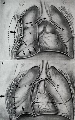
- Flail Chest: Say you perform aggressive, life-saving CPR. You might crack a few ribs in a row. Paradoxical chest movement indicates a flail chest complication following aggressive cardiopulmonary resuscitation. The broken chest wall segment gets sucked in during inspiration and pushes out during expiration—the exact opposite of normal.
Intravenous Therapies gone Wrong
- Heparin-Induced Thrombocytopenia (HIT): You give heparin to prevent clots. But in HIT, the immune system creates antibodies that activate platelets. It’s a paradoxical immune-mediated decrease in a client's platelet count. Because those platelets are being used up to form tiny clots everywhere, HIT increases the risk of venous thrombosis AND increases the risk of arterial thrombosis. The immediate action? All heparin products must be discontinued immediately upon suspicion of HIT.
- Extravasation: Extravasation occurs when a vesicant medication leaks into surrounding subcutaneous tissue. Vesicants are tissue-destroying chemicals (like chemotherapy). Extravasation of vesicant medications causes severe localized tissue necrosis. The immediate nursing action for medication extravasation is to stop the intravenous infusion. But wait—don't pull the IV out yet! Aspiration of the remaining vesicant from the catheter is required before removing the intravenous line. Pull out as much poison as you can first!
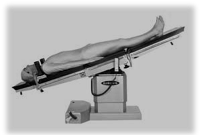
- Central Venous Catheters (CVC) & Air Embolisms: When putting a large line into the chest, you can puncture the lung. A sudden drop in oxygen saturation during central venous catheter insertion indicates a possible pneumothorax. What if a giant air bubble accidentally enters the IV line? Suspected air embolism during intravenous therapy requires positioning the client in the left lateral Trendelenburg position. Why this specific gymnastics routine? Because of gravity! The left lateral Trendelenburg position traps air in the right atrium to prevent systemic circulation of an air embolus. The bubble floats up to the highest point (the right atrium) and stays there instead of shooting into the lungs and causing a fatal pulmonary embolism.
Finally, let's talk about what happens when fluid builds up inside spaces that absolutely refuse to stretch.
Compartment Syndrome
Muscles are wrapped in tight sheaths of connective tissue called fascia. Compartment syndrome occurs when increased pressure within a muscle compartment compromises regional blood circulation. The fluid builds up, the fascia doesn't give, and the pressure starts choking the blood vessels and nerves.
Because nerves are incredibly sensitive to oxygen deprivation, the earliest sign of compartment syndrome is severe pain unrelieved by standard analgesic medications. You give them morphine, and they are still screaming. That’s a red flag. Eventually, the pressure gets so high it stops arterial blood flow entirely, which is why the loss of a palpable distal pulse is a late sign of compartment syndrome.
The Positioning Paradox: Normally, if a limb is swollen, we elevate it. DO NOT DO THIS HERE. Elevating an extremity with compartment syndrome above heart level worsens localized tissue ischemia. Why? Because you are forcing arterial blood to fight gravity to get into a space that is already highly pressurized! Instead, the affected extremity in suspected compartment syndrome should be positioned at the level of the heart. This maximizes arterial perfusion pressure without promoting excessive dependent swelling.
_(annotated).jpg)
Cardiac Tamponade
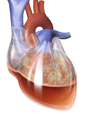
If the heart is bleeding or leaking fluid into the pericardial sac (the tough bag holding the heart), the heart gets squished. It can't fill with blood, so it can't pump blood.
If a client has a chest tube draining blood, and suddenly the tube clots off, where does that blood go? Inside. A sudden decrease in chest tube drainage can indicate an unexpected cardiac tamponade complication.
Because there is a layer of fluid physically separating the heart from your stethoscope, muffled heart sounds are a classic clinical manifestation of cardiac tamponade. And because the blood is trying to get into the heart but faces massive resistance, it backs up into the neck veins. Therefore, jugular venous distention is a clinical sign of cardiac tamponade.
The Feynman Takeaway
Nature plays by rules, and anatomy is just applied physics and chemistry. When therapies cause unexpected reactions, your job is to recognize the mechanism of the failure. Is it a pressure problem? A plumbing problem? A chemical misfire?
Look at the vital signs. Understand the 'why' behind the symptom. Trapped air? Use gravity. Tight space? Maximize blood flow. Immune reaction? Stop the trigger. By mastering the underlying principles of these complications, you don't just memorize the interventions—you invent them logically in your mind when the alarms start ringing.