Pathophysiology
Not sure you’re ready?
Take the ~3-minute readiness diagnostic and see where you stand.
The NCLEX-RN Pathophysiology Masterclass: From Cells to Systems
Welcome. If you want to really understand the human body—not just memorize a list of diseases for an exam, but truly get it—you have to start small. I mean, microscopically small.
Pathophysiology isn’t just a catalog of things going wrong. It is a logical, predictable chain of physical events. When a patient goes into shock or heart failure, it is the macro-level result of billions of tiny cellular machines breaking down. In this guide, we’re going to build your understanding of the body from the ground up: starting with the cell, moving to how tissues respond to injury, and finally exploring what happens when entire organ systems fail.
Let’s dive in.
Before we can talk about disease, we must talk about the machinery of life. Imagine a single human cell as a bustling, highly secure factory.
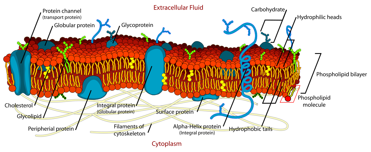
The outer wall of this factory is the cell membrane. Structurally, the cell membrane consists of a phospholipid bilayer containing embedded proteins. This isn't just a passive wall; it is a highly intelligent gatekeeper. The cell membrane regulates the selective passage of substances into and out of the cell, keeping the chaos of the outside world at bay.
Inside the factory, we have the power plants. Mitochondria generate the majority of cellular adenosine triphosphate (ATP) through oxidative phosphorylation. ATP is the cash currency of the cell. Without it, work stops. Finally, we have the janitorial crew: Lysosomes contain digestive enzymes to break down cellular debris and foreign substances, keeping the factory floor clean.
Cellular Adaptation: Survival of the Fittest
Cells are incredibly adaptable. If the environment changes, the cell alters its structure to survive.
- Atrophy: If a muscle cell is stuck in a cast, or a tissue loses its blood flow, the cell shrinks to conserve energy. Cellular atrophy is a decrease in cellular size, and importantly, cellular atrophy occurs in response to reduced use or decreased blood supply.
- Hypertrophy: Conversely, if you force a cell to work harder (like lifting weights or a heart pumping against high blood pressure), it beefs up. Cellular hypertrophy is an increase in cellular size, and it occurs in response to an increased mechanical workload.
- Hyperplasia: Sometimes tissues need more workers, not just bigger ones. Hyperplasia is an increase in the total number of cells within an organ or tissue.
- Metaplasia: If a tissue faces chronic irritation—like a smoker’s windpipe—the delicate cells will tag out for tougher ones. Metaplasia is the reversible replacement of one mature cell type by another less mature cell type.
- Dysplasia: But if the stress continues too long, the cells go haywire. Dysplasia involves abnormal changes in the size, shape, and organization of mature cells. This is cellular chaos and a major red flag for cancer.
So, what actually kills a cell?
By far, the most dangerous threat is a lack of oxygen. Hypoxia is the most common cause of cellular injury.
Let’s walk through the exact physical cascade of what happens when a cell suffocates. It is a beautiful, tragic chain reaction:
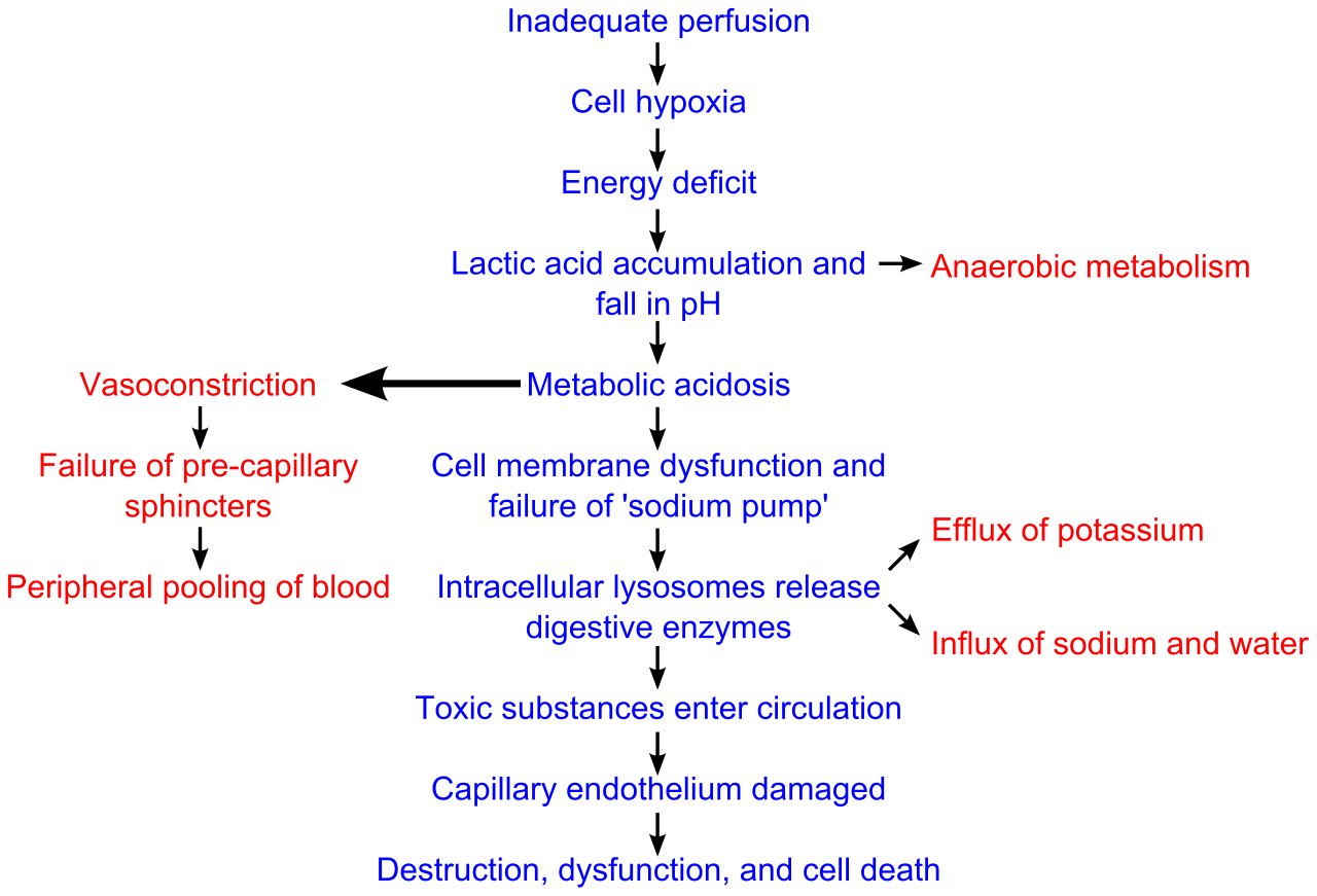
- Without oxygen, the mitochondrial power plants shut down. Cellular hypoxia decreases mitochondrial adenosine triphosphate (ATP) production.
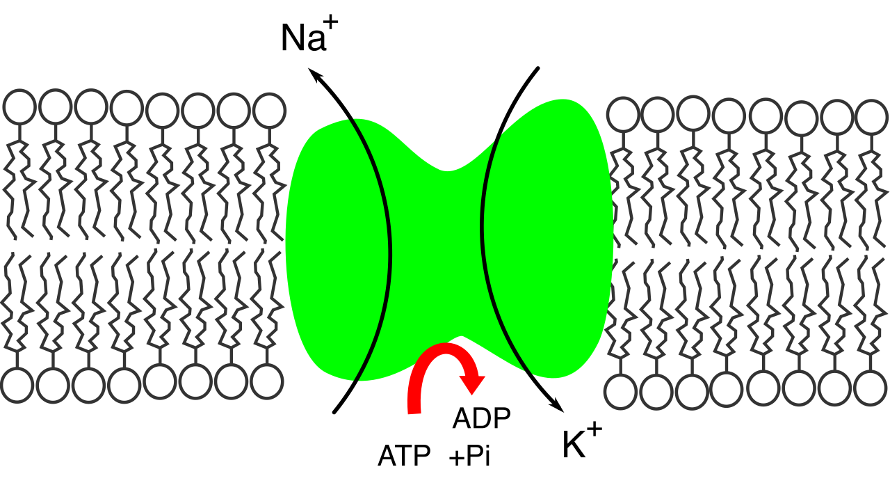
- Without ATP, the cell's essential machinery stops. Specifically, decreased adenosine triphosphate production impairs the cellular sodium-potassium pump.
- Because this pump isn't pushing sodium out anymore, impairment of the sodium-potassium pump allows sodium to rapidly accumulate inside the cell.
- Remember the golden rule of osmosis: water always follows sodium. Intracellular sodium accumulation draws extracellular water into the cell.
- The cell fills up like a water balloon until—pop! The influx of water into the cell causes cellular swelling and potential lysis (rupture).
The Two Faces of Cell Death
When a cell dies, how it dies matters immensely to the rest of the body.
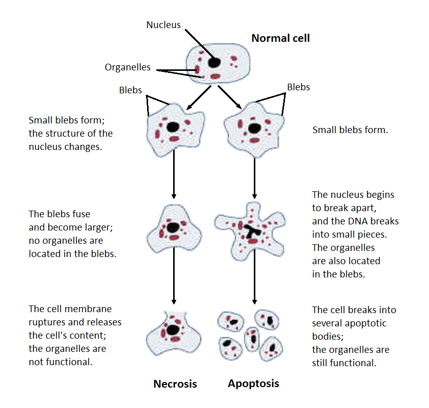
- Apoptosis is a programmed cell death mechanism. Think of it as polite cellular suicide. Apoptosis eliminates aged or damaged cells without triggering an inflammatory response. The cell quietly dismantles itself.
- Necrosis, on the other hand, is a messy murder. Necrosis is unregulated cell death resulting from severe injury. When the cell explodes, necrotic cell death releases intracellular contents and triggers an inflammatory response.
When necrosis happens, the body’s alarm systems go off. This triggers inflammation and the immune response.
The Inflammatory Cascade
Acute inflammation is the body calling 911. It happens in two distinct phases:
- The Vascular Phase: The moment tissue is injured, local sirens (mast cells) release chemical mediators. Mast cell histamine release causes immediate vasodilation during acute inflammation. This vascular phase of acute inflammation involves localized vasodilation and increased capillary permeability. The blood vessels open wide and become leaky, bringing blood to the site (redness and heat). The increased capillary permeability allows fluid and proteins to leak into the interstitial space, causing localized swelling (edema).
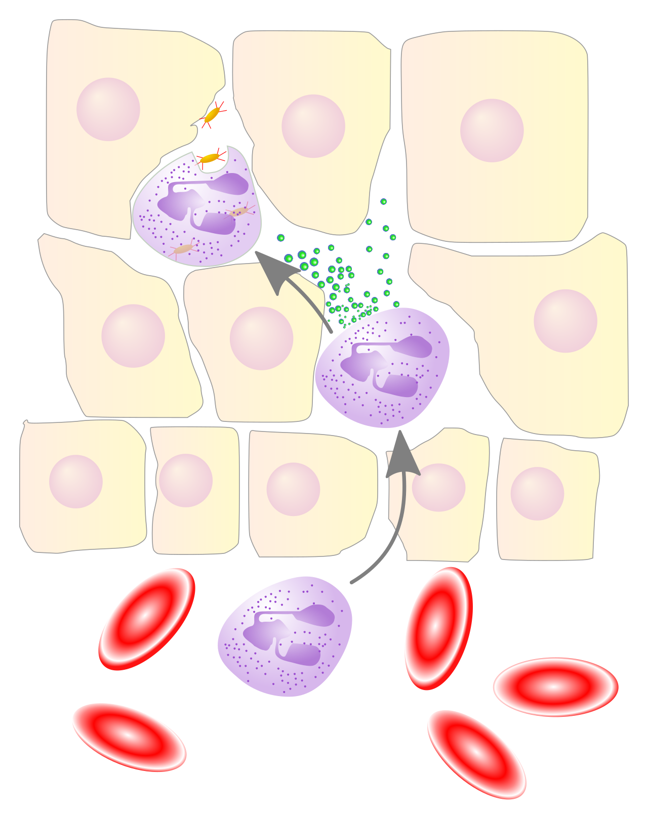
- The Cellular Phase: Now the first responders arrive. The cellular phase of acute inflammation involves leukocyte migration to the site of injury. The absolute quickest paramedics on the scene? The neutrophils. Neutrophils are the first leukocytes to arrive at the site of acute inflammation, where they begin eating up the debris.
Wound Healing
Once the mess is cleared, rebuilding begins. The goal is to stitch the tissue back together.
- If you have a clean surgical cut, primary intention wound healing occurs when wound edges are cleanly approximated with minimal tissue loss.
- If you have a gaping ulcer, secondary intention healing occurs in open wounds requiring tissue granulation and epithelialization.
To fill that gap, granulation tissue forms during the proliferative phase of wound healing. Then, specialized builder cells get to work: fibroblasts synthesize collagen to provide structural tensile strength to healing wounds.
The Immune System
The immune system is your military. It operates in two divisions:
| Immune Division | Characteristics |
|---|---|
| Innate Immunity | The broad, blunt weapon. Innate immunity provides immediate, non-specific protection against a broad array of pathogens. |
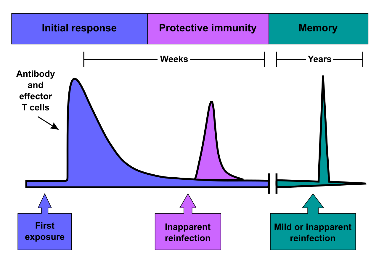
| Adaptive Immunity | The sniper rifle. Adaptive immunity provides delayed, pathogen-specific protection. Crucially, adaptive immunity generates immunologic memory for faster responses upon subsequent pathogen exposures. |
You can acquire adaptive immunity in two ways. Active immunity develops when the host immune system produces intrinsic antibodies in response to an antigen (like fighting off a virus or getting a vaccine). Conversely, passive immunity involves the transfer of pre-formed exogenous antibodies to a recipient (like a baby getting antibodies through breast milk).
Hypersensitivity: When the Military Mutinies
Sometimes, the immune system overreacts or attacks the wrong target. We classify these into four types of hypersensitivity:
- Type I: The classic allergy. Type I hypersensitivity is an immunoglobulin E (IgE)-mediated allergic reaction. (Think anaphylaxis or pollen allergies).
- Type II: Tissue-specific attacks. Type II hypersensitivity involves antibodies directly attacking antigens on the surface of specific target cells. (Think mismatched blood transfusions).
- Type III: Collateral damage. Type III hypersensitivity is mediated by the formation and tissue deposition of antigen-antibody immune complexes. These clumps lodge in tissues and cause systemic inflammation (think Lupus or Rheumatoid Arthritis).
- Type IV: The slow burn. Type IV hypersensitivity is a delayed cell-mediated immune response involving T lymphocytes. (Think Poison Ivy or a TB skin test).
Now let's zoom out. What happens when these cellular and inflammatory concepts happen on a massive, organ-wide scale?
The Cardiovascular System: Plumping and Pumping
Let's look at the heart and vessels.
Heart failure occurs when the heart cannot pump sufficient blood to meet the metabolic needs of the body. It’s a pump problem, and the backlog depends on which side of the pump fails:
- Left-sided heart failure causes pulmonary edema due to hydrostatic fluid back-up into the pulmonary circulation. The left ventricle can't push blood out to the body, so it backs up into the lungs. The patient drowns in their own fluids.
- Right-sided heart failure causes systemic venous congestion leading to peripheral edema and hepatomegaly (an enlarged liver). The right ventricle can't push blood into the lungs, so fluid backs up into the legs and abdomen.
What causes the heart to fail in the first place? Often, it's a piping issue. Atherosclerosis involves the formation of lipid-laden plaques within the arterial intima (the inner lining of the blood vessel). Over time, these atherosclerotic plaques narrow the arterial lumen resulting in reduced distal blood flow.
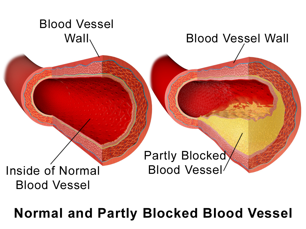
But a narrowed artery isn't the real widow-maker. The lethal event is when a plaque pops. Rupture of an atherosclerotic plaque exposes highly thrombogenic core materials to the bloodstream. The moment blood touches this core, an explosive clot forms. Atherosclerotic plaque rupture triggers rapid platelet aggregation and thrombus formation.
If this clot forms in the coronary arteries feeding the heart, blood flow stops. A myocardial infarction results from prolonged myocardial ischemia. And because heart muscle cells are starving for oxygen, prolonged myocardial ischemia causes irreversible necrosis of heart muscle cells.
The Respiratory System: Airflow Obstruction
When we talk about lung diseases, we are mostly dealing with blocked pipes or destroyed tissues.
- Asthma involves recurrent episodes of reversible airway obstruction. Notice the word reversible. An asthmatic's airways are twitchy. The asthma airway obstruction is driven by bronchial hyperresponsiveness and chronic airway inflammation. Give them a bronchodilator, and the airways open up again.
- Chronic obstructive pulmonary disease (COPD) involves progressive airflow limitation. Unlike asthma, the airflow limitation in chronic obstructive pulmonary disease is largely irreversible.
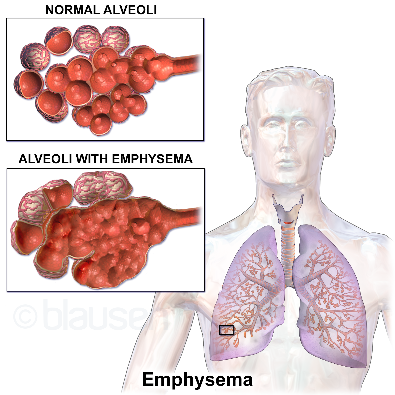
- Emphysema is a form of chronic obstructive pulmonary disease characterized by the destruction of alveolar walls. Imagine a bunch of tiny grapes (alveoli) fusing into one giant, flabby balloon. This is a disaster for oxygen exchange, because alveolar wall destruction in emphysema reduces the total surface area available for pulmonary gas exchange.
The Renal System: The Filter Fails
The kidneys filter our blood. When they fail, toxins back up. Acute kidney injury (AKI) is a sudden decline in kidney function, which directly results in the systemic accumulation of nitrogenous waste products (like BUN and Creatinine).
To figure out what caused the AKI, we look at the location of the problem:
- Prerenal AKI: A blood flow problem before the kidney. Prerenal acute kidney injury is caused by decreased systemic blood flow leading to reduced renal perfusion. (E.g., extreme dehydration or shock).
- Intrarenal AKI: A problem inside the kidney. Intrarenal acute kidney injury involves direct ischemic or toxic damage to the kidney parenchyma. (E.g., nephrotoxic drugs or prolonged hypoxia).
- Postrenal AKI: A plumbing backup after the kidney. Postrenal acute kidney injury results from a mechanical obstruction of urine flow out of the urinary tract. (E.g., kidney stones or an enlarged prostate).
Unlike the sudden onset of AKI, chronic kidney disease involves the progressive and irreversible loss of functioning nephrons over months or years.
The Endocrine System: The Insulin Story
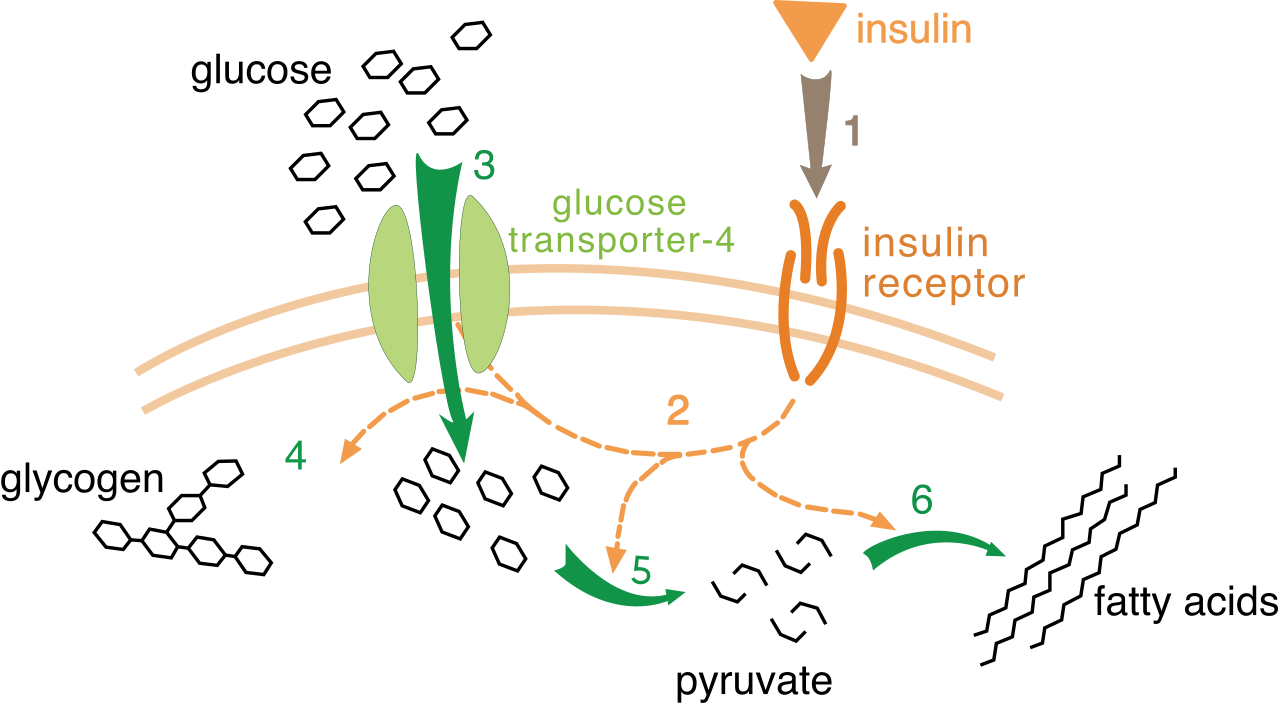
Diabetes mellitus is fundamentally an issue of glucose not being able to enter cells due to an insulin problem.
- Type 1 diabetes mellitus results from the autoimmune destruction of pancreatic beta cells. The factory is destroyed. The destruction of pancreatic beta cells in Type 1 diabetes causes an absolute insulin deficiency.
- Type 2 diabetes mellitus is characterized by peripheral cellular insulin resistance. The body makes insulin, but the cellular "locks" are jammed. Over time, the pancreas exhausts itself trying to crank out more insulin, meaning the peripheral cellular insulin resistance in Type 2 diabetes eventually leads to a relative insulin deficiency.
The Neurological System: Brain Ischemia
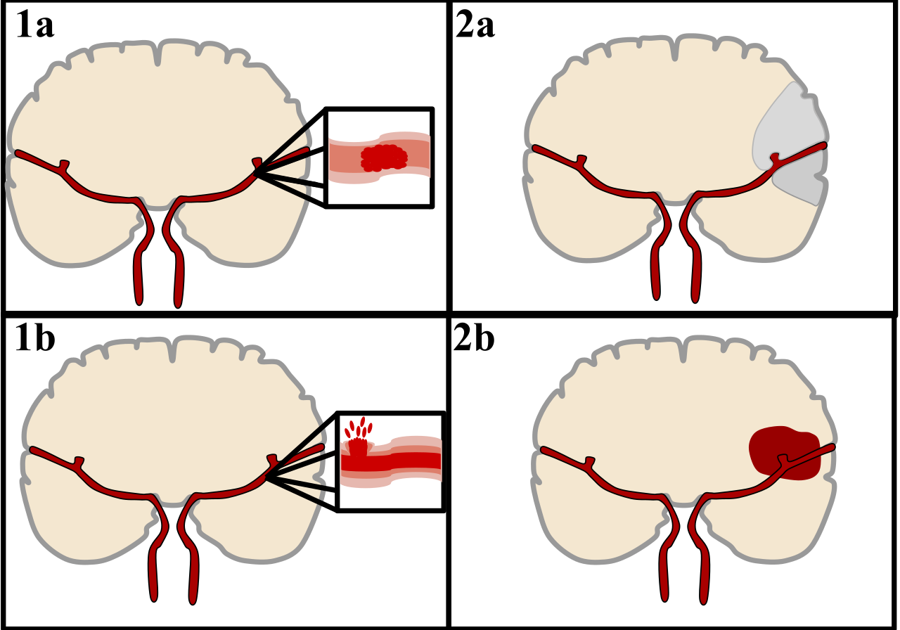
Strokes are like heart attacks of the brain, and they come in two distinct pathophysiologies:
- Ischemic stroke is caused by the thrombotic or embolic occlusion of a cerebral artery. A clot blocks the pipe, and just like in the heart, cerebral artery occlusion rapidly leads to downstream brain tissue infarction (necrosis).
- Hemorrhagic stroke is caused by the spontaneous rupture of a cerebral blood vessel. The pipe bursts. This cerebral vessel rupture leads to acute bleeding directly within the brain parenchyma or subarachnoid space, crushing delicate brain tissue under high pressure.
The Hepatic System: Liver Scarring
The liver is an incredible chemical processing plant, but chronic damage destroys it. Cirrhosis is the end stage of chronic liver disease. Instead of smooth, functional tissue, cirrhosis is characterized by the diffuse replacement of functional hepatic tissue with fibrous scar tissue.
Because the liver becomes a hard knot of scars, blood from the intestines struggles to pass through it. This creates a massive traffic jam called portal hypertension. Hepatic portal hypertension results from increased resistance to venous blood flow through a scarred cirrhotic liver.
We end where life ends.
If you understand everything we’ve talked about—from ATP production to blood flow—you will understand Shock. Shock is a critical state of widespread inadequate tissue perfusion. The heart isn't getting oxygen to the tissues. And what did we learn about cells without oxygen? Widespread inadequate tissue perfusion during shock results in global cellular hypoxia. The sodium-potassium pumps fail, cells swell, cells pop, massive necrosis occurs, and multi-organ failure follows.
But why does tissue perfusion fail? We can categorize shock by identifying which part of the circulatory plumbing system broke:
- Hypovolemic Shock (Not enough fluid): Hypovolemic shock results from a significant loss of intravascular blood volume or total body fluid volume. The pipes are fine, the pump is fine, but there's no fluid to pump (e.g., massive hemorrhage).
- Cardiogenic Shock (Broken pump): Cardiogenic shock occurs when the myocardium fails to pump adequately despite adequate intravascular volume. There is plenty of fluid, but a massive myocardial infarction has destroyed the pump.
- Distributive Shock (Pipes are too big): Distributive shock is characterized by massive systemic vasodilation. All the blood vessels in the body relax at once. This massive systemic vasodilation pathologically expands the size of the vascular compartment relative to blood volume. There is a normal amount of fluid, but the container just got three times bigger, so blood pressure bottoms out.
Distributive shock has two major sub-types you must know:
- Anaphylactic shock is a severe distributive shock caused by a systemic Type I hypersensitivity reaction. IgE causes mast cells everywhere to dump histamine, causing catastrophic whole-body vasodilation.
- Septic shock is a distributive shock caused by a dysregulated systemic inflammatory response to severe infection. The body's immune reaction to a widespread pathogen causes massive, uncontrolled capillary leak and vasodilation.
Final Thoughts
When you look at an NCLEX question, don't just ask "what is the symptom?" Ask why the symptom is happening. If a patient is swelling, is it hydrostatic pressure backing up from heart failure? Is it a loss of protein from a leaky capillary during inflammation? Is it an intracellular sodium pump failure due to hypoxia?
Trace the pathology back to the mechanism. Master the mechanism, and you master the medicine. Keep studying. Keep questioning.