Cell Structure and Transport
Not sure you’re ready?
Take the ~3-minute readiness diagnostic and see where you stand.
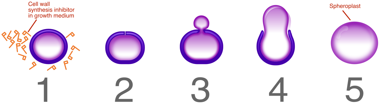
When you administer an intravenous antibiotic to a patient fighting an infection, you are exploiting a fundamental biological asymmetry. You are banking on the fact that the bacteria causing the illness are structurally distinct from the human cells they are invading. Penicillin, for instance, destroys bacterial cell walls—a structure human cells completely lack. If our cells and bacterial cells were built identically, the drugs used to cure infections would invariably kill the patient.
Understanding the architecture of the cell, the distinct types of cellular life, and how matter moves across microscopic borders is not just an academic exercise. It is the molecular foundation of pharmacology, pathology, and fluid resuscitation. Before you can understand how the human body functions as a macroscopic organism, you must understand how it operates at the level of its smallest functional unit: the cell.
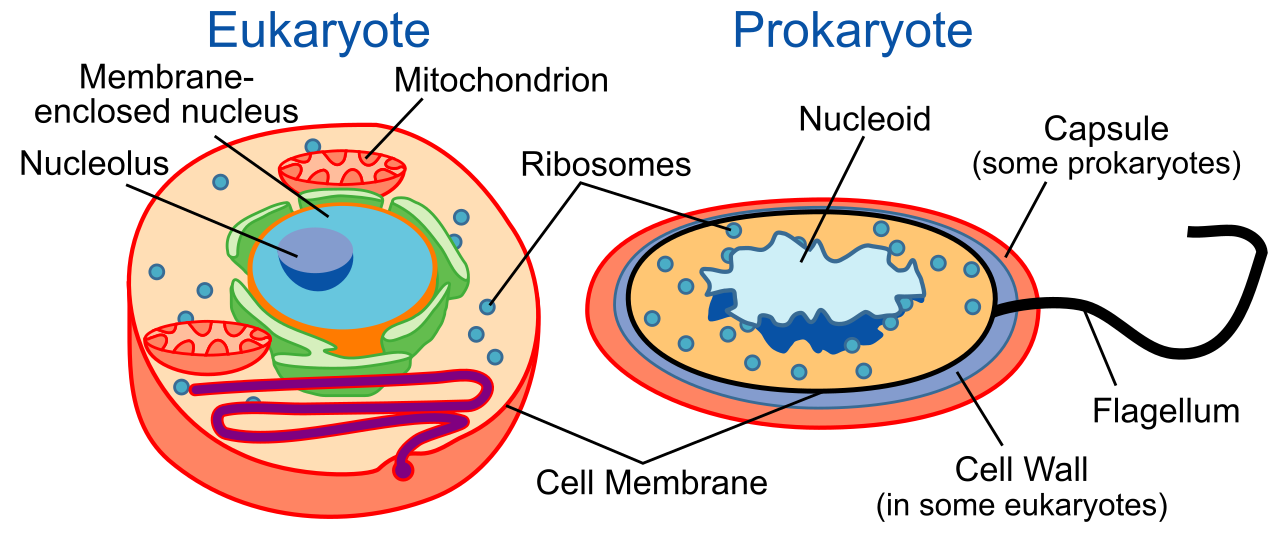
Biologists divide all cellular life into two major categories based on their internal architecture. The distinction is defined by how the cell manages its genetic material and whether it possesses compartmentalized internal machinery.
Prokaryotic cells represent the most ancient and structurally streamlined forms of life. Because they are optimized for rapid replication and survival in diverse environments, prokaryotic cells lack a membrane-bound nucleus and lack membrane-bound organelles. Instead of being housed in a protective vault, prokaryotic DNA is typically circular and floats freely within the cell. Bacteria are examples of prokaryotic cells, as are the extremophiles known as Archaea.
Eukaryotic cells are the structural basis of complex life. Unlike their prokaryotic counterparts, eukaryotic cells contain a membrane-bound nucleus—a secure command center for genetic data—and they contain membrane-bound organelles, which are specialized functional compartments. Furthermore, eukaryotic DNA is typically linear, organized into the complex chromosomes you study in human genetics. Animal cells are eukaryotic cells, as are Plant cells. The eukaryotic classification also extends beyond flora and fauna; Fungi are eukaryotic cells, and microscopic Protists are eukaryotic cells as well.
Comparing Plant and Animal Cells
While plants and animals share a eukaryotic foundation, their evolutionary paths required entirely different architectural features. A plant cannot run away from a predator or walk to a water source; its cells must structurally support the organism and manufacture their own food.
| Feature | Plant Cells | Animal Cells |
|---|---|---|
| Outer Boundary | Have a rigid cell wall composed of cellulose. | Do not have a cell wall; enclosed only by a cell membrane. |
| Energy Production | Contain chloroplasts. Chloroplasts are the site of photosynthesis in plant cells. | Do not contain chloroplasts; rely solely on consuming external nutrients. |
| Vacuoles | Typically have one large central vacuole to maintain internal water pressure (turgor). | Possess multiple small vacuoles or lack vacuoles entirely. |
| Division Structures | Most plant cells lack centrioles. | Contain centrioles, which are involved in animal cell division (mitosis). |
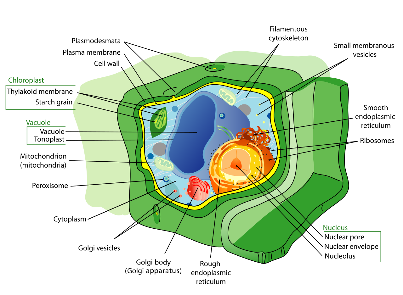
If a cell is a bustling, microscopic city, its organelles are the factories, power plants, and waste management facilities that keep the metropolis alive. All of this infrastructure is suspended in the cytoplasm, which is the jelly-like substance filling the interior of the cell. The purely liquid portion of the cell cytoplasm is known as the cytosol.
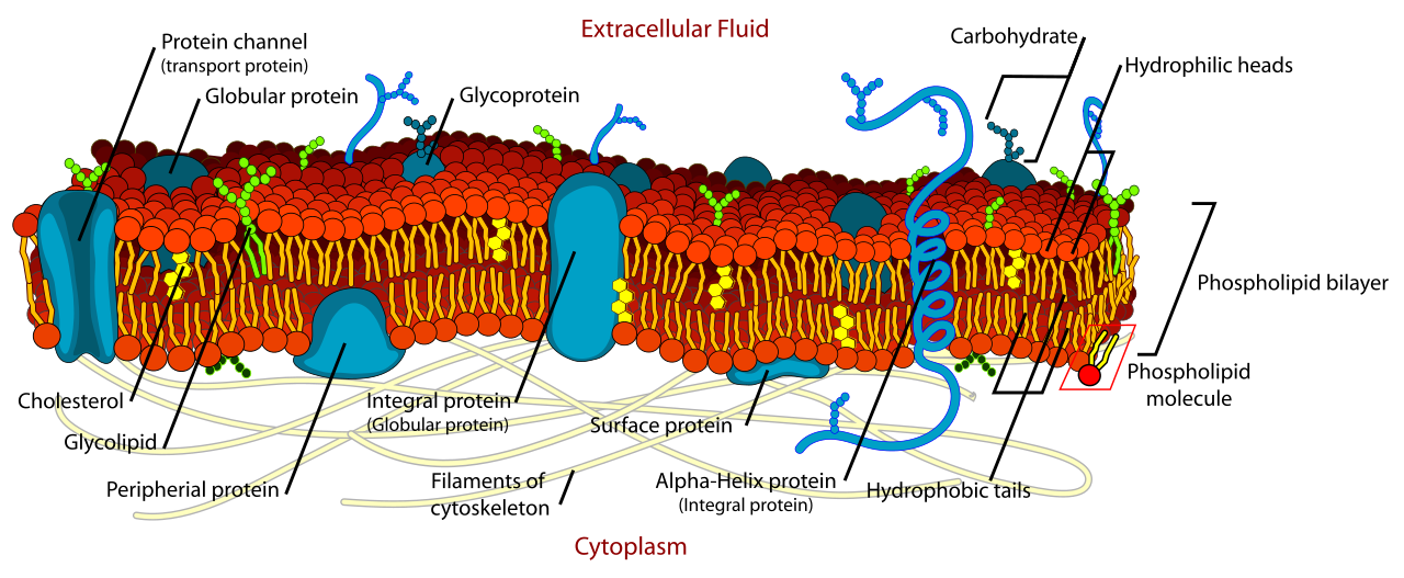
Providing the physical scaffolding for this space is the cytoskeleton. It acts as both structural support and an intricate highway system; the cytoskeleton provides structural support to the cell and facilitates the transport of materials within the cell. Surrounding the entire city is the cell membrane, a sophisticated barrier consisting primarily of a phospholipid bilayer. The cell membrane regulates the passage of substances in and out of the cell.
Information and Production
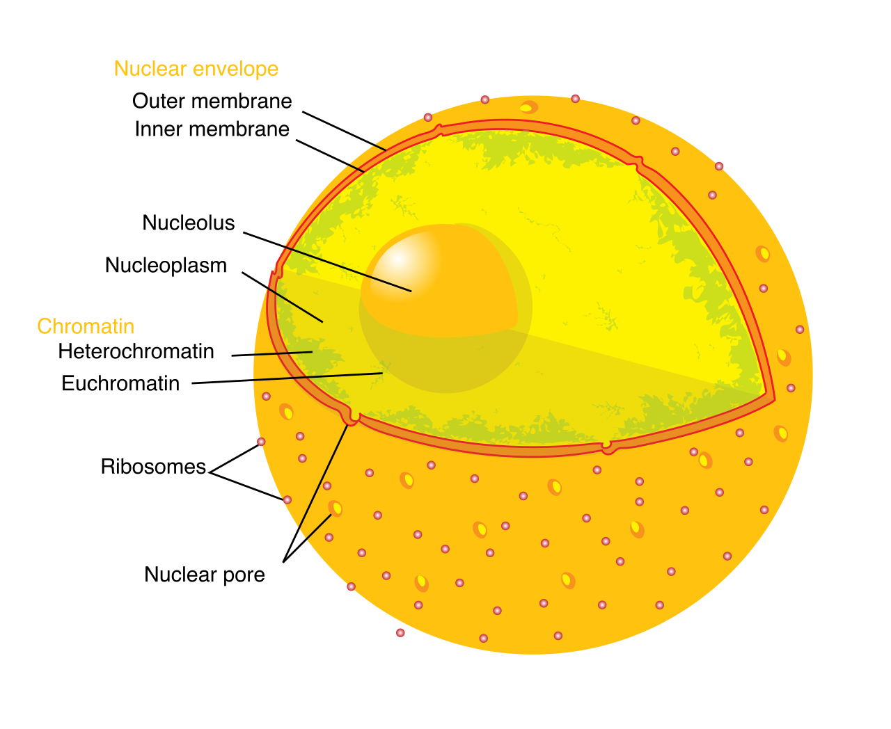
- The Nucleus and Nucleolus: The cell nucleus stores the genetic information of the cell. Deep inside the nucleus sits the nucleolus, a dense structure located inside the cell nucleus. The nucleolus is the site of ribosome synthesis.
- Ribosomes: Once manufactured, ribosomes act as the molecular workhorses of the cell. Ribosomes are the cellular structures responsible for protein synthesis. Depending on what kind of protein is being built, ribosomes can float freely in the cell cytoplasm or they can be physically attached to the rough endoplasmic reticulum.
- Endoplasmic Reticulum (ER): The ER is a continuous manufacturing network. The rough endoplasmic reticulum (studded with ribosomes) modifies newly synthesized proteins, preparing them for their final destination. The smooth endoplasmic reticulum (which lacks ribosomes) serves a different role: it synthesizes lipids and detoxifies harmful substances within the cell. This is why liver cells, responsible for filtering toxins from your blood, are densely packed with smooth ER.
- Golgi Apparatus: Often visualized as a stack of flattened pancakes, the Golgi apparatus is the cell's post office. It modifies proteins for cellular transport and packages proteins into vesicles (tiny shipping containers) for delivery inside or outside the cell.
Energy, Maintenance, and Waste
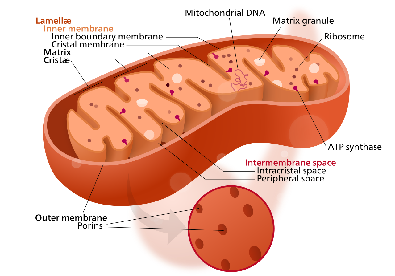
- Mitochondria: To keep the city running, you need energy. Mitochondria generate most of the cell's supply of adenosine triphosphate, which is commonly referred to as ATP—the primary energy currency of biological systems. Interestingly, mitochondria contain their own independent circular DNA, a remnant of their evolutionary history as free-living bacteria.
- Lysosomes: Waste management is critical. Lysosomes are specialized vesicles that contain digestive enzymes. They act as microscopic recycling centers, as lysosomes break down cellular waste materials. Furthermore, when a cell becomes damaged or cancerous, lysosomes play a key role in programmed cell death (apoptosis), intentionally releasing their enzymes to dismantle the cell from the inside.
- Peroxisomes: Operating alongside lysosomes, peroxisomes contain enzymes that break down fatty acids. Crucially, they also contain enzymes that neutralize hydrogen peroxide—a toxic byproduct of cellular metabolism—converting it safely into water and oxygen.
A cell cannot exist in isolation. It must constantly absorb nutrients, exchange gases, and expel waste. The rules governing how molecules cross the cell membrane dictate every physiological process in the human body, from how your lungs absorb oxygen to how your kidneys filter blood.
The fundamental metric of cellular transport is the concentration gradient.
A concentration gradient is the difference in the concentration of a substance between two areas. Transport mechanisms are categorized based on how they interact with this gradient—whether they "roll downhill" or are "pushed uphill."
Passive Transport Mechanisms
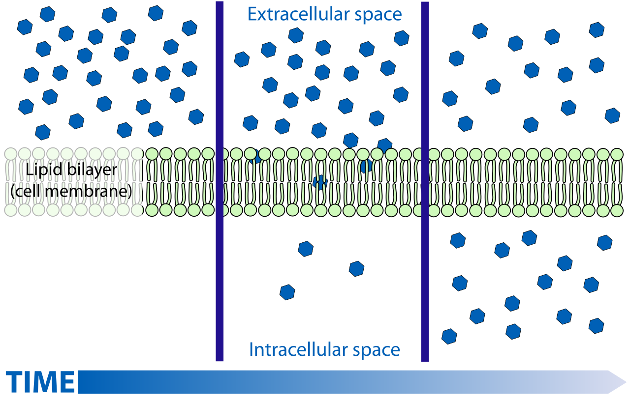
Picture a boulder rolling down a hill; gravity does the work. Passive transport mechanisms do not require the expenditure of cellular energy. Because they follow the natural laws of entropy, substances in passive transport move down a concentration gradient.
- Diffusion: This is the simplest form of transport. Diffusion is the passive movement of particles from an area of higher concentration to an area of lower concentration. When you inhale, oxygen naturally diffuses from the high-concentration environment of your lungs into the lower-concentration environment of your bloodstream.
- Facilitated Diffusion: Some molecules, like glucose, are too large or too highly charged to slip directly through the phospholipid bilayer. They still want to move down their gradient, but they need a doorway. Facilitated diffusion utilizes specific transmembrane proteins to move molecules across the cell membrane. Because it still moves molecules from high to low concentration, facilitated diffusion does not require cellular energy.
Osmosis and Tonicity: The Movement of Water
Water plays by a specific set of transport rules. Osmosis is the passive transport of water across a selectively permeable membrane.
When solutes (like salt or sugar) cannot cross the membrane to balance out a concentration gradient, water will move instead to dilute the side with more solutes. Therefore, water moves during osmosis from an area of lower solute concentration to an area of higher solute concentration.
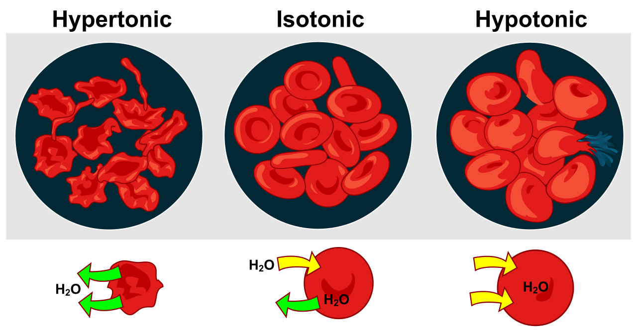
As a healthcare professional, mastering osmosis is paramount because it dictates how intravenous (IV) fluids affect red blood cells and tissues. We classify these fluid environments by their tonicity:
- Isotonic Solutions: Isotonic solutions have equal solute concentrations on both sides of the cell membrane. When a cell is in an isotonic solution (like 0.9% Normal Saline), water moves equally in both directions across the cell membrane. The cell maintains its normal shape and function.
- Hypotonic Solutions: A hypotonic solution has a lower solute concentration than the inside of the cell. Because water chases the higher solute concentration, water enters the cell when the cell is placed in a hypotonic solution. If too much fluid rushes in, animal cells may swell and burst (lyse).
- Hypertonic Solutions: A hypertonic solution has a higher solute concentration than the inside of the cell. If you place a cell in a highly concentrated saltwater solution, water leaves the cell. Consequently, cells shrink and shrivel when placed in a hypertonic solution.
Active Transport Mechanisms
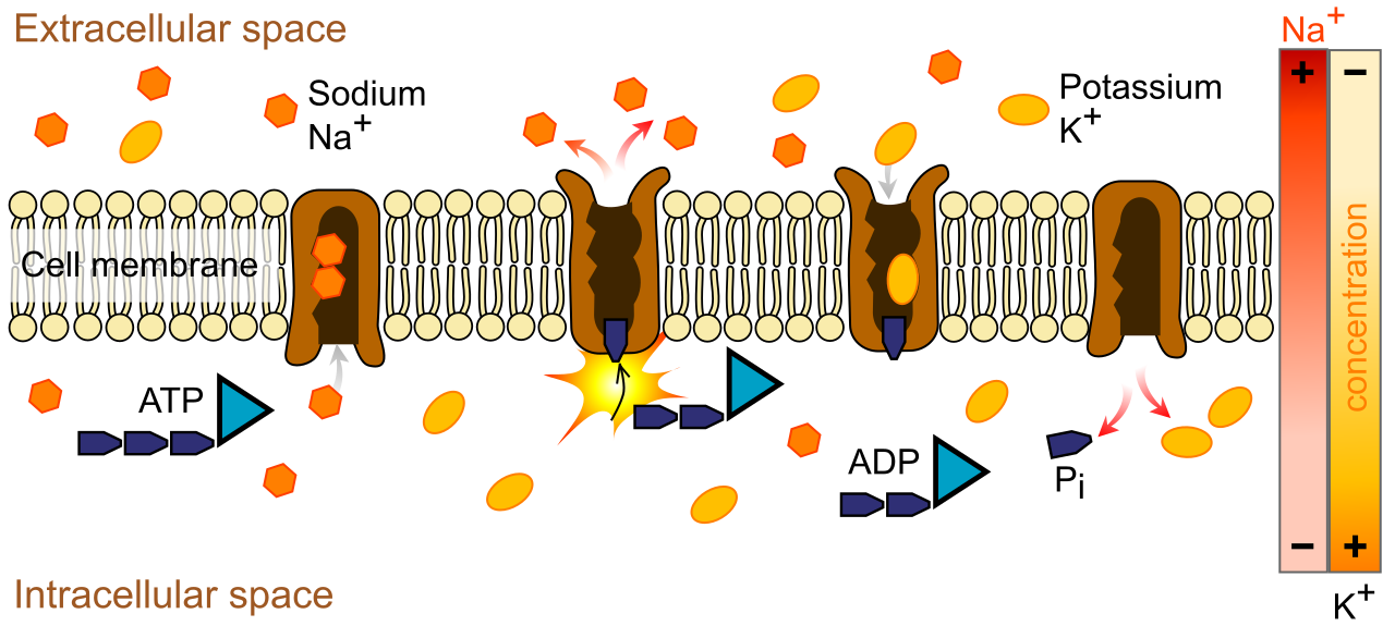
Now, imagine pushing that boulder back up the hill. You are working against natural forces, which requires an investment of energy. Active transport requires cellular energy in the form of adenosine triphosphate (ATP).
Because it utilizes power, active transport moves substances against a concentration gradient. Therefore, substances in active transport move from an area of lower concentration to an area of higher concentration.
A vital example in human physiology is the sodium-potassium pump, which is an active transport mechanism. This pump constantly expends ATP to shove sodium out of neurons and pull potassium in—against their respective gradients—to maintain the electrical charge necessary for nerve impulses and heartbeat regulation.
Bulk Transport: Endocytosis and Exocytosis
Sometimes, a cell needs to move massive molecules, or large quantities of fluid, that are simply too big for a transmembrane protein doorway. To accomplish this, the cell physically manipulates its own membrane.
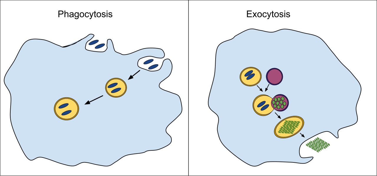
Endocytosis is an active transport process that brings large molecules into the cell by engulfing the molecules in a cell membrane vesicle. There are two primary variations of this process:
- Phagocytosis (Cell Eating): This is a form of endocytosis involving the ingestion of large solid particles. Your white blood cells use phagocytosis to hunt, engulf, and destroy pathogenic bacteria.
- Pinocytosis (Cell Drinking): This is a form of endocytosis involving the ingestion of extracellular fluid, allowing the cell to sample its immediate environment.
Conversely, exocytosis is an active transport process moving material out. Exocytosis expels materials from the cell using intracellular vesicles that fuse with the cell membrane, spilling their contents into the extracellular space. This is precisely how neurons release neurotransmitters to communicate with one another across synapses.