Urinary, Immune, and Skeletal Systems
Not sure you’re ready?
Take the ~3-minute readiness diagnostic and see where you stand.
Every living human body is an autonomous, self-sustaining metropolis. For this biological city to function, three fundamental infrastructural requirements must be met: a masterful sanitation system to filter out chemical waste while preserving precious water, an elite defense force capable of identifying and neutralizing microscopic invaders, and a dynamic, mineral-reinforced framework that simultaneously protects vital machinery and manufactures the very blood that gives the city life. In the clinical environment, a nurse encounters the failure of these systems every day. Edema in a patient's lower extremities reveals a faltering filtration plant; a sudden, spiking fever signals a biological war waged by the defense force; and a fractured hip in an elderly patient demonstrates the catastrophic loss of structural integrity. To accurately assess and treat a patient, you must intimately understand the intricate engineering of the urinary, immune, and skeletal systems.
At a glance, the urinary system is composed of the kidneys, ureters, urinary bladder, and urethra. While it is common to think of this system merely as a waste-disposal mechanism, its true physiological genius lies in what it keeps, not just what it throws away.
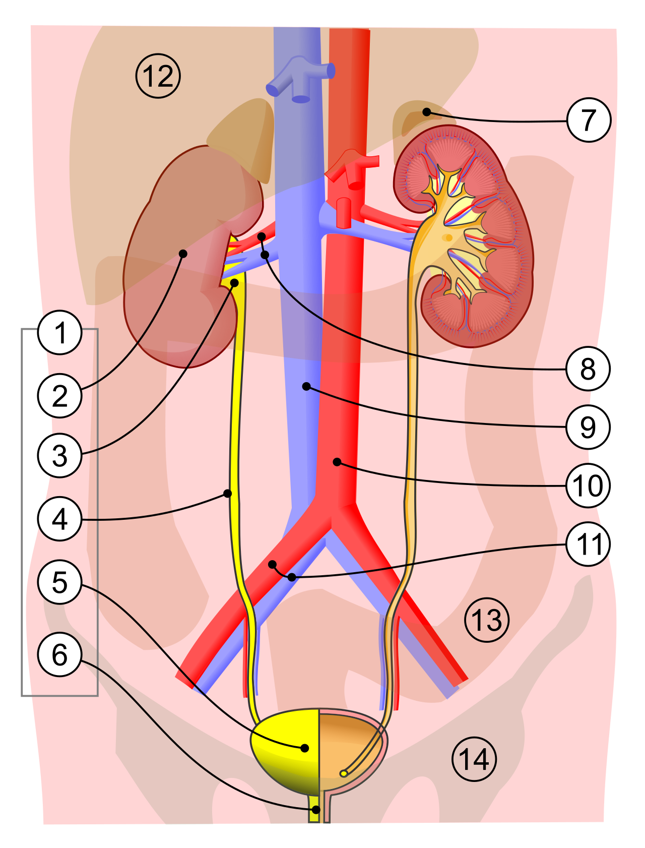
The kidneys filter blood to produce urine, but in doing so, they make profound, life-or-death adjustments to the body's cardiovascular status. Because water follows sodium, the urinary system regulates overall blood volume in the human body. By dictating how much fluid remains in the vascular space, the urinary system plays a major role in regulating systemic blood pressure. Furthermore, when systemic blood pressure drops, the kidneys release the enzyme renin to help regulate systemic blood pressure through a cascading chemical pathway that ultimately forces blood vessels to constrict.
The Nephron: The Microscopic Factory
To understand the kidney, you must zoom in. The structural and functional unit of the kidney is the nephron. Each human kidney contains approximately one million nephrons, working in parallel to process your entire blood volume dozens of times a day.
Think of nephron filtration like cleaning out a messy closet. It is often faster to throw everything out into the hallway, and then carefully carry back only the items you actually want to keep, leaving the true garbage outside. The nephron does exactly this.
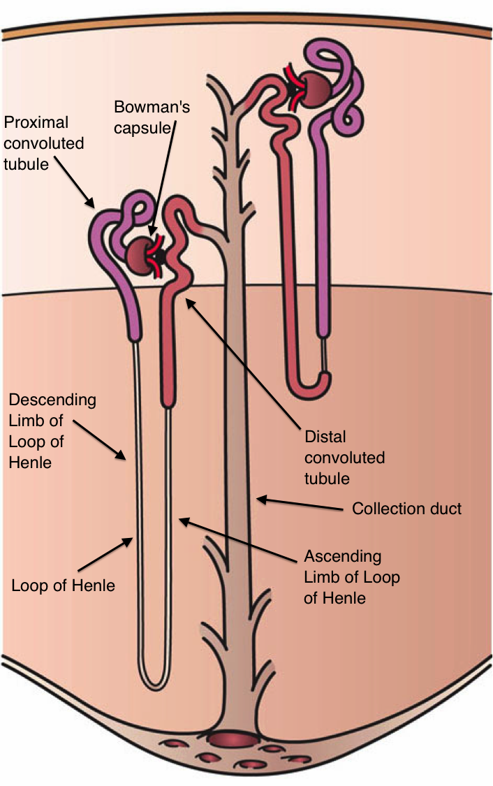
- The Glomerulus and Bowman's Capsule: The glomerulus is a network of capillaries where blood filtration begins in the kidney. Blood enters under high pressure, forcing water, ions, and small molecules out of the bloodstream. Bowman's capsule is a cup-like anatomical structure surrounding the glomerulus; it acts as a catcher's mitt. Bowman's capsule collects the initial blood filtrate produced by the glomerulus.
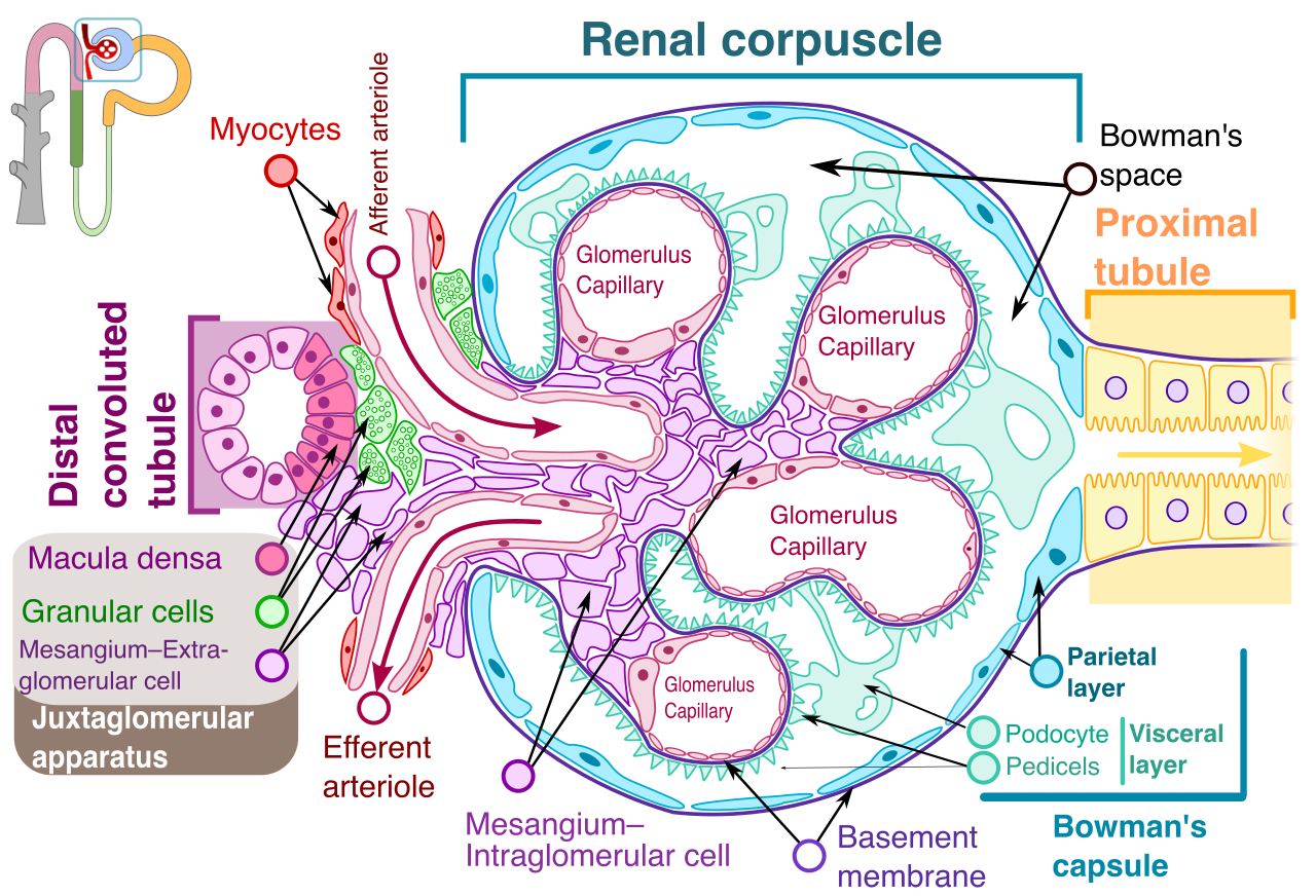
- The Proximal Convoluted Tubule (PCT): This is where we bring the "good stuff" back into the closet. The proximal convoluted tubule is the primary kidney site for the reabsorption of water into the bloodstream. Furthermore, the proximal convoluted tubule reabsorbs essential ions and nutrients back into the blood, ensuring you do not lose vital glucose or amino acids.
- The Loop of Henle: Plunging deep into the kidney's center, the Loop of Henle creates an osmotic concentration gradient within the kidney medulla. This salty gradient is the physical mechanism that allows the kidney to concentrate urine later in the process.
- The Distal Convoluted Tubule (DCT): Here, fine-tuning occurs. The distal convoluted tubule regulates potassium concentrations in the blood and simultaneously regulates sodium concentrations in the blood.
- The Collecting Duct: Finally, the collecting duct carries concentrated urine from the nephron to the renal pelvis, where it prepares to exit the kidney entirely.
Clinical Correlation: Hormonal Control The kidney's fine-tuning is governed by hormones you will administer or manipulate pharmaceutically every day. Antidiuretic hormone (ADH) increases water reabsorption within the collecting ducts of the kidneys, preventing dehydration. Meanwhile, aldosterone increases sodium reabsorption within the distal convoluted tubule, which pulls more water back into the blood, raising blood pressure.
The Plumbing: Transport and Elimination
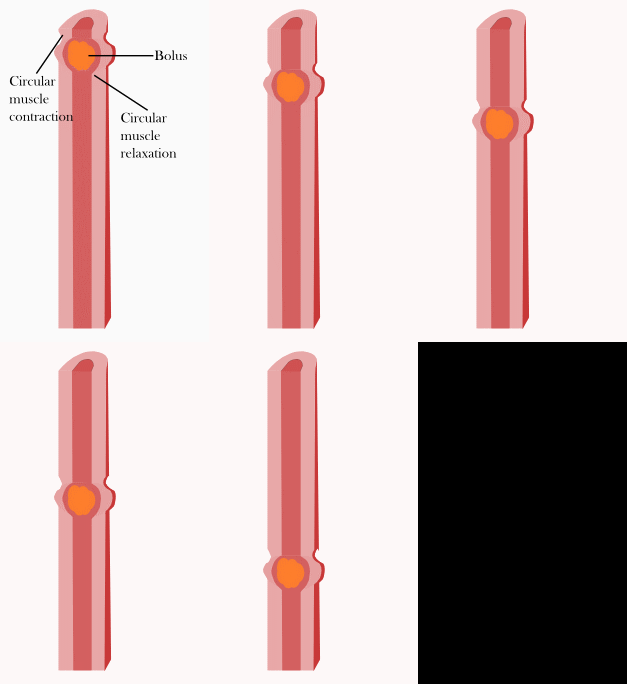
Once urine is formed, it must be safely removed. Ureters are muscular anatomical tubes. They do not rely on gravity; rather, ureters actively transport urine from the kidneys down to the urinary bladder via peristalsis. The urinary bladder is a hollow muscular pelvic organ. Because of its highly specialized tissue, the urinary bladder expands to store urine prior to physiological excretion. Finally, the urethra is the anatomical tube through which urine exits the human body.
A perfect filtration system is useless if the host is destroyed by foreign pathogens. The human immune system is broadly divided into innate immunity and adaptive immunity. They operate like a medieval castle's defense strategy: innate immunity is the moat and the stone walls, while adaptive immunity represents the highly trained archers who recognize the exact faces of their enemies.
Innate Immunity: The First Responders
Innate immunity provides non-specific physiological defense mechanisms against foreign pathogens. It does not care what the invader is; it simply knows the invader does not belong. Because it is pre-programmed, innate immune responses occur almost immediately following initial pathogen exposure.
The First Line of Defense Before a pathogen can infect you, it must breach the perimeter. The first line of innate immune defense includes physical barriers like the human skin, which is tough and slightly acidic. Inside the body's cavities, mucous membranes act as physical barriers in the first line of innate immune defense, trapping dust and bacteria. If a pathogen is swallowed, stomach acid acts as a harsh chemical barrier in the first line of innate immune defense, incinerating most microbes before they reach the intestines.
The Second Line of Defense If the perimeter is breached (e.g., a paper cut), the alarms sound. The second line of innate immune defense includes the generalized inflammatory response—bringing heat, swelling, and fluid to the breach site.
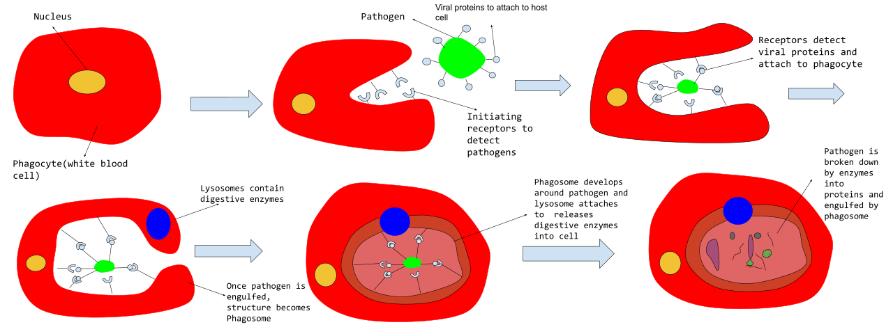
- Phagocytes: Phagocytic white blood cells are a core component of the second line of innate immune defense. Specifically, macrophages are large phagocytic white blood cells. Like cellular Pac-Men, macrophages engulf and destroy pathogens through a cellular process called phagocytosis.
- Chemical Warfare: When a cell is hijacked by a virus, it sends out an SOS. Interferons are specific signaling proteins secreted by virus-infected host cells. In a final act of sacrifice, interferons protect neighboring healthy cells by inhibiting active viral replication.
- The Complement System: The complement system is a specialized group of circulating blood proteins. Floating passively until activated, the complement system enhances the ability of antibodies and phagocytic cells to clear microbes, often by literally punching holes in bacterial cell walls.
Adaptive Immunity: The Special Forces
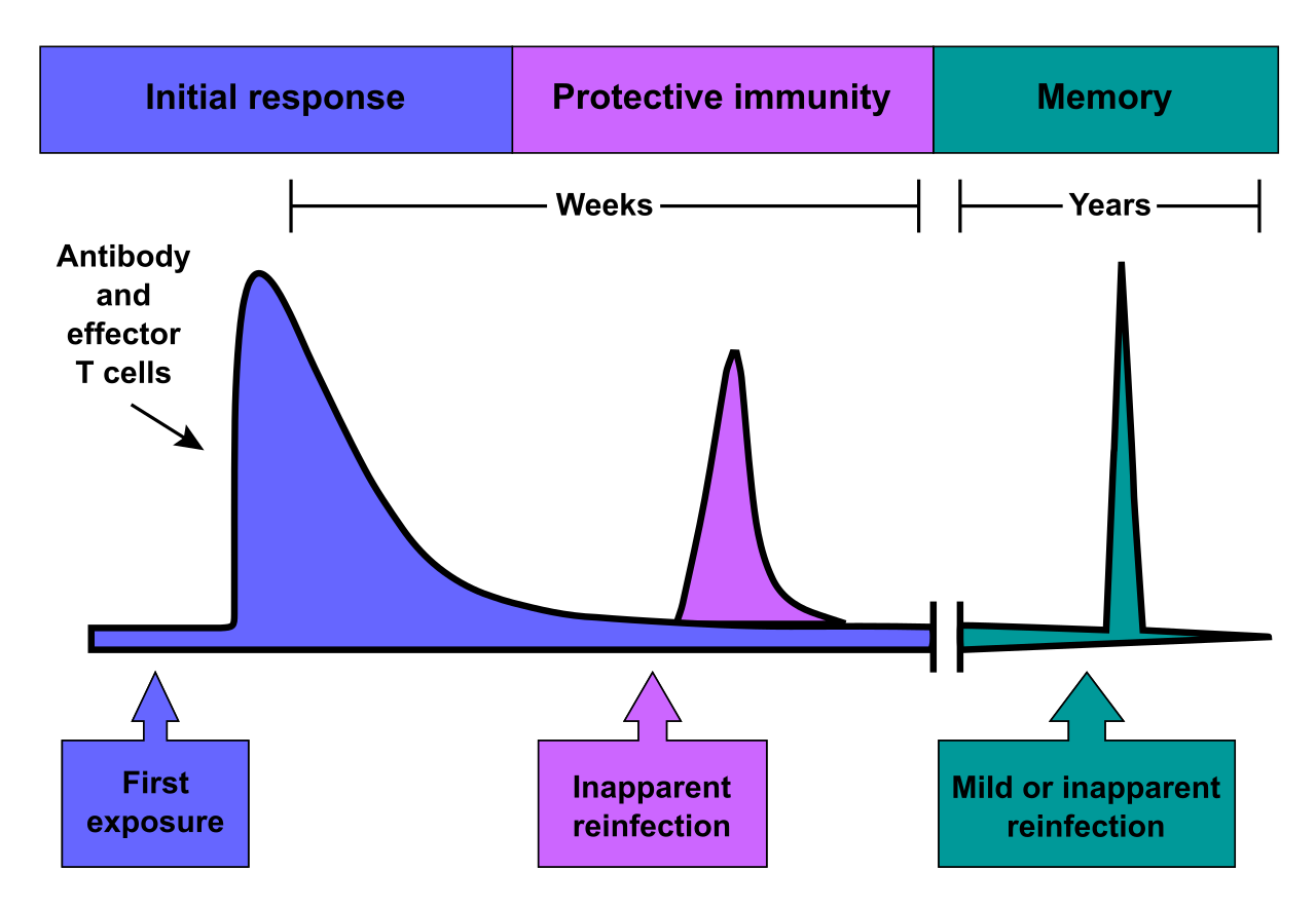
When innate defenses are overwhelmed, the heavy artillery arrives. Adaptive immunity provides highly specific defense mechanisms against individual pathogen strains. It takes days to deploy, but it has a superpower: adaptive immunity generates long-term immunological memory after an initial pathogen exposure.
Adaptive immunity is driven by lymphocytes.
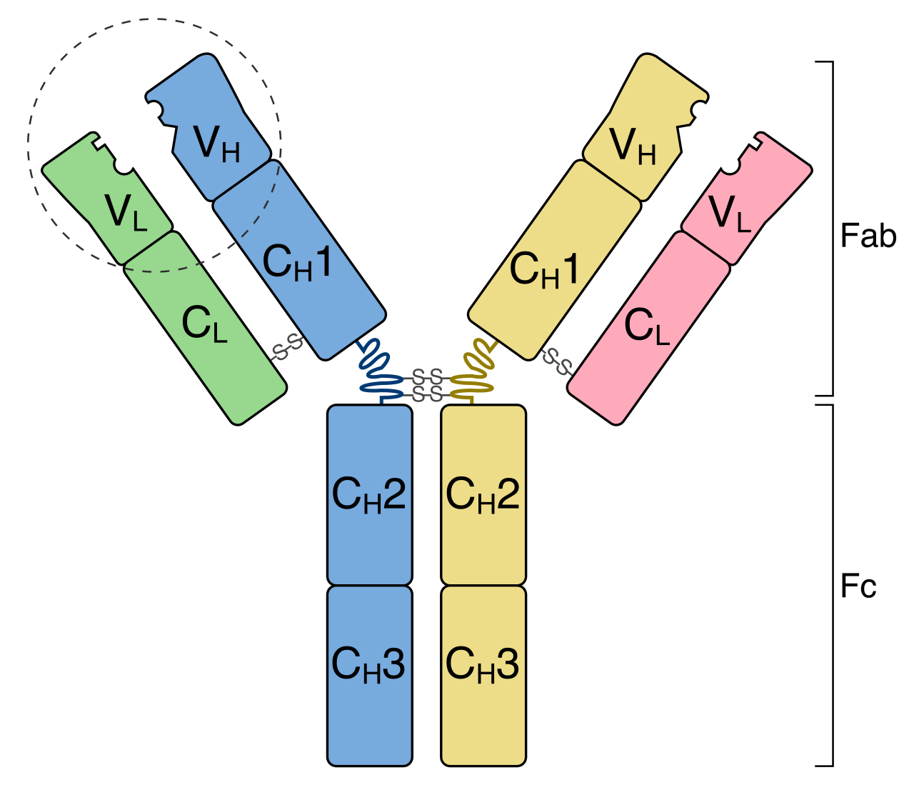
- B Cells: B cells are a specialized type of white blood cell called a lymphocyte. B cells undergo their primary physiological maturation within the bone marrow. Upon detecting an enemy, B cells produce target-specific antibodies to neutralize extracellular pathogens.
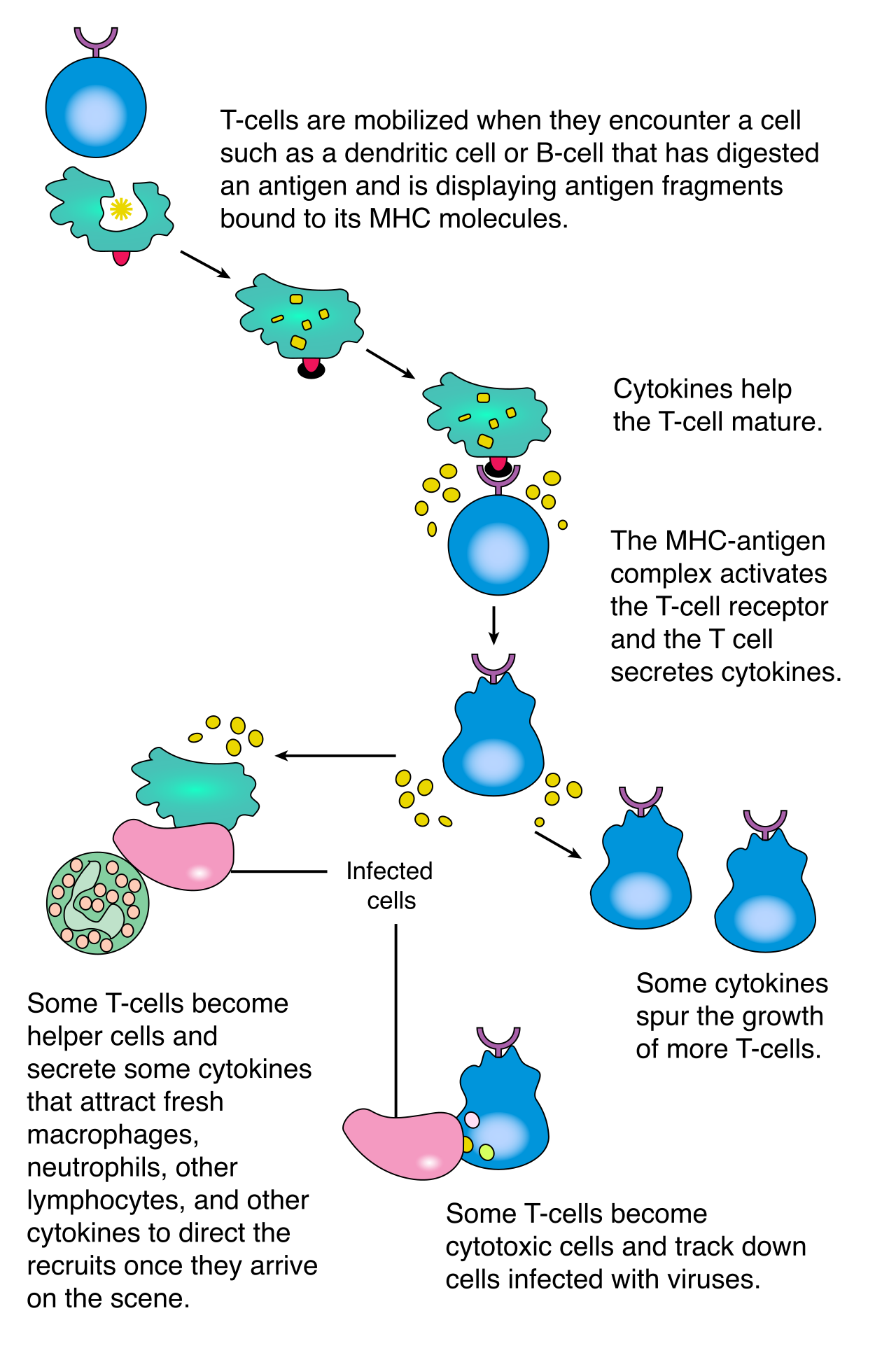
- T Cells: T cells are a specialized type of white blood cell called a lymphocyte, but unlike B cells, T cells undergo their primary physiological maturation within the thymus gland. They engage in close-quarters combat. Cytotoxic T cells directly bind and destroy infected host cells. Furthermore, cytotoxic T cells possess the ability to directly destroy cancerous host cells before tumors can form. To coordinate all of this, helper T cells secrete crucial signaling proteins called cytokines. In turn, cytokines coordinate the localized and systemic activity of various immune cells, acting as the battlefield communicators.
Acquiring Immunity
Immunity is acquired in two ways:
- Active Immunity: Active immunity develops when a host body produces native antibodies in response to a specific antigen. Because the body does the work, memory is formed. Clinical vaccination is a prime example of artificially acquired active immunity.
- Passive Immunity: Passive immunity occurs when an individual physically receives pre-made antibodies from an external source. It is immediate but temporary, as no memory is formed. Maternal antibodies passed through breast milk provide naturally acquired passive immunity.
The Lymphatic System: The Biological Highway
The immune system requires a physical network to transport cells and filter fluids. The lymphatic system collects leaked interstitial fluid and returns it back to the bloodstream, preventing massive tissue edema.
As this fluid travels, it passes through security checkpoints. Lymph nodes biologically filter lymph fluid to trap foreign infectious particles. To ensure these particles are destroyed, lymph nodes house high internal concentrations of defensive lymphocytes.
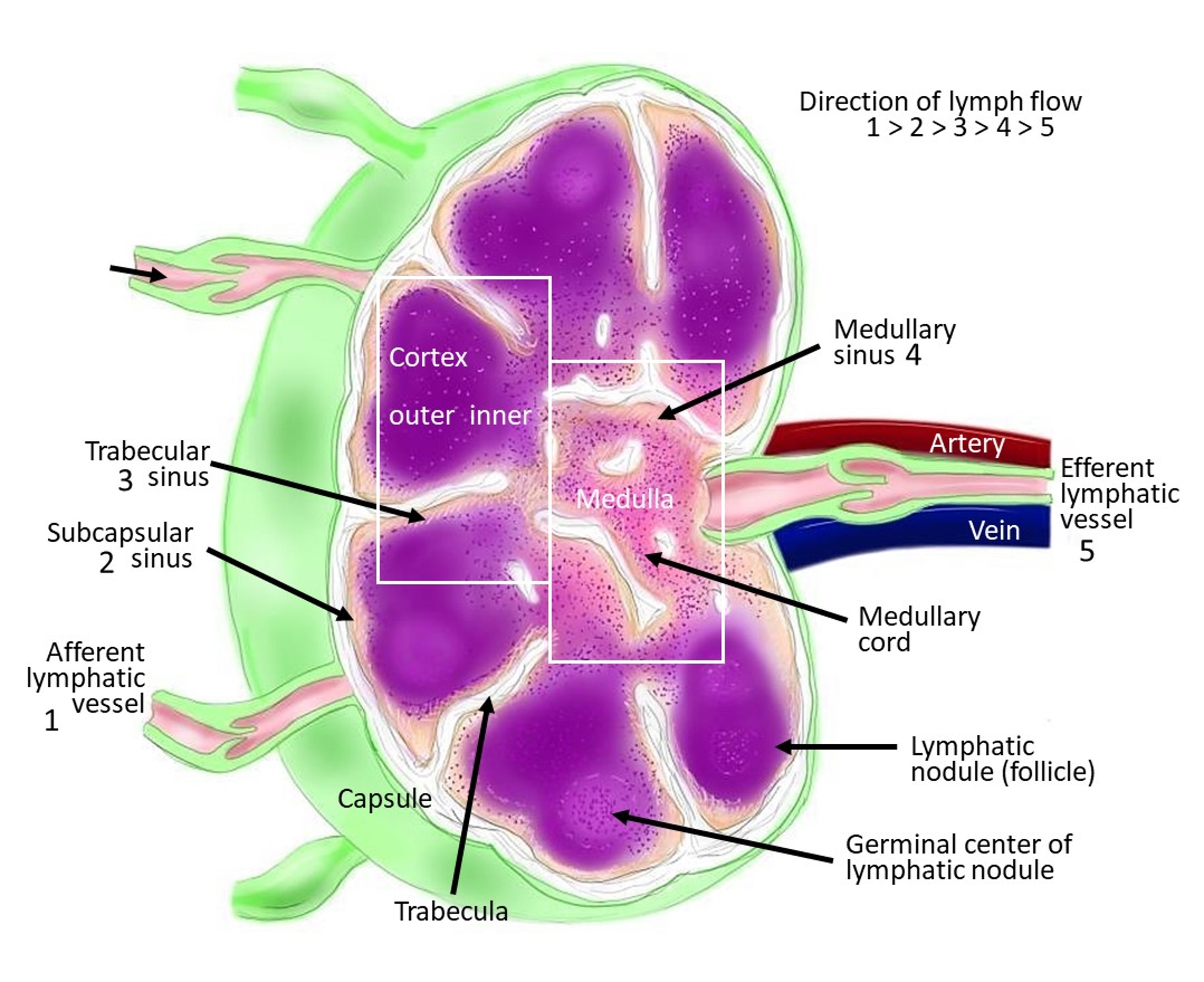
Other crucial lymphatic organs include:
- The Spleen: The spleen is a specialized lymphatic organ. Aside from storing white blood cells, the spleen physiologically filters circulating blood to remove old or damaged red blood cells.
- The Tonsils: Tonsils are protective masses of lymphatic tissue situated in the back of the human throat, standing guard against inhaled or ingested pathogens.
Far from being a static, dried-out scaffold, the skeleton is a bustling, heavily vascularized construction site. The adult human skeletal system naturally consists of 206 individual bones.
Beyond merely holding the body upright, the skeleton acts as a vital metabolic bank vault. The skeletal system acts as a physiological reservoir for essential minerals like calcium, and the skeletal system physically stores essential biological minerals like phosphorus. When blood calcium drops, the body literally dissolves a microscopic fraction of its own skeleton to keep the heart beating.
Architectural Divisions
We divide this framework into two distinct regions:
- The Axial Skeleton: The axial skeleton structurally forms the central vertical axis of the human body. Its primary job is protection and central support. The axial skeletal division includes the bones of the skull, the bones of the vertebral column, and the bones of the rib cage.
- The Appendicular Skeleton: This handles mobility and interaction with the environment. The appendicular skeleton includes the individual bones of the upper and lower limbs. It also includes the mounting hardware that attaches those limbs to the core: the appendicular skeleton includes the bones of the pectoral girdle (shoulders) and the bones of the pelvic girdle (hips).
Bone Microanatomy and Physiology
Bone tissue comes in two distinct densities.
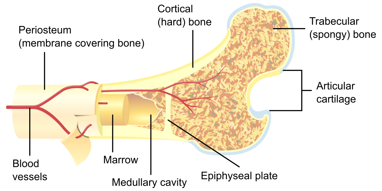
Compact Bone Compact bone is incredibly dense connective tissue. Because of its strength, compact bone physically forms the hard outer protective layer of skeletal bones. If you look at compact bone under a microscope, it looks like a cross-section of a dense forest of tree trunks. Osteons are the basic cylindrical microscopic structural units of compact bone. Running directly down the middle of each "tree trunk" is a pathway: Haversian canals are central anatomical channels running lengthwise within bone osteons. Because bone is living tissue, Haversian canals contain critical blood vessels and nerve fibers for bone maintenance.
Spongy Bone Beneath the dense exterior lies a highly engineered shock absorber. Spongy bone is highly porous internal bone tissue. Spongy bone is anatomically concentrated at the flared ends of long bones, and spongy bone is commonly located inside the interior cross-section of flat bones. The microscopic struts that form this porous network are called trabeculae. Trabeculae are the specific structural lattice networks found within spongy bone architecture.
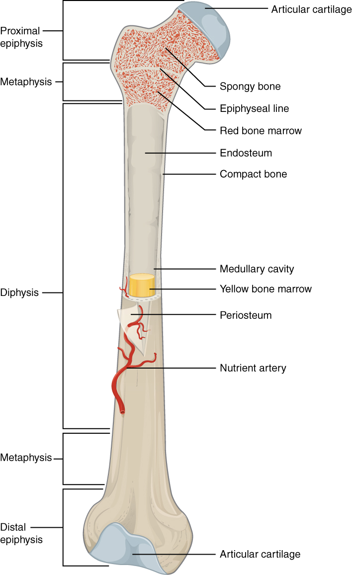
This porous space is where the magic of blood production occurs. Red bone marrow is physically located within the porous open spaces of spongy bone. Crucially, red bone marrow is the primary physiological site of hematopoiesis, and hematopoiesis is the essential biological process of new blood cell production. Conversely, yellow bone marrow primarily stores energy-rich adipose tissue. Yellow bone marrow is located within the central medullary cavity of long bones.
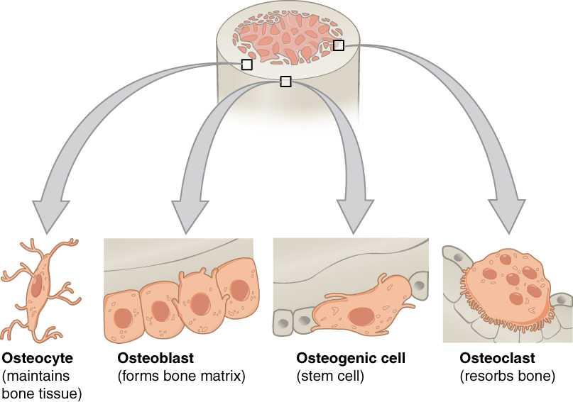
The Cellular Workforce Bone is constantly being demolished and rebuilt by three distinct cell types:
- Osteoclasts: Osteoclasts are specialized multinucleated bone cells. Acting as a demolition crew, osteoclasts enzymatically break down and resorb existing bone tissue (releasing calcium into the blood).
- Osteoblasts: Osteoblasts are specialized mononuclear bone-forming cells. Following the demolition crew, osteoblasts actively synthesize and mineralize new bone matrix.
- Osteocytes: Once osteoblasts finish pouring the concrete, they get trapped inside it. Osteocytes are mature bone cells physically trapped within the calcified bone matrix. From their trapped positions, osteocytes chemically maintain the mineral concentration of their surrounding bone matrix.
Bone Morphologies
Bones are categorized by their physical geometry, which dictates their mechanical function:
- Long Bones: Designed for leverage. Long bones structurally feature a long central tubular shaft called the diaphysis and rounded distal and proximal ends called epiphyses. The human femur is an anatomical example of a standard long bone.
- Short Bones: Designed for stability and weight distribution. Short bones are roughly cube-shaped skeletal structures. Short bones are anatomically located in the human wrists and ankles.
- Flat Bones: Designed for shielding. Flat bones physically protect vital underlying internal body organs, and structurally provide broad anatomical surfaces for muscle attachment. The human sternum is an anatomical example of a flat bone.
- Irregular Bones: Irregular bones possess complex shapes that strictly do not fit other bone geometric categories. Human spinal vertebrae are anatomical examples of irregular bones.
- Sesamoid Bones: These are mechanical pulleys. Sesamoid bones physically develop entirely within muscle tendons. By holding the tendon slightly away from the joint center, sesamoid bones mechanically act to reduce physical friction across adjacent joints. The human patella is a classic anatomical example of a sesamoid bone.
Joints and Connective Tissue
Where two bones meet, an articulation—or joint—is formed.
| Joint Type | Characteristics | Example |
|---|---|---|
| Fibrous Joints | Fibrous joints are structurally connected by dense collagen-rich connective tissue. Fibrous joints are generally entirely immovable within the adult human body. | The cranial sutures of the skull are prime examples of fibrous joints. |
| Cartilaginous Joints | Cartilaginous joints are structurally connected entirely by flexible cartilage. Cartilaginous joints physically allow for only limited biomechanical movement. | Intervertebral discs of the spine. |
| Synovial Joints | Synovial joints fundamentally feature a fully enclosed fluid-filled joint cavity. This lubrication means synovial joints physically allow for a very wide range of biomechanical motion. | See breakdown below. |
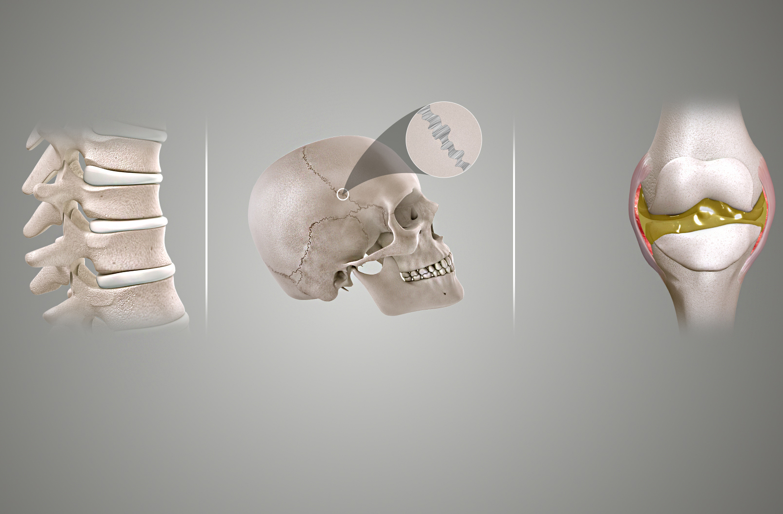
Synovial joints are further classified by the specific type of movement they permit:
- Hinge Joints: Hinge joints are synovial joints that only allow movement along a single anatomical plane (like a door). The human elbow is an anatomical example of a synovial hinge joint.
- Ball-and-Socket Joints: Ball-and-socket joints biomechanically allow for the greatest range of motion across multiple planes. The human shoulder is an anatomical example of a ball-and-socket joint.
- Pivot Joints: Pivot joints strictly allow for rotational movement around a single central axis. The specific joint between the first and second cervical vertebrae acts as a pivot joint, allowing you to shake your head "no."
Finally, this skeletal framework is lashed together and operated via incredibly strong connective tissues. Ligaments are incredibly tough structural bands of dense connective tissue. Their job is skeletal stability; ligaments physically attach one bone to another bone at human joints. Tendons are exceptionally strong structural cords of dense connective tissue. Their job is biological leverage; tendons physically attach skeletal muscle tissue to bone surfaces, turning muscle contraction into actual human movement.