Biological Molecules and Enzymes
Not sure you’re ready?
Take the ~3-minute readiness diagnostic and see where you stand.
A human cell is not a mysterious, indivisible blob of protoplasm; it is an incredibly busy, violently active chemical factory. Every second, millions of precise chemical reactions occur within your cells to keep you breathing, thinking, and reading these words. As a future healthcare professional, your entire career will revolve around manipulating and preserving this chemical factory. When you administer insulin, measure blood pH, or try to bring down a dangerous fever, you are not treating an abstract disease—you are directly intervening in the interactions of microscopic biological molecules. To understand human physiology, pharmacology, and pathology, you must first understand the structural materials that build the body and the molecular machines that do the work.
Every living organism on Earth is constructed from just four major classes of biological macromolecules: carbohydrates, lipids, proteins, and nucleic acids. These molecules are primarily built by linking smaller, repeatable subunits called monomers into long chains called polymers. Let us examine how the structure of each molecule perfectly dictates its function in the human body.
When a patient arrives in the emergency department weak and disoriented, one of the first things you will check is their blood glucose. You are checking their carbohydrate levels.
Carbohydrates are macromolecules composed entirely of carbon, hydrogen, and oxygen atoms. If you look closely at the name—carbo (carbon) and hydrate (water, H2O)—it perfectly describes their chemical makeup. In fact, the ratio of carbon to hydrogen to oxygen atoms in simple carbohydrates is approximately 1:2:1.
Carbohydrates function as the primary source of immediate energy for cellular processes. When a cell needs to do work right now, it burns carbohydrates.
Monomers and Polymers of Carbohydrates
The simplest single-sugar monomer units of carbohydrates are called monosaccharides. The most vital of these is glucose, which is a primary monosaccharide utilized by cells for energy. It is the molecular fuel that powers the brain and muscles.
When cells need to store this fuel for later, they link thousands of glucose molecules together to form polysaccharides, which are long polymer chains consisting of many linked monosaccharides. Nature uses polysaccharides in a few brilliantly distinct ways depending on the organism:
- Glycogen: A highly branched polysaccharide used for glucose storage in animal cells. When your patient’s blood sugar drops, their liver breaks down glycogen to release glucose back into the blood.
- Starch: A polysaccharide used for glucose storage in plant cells. When you eat a potato, you are consuming plant energy reserves.
- Cellulose: A structural polysaccharide that forms the primary component of plant cell walls. Humans cannot digest cellulose, but we rely on it as dietary fiber.
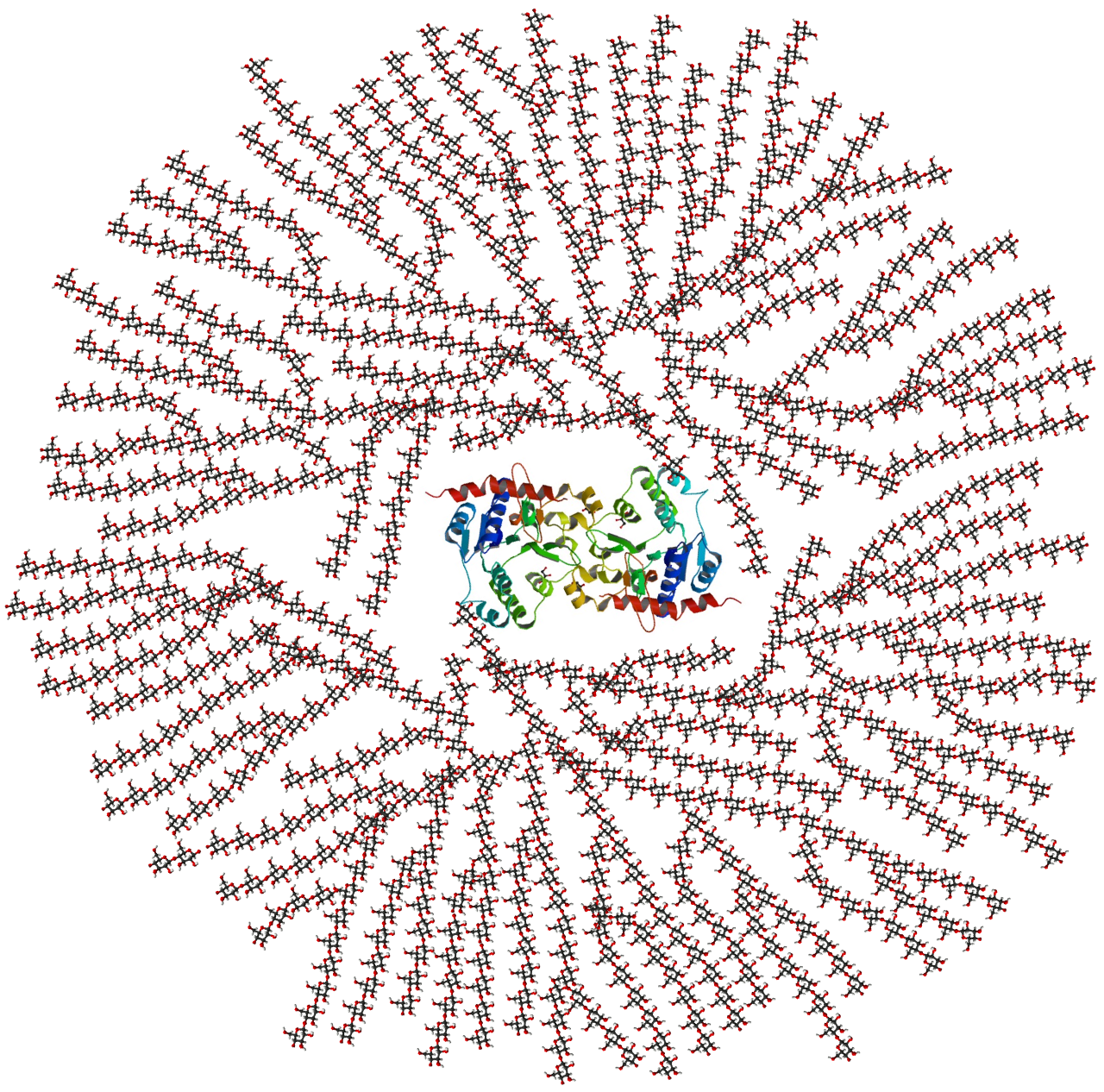
If carbohydrates are the immediate cash in a cell's wallet, lipids are the gold bars locked deep in the vault. Lipids are a diverse group of macromolecules unified by one glaring chemical trait: they are highly hydrophobic and insoluble in water. They repel water completely.
Because the human body is mostly water, lipids play specialized roles. The main biological categories of lipids are fats, phospholipids, and steroids.
Fats (Triglycerides)
While carbohydrates provide quick energy, fats primarily function to provide long-term energy storage for organisms. A standard fat molecule consists of a single glycerol backbone covalently bonded to three fatty acid chains.
The shape of these fatty acid chains determines how the fat behaves:
- Saturated fatty acids contain exclusively single bonds between their constituent carbon atoms. Because they are straight, they pack tightly together and form solids at room temperature (like butter).
- Unsaturated fatty acids contain one or more double bonds between their constituent carbon atoms. These double bonds create "kinks" in the chain, preventing tight packing and making them liquid at room temperature (like olive oil).
Phospholipids
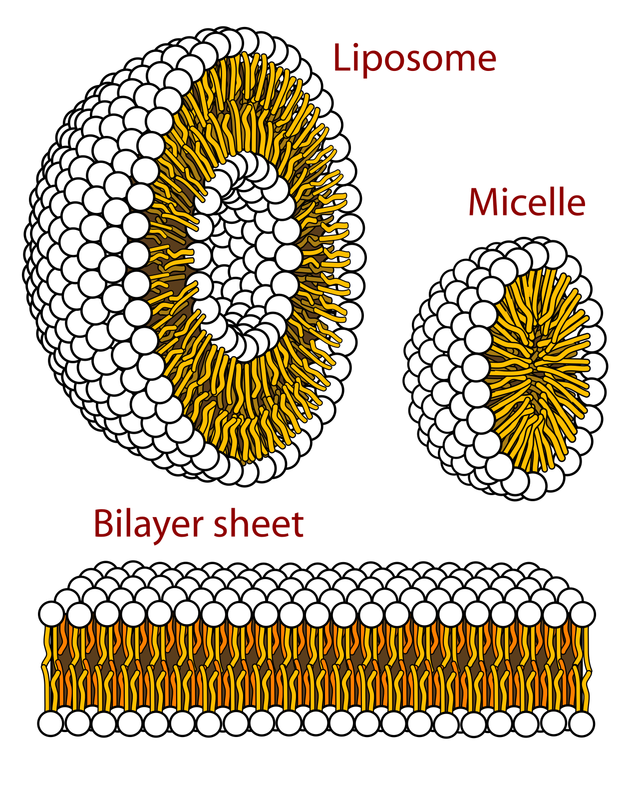
Life requires a border. A cell cannot exist if its internal machinery washes away into the surrounding fluid. Enter the phospholipid. Phospholipids consist of two fatty acid tails and a phosphate group attached to a glycerol backbone.
This creates a molecular split personality: the phosphate "head" loves water, while the two fatty acid "tails" hate water. If you throw them in water, they instantly arrange themselves tail-to-tail to hide from the moisture. Because of this, the structure of phospholipids allows them to form the selectively permeable bilayers of cellular membranes. They act as the physical walls of the cell, dictating exactly what can enter and exit.
Steroids
Unlike the long chains of fats, steroids are lipid molecules characterized by a carbon skeleton consisting of four fused molecular rings. They act as structural components and powerful chemical messengers (hormones). Cholesterol is a prominent steroid molecule that modulates the fluidity of animal cell membranes, preventing them from becoming too rigid in the cold or too floppy in the heat.
If carbohydrates are the fuel, and lipids are the walls, proteins are the workers, the scaffolding, the sensors, and the machinery.
Amino acids are the fundamental monomer building blocks of proteins. In all of biology, there are exactly twenty standard amino acids utilized by living organisms to construct proteins. You can think of these twenty amino acids like a 20-letter alphabet. By stringing these letters together in different combinations, you can write thousands of different "words."
Amino acids are covalently linked together by peptide bonds to form long polypeptide chains. But a long string of amino acids isn't a functional protein yet. It must fold.
The Golden Rule of Proteins: The specific sequence of amino acids directly dictates the final three-dimensional folded shape of a protein. Consequently, the unique three-dimensional shape of a protein strictly determines the biological function of that protein.
If you change even one amino acid out of a chain of hundreds, the chain folds differently. Because its three-dimensional shape is altered, its biological function is destroyed. This is exactly what happens in sickle cell anemia: a single microscopic typo in an amino acid sequence causes hemoglobin proteins to fold incorrectly, leading to devastating clinical consequences.
How does the cell know exactly which sequence of amino acids to string together to build a functional protein? That information is stored in the nucleic acids, which function primarily to store and transmit genetic information within cells.
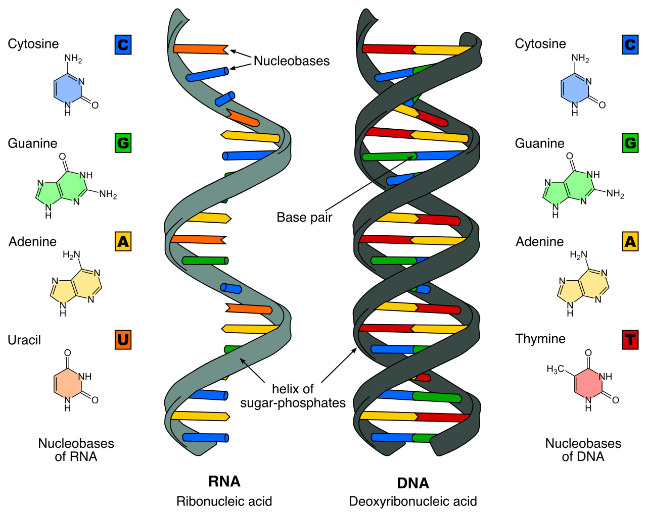
Nucleotides are the specific monomer units that make up nucleic acid polymers. To build a single nucleotide, you need three pieces: a pentose sugar, a phosphate group, and a nitrogenous base.
There are two primary types of nucleic acids in biological systems:
- Deoxyribonucleic acid (DNA): The master blueprint safely locked in the nucleus.
- Ribonucleic acid (RNA): The temporary, mobile copy of the blueprint used to actually build the proteins.
Macromolecule Summary Matrix
| Macromolecule | Monomer | Primary Functions | Key Examples |
|---|---|---|---|
| Carbohydrates | Monosaccharides | Immediate energy | Glucose, Glycogen, Starch |
| Lipids | Varies (Fatty acids/Glycerol) | Long-term energy, membranes | Fats, Phospholipids, Cholesterol |
| Proteins | Amino Acids | Machinery, structure, transport | Hemoglobin, Antibodies, Enzymes |
| Nucleic Acids | Nucleotides | Genetic information storage | DNA, RNA |
Now we arrive at the most dynamic proteins in the body: the enzymes. Enzymes act as biological catalysts in cellular metabolism. While some rare RNA molecules have catalytic properties, the vast majority of biological enzymes are specialized protein molecules.
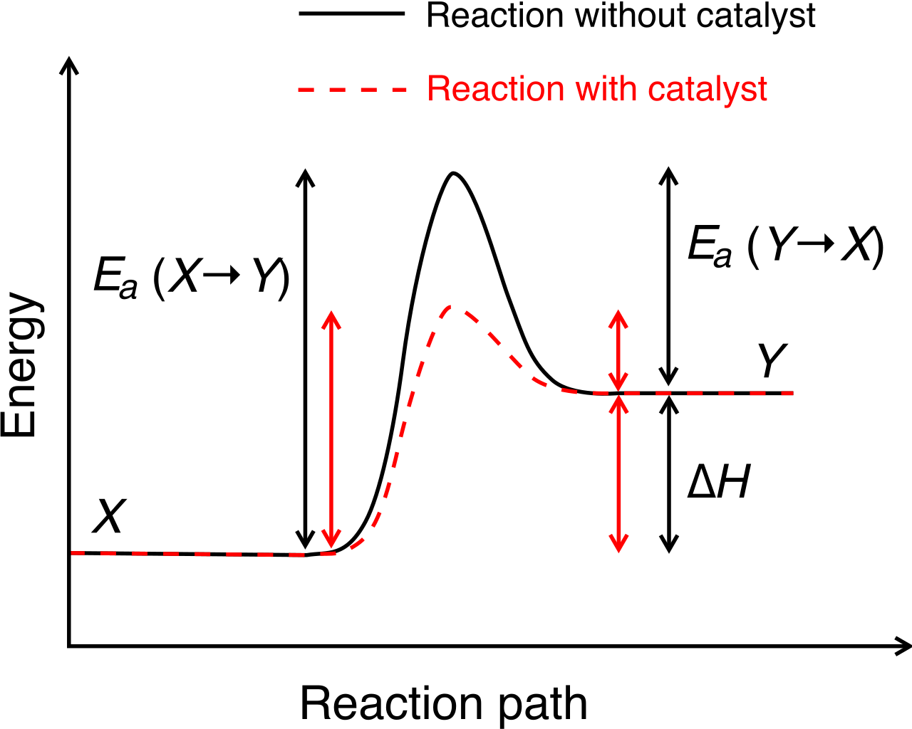
In chemistry, a catalyst speeds up a reaction. Without enzymes, the chemical reactions required to keep you alive would happen so incredibly slowly that life would be impossible. Enzymes accelerate specific chemical reactions by lowering the required activation energy—the initial "push" of energy required to get a chemical reaction started. Think of it like a boulder resting in a valley at the top of a mountain. To roll the boulder down the mountain (the reaction), you first have to push it up and out of the valley (the activation energy). An enzyme simply digs a tunnel through the side of the valley, requiring far less energy to get the boulder rolling.
Crucially, enzymes are neither consumed nor permanently altered during the chemical reactions they facilitate. Like a wrench tightening a bolt, the tool does the work and is immediately ready to be used again.
The Lock, the Key, and the Handshake
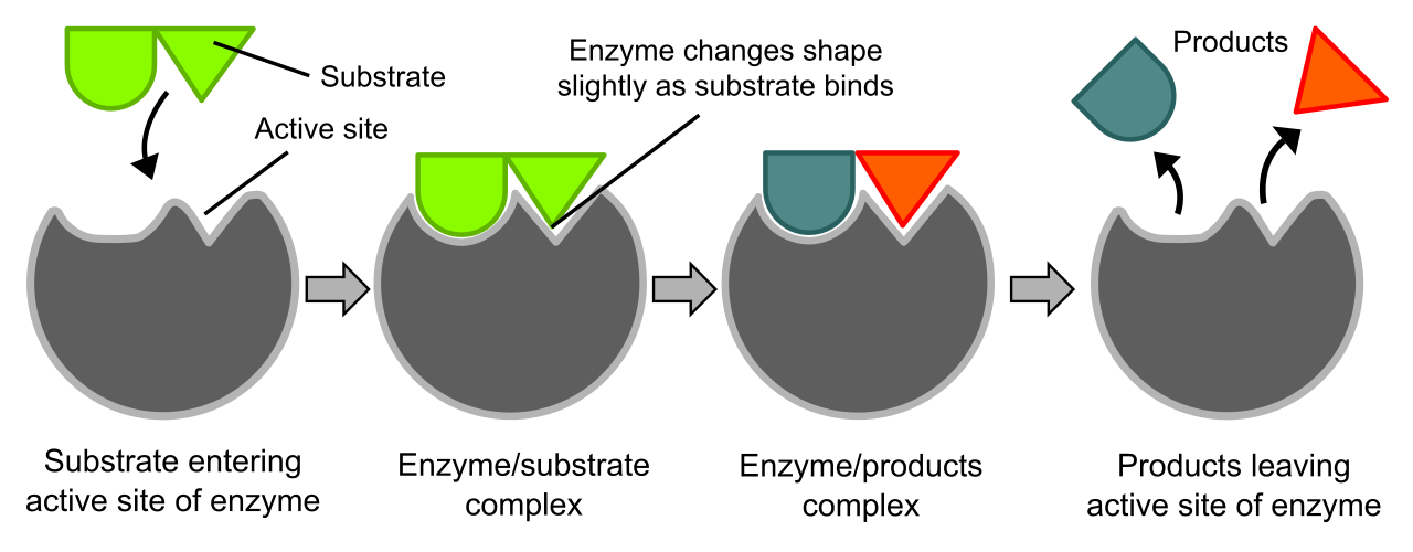
How do enzymes know what to work on? A substrate is the specific reactant molecule upon which a particular enzyme acts.
Enzymes have a active site, which is a specialized geometric pocket on the enzyme where the substrate physically binds. Remember the Golden Rule of Proteins? Shape dictates function. The unique physical geometry of an active site severely restricts an enzyme to bind only with specific substrates. A protein-digesting enzyme cannot digest a lipid; the shapes simply do not fit.
When the substrate slides into this pocket, the physical binding of a substrate to an enzyme creates an intermediate structure called the enzyme-substrate complex.
Originally, scientists thought this was a rigid "lock and key" mechanism. We now know it is far more dynamic. The induced fit model dictates that an enzyme will slightly alter its structural shape to more tightly grasp the bound substrate, much like a baseball glove naturally wrapping around a ball the moment it hits the leather. Once the reaction is complete, the enzyme releases the new products and returns to its original shape.
As a clinician, you will constantly monitor your patient's temperature and blood pH. Why? Because you are guarding their enzymes.
The Impact of Temperature
Changes in environmental temperature can significantly alter the functional rate of enzyme-catalyzed chemical reactions. Molecules move faster as they warm up. Therefore, increasing temperatures generally elevate enzyme activity rates until an optimal temperature threshold is reached. Up to roughly 37°C (98.6°F) in humans, the biochemical factory hums along beautifully.
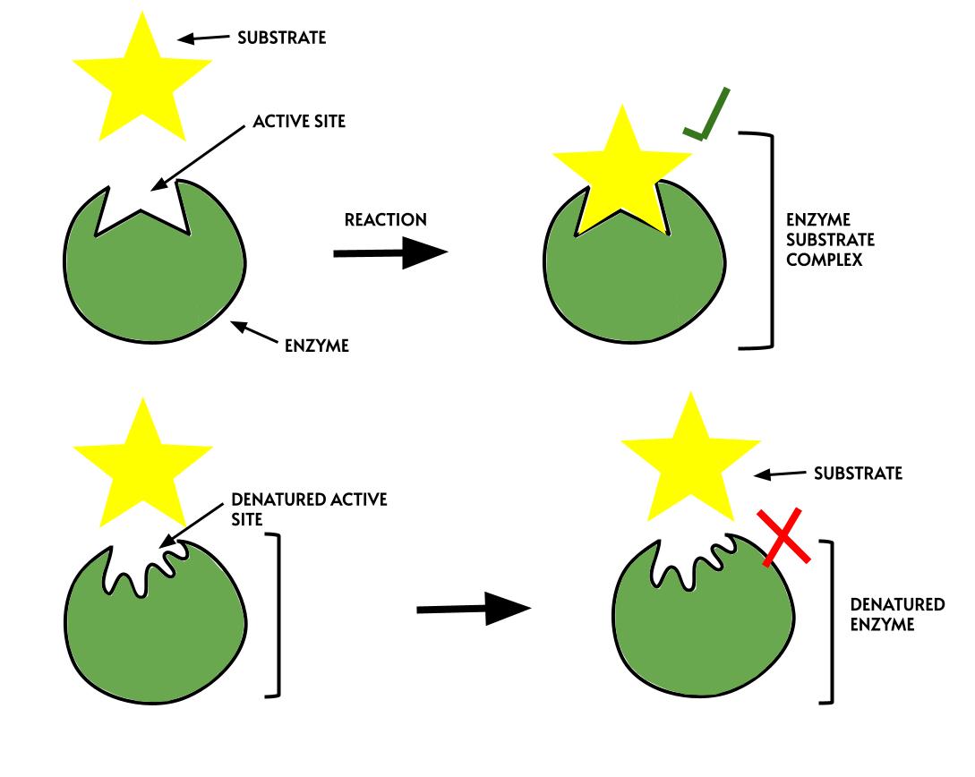
But if a patient develops a severe, uncontrolled fever, disaster strikes at the molecular level. Environmental temperatures exceeding an enzyme's optimal range disrupt the weak chemical bonds maintaining the enzyme's structure. The heat literally shakes the protein apart.
The loss of a protein's functional three-dimensional physical structure is defined as denaturation. A completely denatured enzyme possesses a deformed active site and cannot bind its specific substrate. If the active site is gone, the chemical reactions of life stop.
The Impact of pH
Just as temperature can shake a protein apart, acids and bases can rip a protein apart electrically.
Every enzyme operates at an optimal pH (for example, enzymes in the highly acidic stomach prefer a pH of 2, while enzymes in the blood prefer a pH around 7.4). Fluctuations in environmental pH alter the electrical charges of amino acid side chains within an enzyme.
Because the folding of a protein relies heavily on positive and negative magnetic-like attractions between these side chains, changing the charges throws the whole system into chaos. Specifically, altered amino acid electrical charges disrupt the specific ionic bonds required to maintain the shape of an active site.
If the pH shifts too drastically—such as in severe diabetic ketoacidosis—the damage becomes irreversible. Extreme deviations from an enzyme's optimal pH level result in permanent enzyme denaturation.
This is the profound beauty and fragility of life. A patient's survival relies entirely on the precise geometry of their molecules. By understanding macromolecules and enzymes, you are no longer just memorizing lab values; you are learning how to actively protect the molecular machinery that sustains human life.