Cell Transport
Not sure you’re ready?
Take the ~3-minute readiness diagnostic and see where you stand.
A biological cell is not a passive balloon filled with fluid; it is a highly secure, bustling metropolis. Just as a hospital regulates who enters the lobby, who requires an escort to the surgical wing, and who is escorted out, the cell membrane regulates the movement of substances into and out of the cell. For a future nurse or allied health professional, understanding this microscopic border control is not just an academic exercise. Every time you administer an IV fluid, push a medication, or monitor a patient’s blood oxygen, you are manipulating cellular transport. If you do not understand the rules of this boundary, you cannot understand how life sustains itself—or how medical interventions save it.
To master cell transport for the HESI A2 exam, we must look closely at the architecture of the membrane, the physical forces that drive molecules across it, and the specialized machinery the cell uses to move cargo against the tide.
To understand how things cross the membrane, you must first understand what the membrane is made of. The cell membrane is composed primarily of a phospholipid bilayer.
Imagine a vast sea of millions of molecules, each shaped roughly like a balloon with two strings attached. The "balloon" is the phosphate head, and the "strings" are lipid (fat) tails.
- Hydrophilic heads: The phosphate heads love water (hydrophilic) and face outward toward the watery environments inside and outside the cell.
- Hydrophobic tails: The lipid tails fear water (hydrophobic) and turn inward, facing each other to hide from the moisture, creating a dense, oily core.
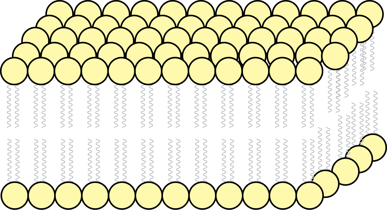
Selective permeability allows certain molecules to pass through the cell membrane while blocking other molecules.
Because the core of this membrane is a thick layer of fats, it dictates a strict entry policy based on size and chemical charge. Small, nonpolar molecules can easily pass directly through the phospholipid bilayer because they dissolve right into that oily core and slip through to the other side. Conversely, large or charged molecules require transport proteins to cross the cell membrane. They cannot pass through the lipid core on their own; they need a specialized door.
Nature favors equilibrium. If you spray perfume in the corner of a room, the molecules randomly collide and spread out until they fill the space evenly. In biology, this physical reality is harnessed as passive transport, which is the movement of substances across a cell membrane without the use of cellular energy.
Passive transport relies entirely on a concentration gradient, which is the difference in the concentration of a substance between two regions. In passive transport, molecules behave like boulders rolling down a hill: passive transport moves substances down the substance's concentration gradient, from an area of high crowding to an area of low crowding.
Simple Diffusion
Diffusion is the net movement of particles from an area of higher concentration to an area of lower concentration. When this happens right through the physical boundary of the cell, we call it simple diffusion.
Simple diffusion occurs directly across the phospholipid bilayer without the aid of transport proteins. This is the exclusive pathway for small, nonpolar molecules. For example, when a patient breathes in, oxygen fills the alveoli of their lungs. Because the concentration of oxygen is higher in the lungs than in the bloodstream, oxygen crosses the cell membrane via simple diffusion. The reverse is true for cellular waste: carbon dioxide crosses the cell membrane via simple diffusion, moving out of the tissues and into the blood to be exhaled.
Facilitated Diffusion
But what about the molecules that want to go down their concentration gradient, but are too large or too charged to squeeze through the lipid bilayer? They require a bridge.
Facilitated diffusion is a type of passive transport that requires specific transmembrane proteins. Like simple diffusion, facilitated diffusion moves molecules from an area of higher concentration to an area of lower concentration, meaning it still costs the cell zero energy.
There are two primary types of protein structures that facilitate this movement:
- Channel proteins provide a hydrophilic passageway for specific ions or molecules to cross the cell membrane. Think of these as open tunnels. Because the inside of the tunnel is hydrophilic (water-loving), charged ions can zip right through without touching the hydrophobic lipid core.
- Carrier proteins undergo a conformational change to transport specific molecules across the cell membrane. Think of these as revolving doors. A molecule binds to the protein, causing the protein to physically change its shape, dumping the molecule out on the other side.
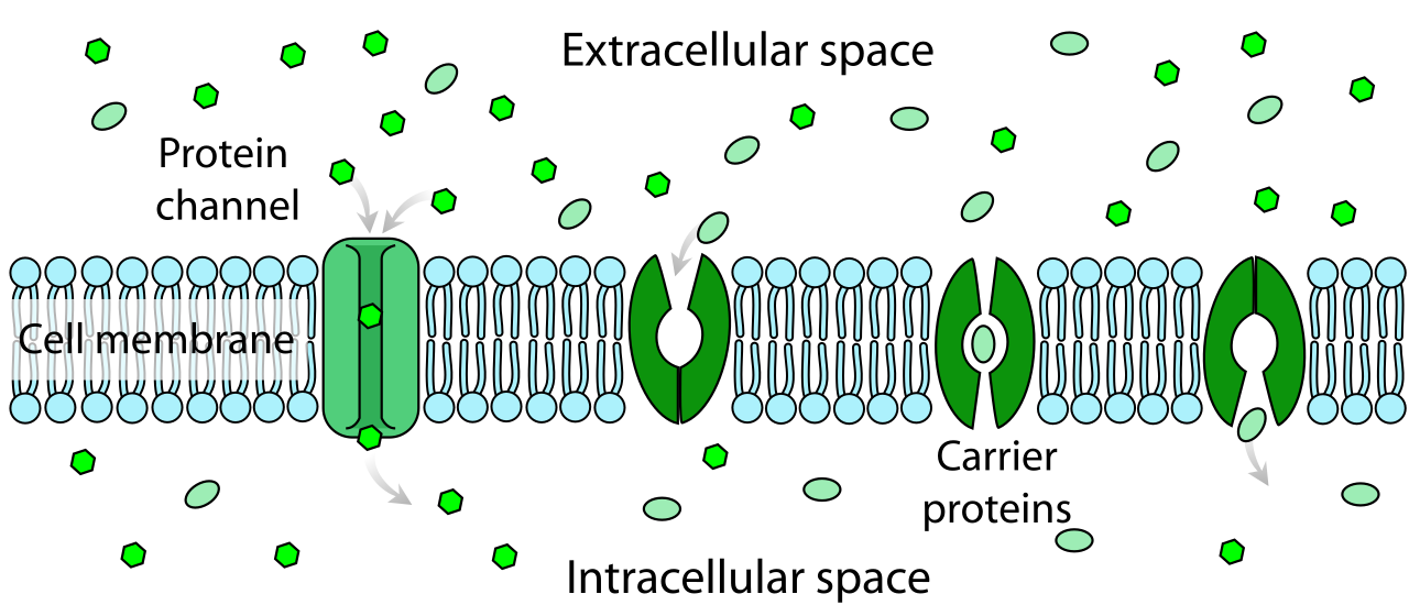
A vital clinical example is blood sugar. Glucose typically enters cells via facilitated diffusion. It is a large molecule that builds up in the blood after a meal. With the help of insulin, carrier proteins are inserted into the cell membrane, grabbing the glucose and folding inward to release it into the cell for fuel.
We have discussed the movement of solutes (the stuff dissolved in the water). But what happens if the solutes are trapped and cannot cross the membrane? In that case, the water itself will move to reach equilibrium.
Osmosis is the specific diffusion of water molecules across a selectively permeable membrane.
Here is the golden rule of osmosis that you must commit to memory: During osmosis, water moves from an area of lower solute concentration to an area of higher solute concentration. Water is drawn to the salt, the sugar, the proteins. It wants to dilute the most concentrated, crowded area.
While some water can slowly sneak through the lipid bilayer, the vast majority of it moves through specialized tunnels. Aquaporins are specialized channel proteins that facilitate the rapid transport of water across the cell membrane.
Tonicity in Clinical Practice
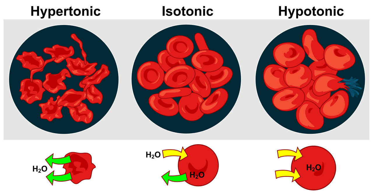
Tonicity describes how an extracellular solution can change the volume of a cell by affecting osmosis. This is the exact physics governing intravenous (IV) fluid administration.
| Solution Type | Definition | Cellular Effect | Clinical Application |
|---|---|---|---|
| Isotonic | An isotonic solution has an equal concentration of solutes compared to the inside of a cell. | Water moves equally in both directions across the cell membrane when a cell is in an isotonic solution. The cell remains stable. | 0.9% Normal Saline is isotonic. It is used to expand blood volume without shifting water into or out of the patient's cells. |
| Hypertonic | A hypertonic solution has a higher concentration of solutes compared to the inside of a cell. | Cells placed in a hypertonic solution will lose water and shrink. Water flees the cell to dilute the heavily concentrated fluid outside. | 3% Saline is hypertonic. It can be used carefully to draw excess fluid out of swollen brain tissue (cerebral edema). |
| Hypotonic | A hypotonic solution has a lower concentration of solutes compared to the inside of a cell. | Cells placed in a hypotonic solution will gain water and swell. The water rushes inside to dilute the cell's internal machinery. | 0.45% Saline is hypotonic. It is used to hydrate severely dehydrated cells. |
Clinical Warning: Never infuse pure, sterile water directly into a patient's veins. Cell lysis can occur if an animal cell is placed in a highly hypotonic solution. Because pure water has zero solutes, it will rapidly rush into the patient's red blood cells, causing them to swell and burst (lyse), which can be fatal.
Up to this point, all movement has been downhill, driven by the natural, free energy of the concentration gradient. But a cell cannot survive simply by letting nature take its course. Sometimes, a cell needs to stockpile nutrients or aggressively expel toxins. It must push the boulders back up the hill.
Active transport is the movement of substances against the substance's concentration gradient. This means active transport moves substances from an area of lower concentration to an area of higher concentration.
Because this defies the natural law of diffusion, it is not free. Active transport requires the expenditure of cellular energy.
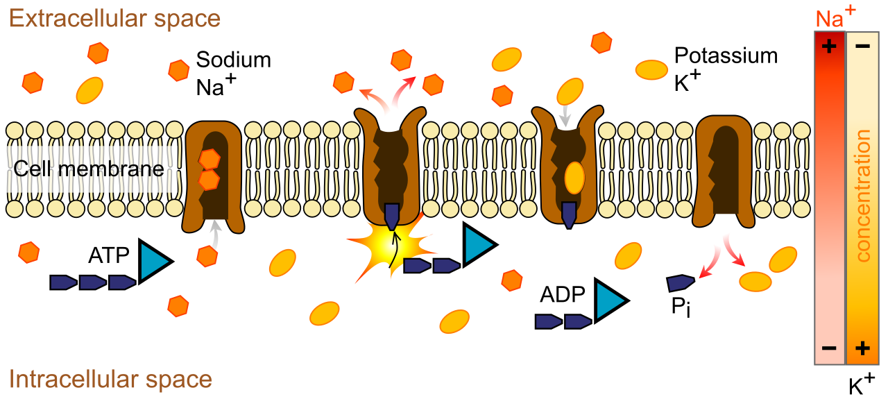
Adenosine triphosphate (ATP) is the primary energy source for active transport. When ATP breaks one of its chemical bonds, it releases a burst of energy that acts like a cellular motor. Furthermore, because these substances cannot slip through the membrane on their own, active transport requires specific carrier proteins often referred to as membrane pumps.
The Sodium-Potassium Pump
The most famous and physiologically critical example of active transport is the sodium-potassium pump. This protein complex is embedded in the membranes of nearly all animal cells, and it is the reason your heart beats and your brain fires thoughts.
The sodium-potassium pump is an example of active transport that tirelessly works to maintain a sharp electrical and chemical gradient across the membrane. Using ATP for power, the sodium-potassium pump moves three sodium ions out of the cell for every two potassium ions moved into the cell.
Because both sodium (Na+) and potassium (K+) are positively charged, pushing three positive charges out while only bringing two in creates a net negative charge inside the cell. This resting electrical potential is the loaded spring that allows nerve impulses to fire and cardiac muscle to contract.
Pumps and channels are excellent for handling individual ions and small molecules like glucose. But what if the cell needs to swallow an entire bacterium? What if it needs to dump massive quantities of hormones into the bloodstream all at once?
Bulk transport mechanisms move large molecules or large quantities of substances across the cell membrane. Because it involves physically wrapping and re-wrapping sections of the actual cell membrane, bulk transport requires the expenditure of cellular energy (ATP).
Bulk transport is divided into two major directional categories: bringing things in (Endocytosis) and pushing things out (Exocytosis).
Endocytosis (Moving In)
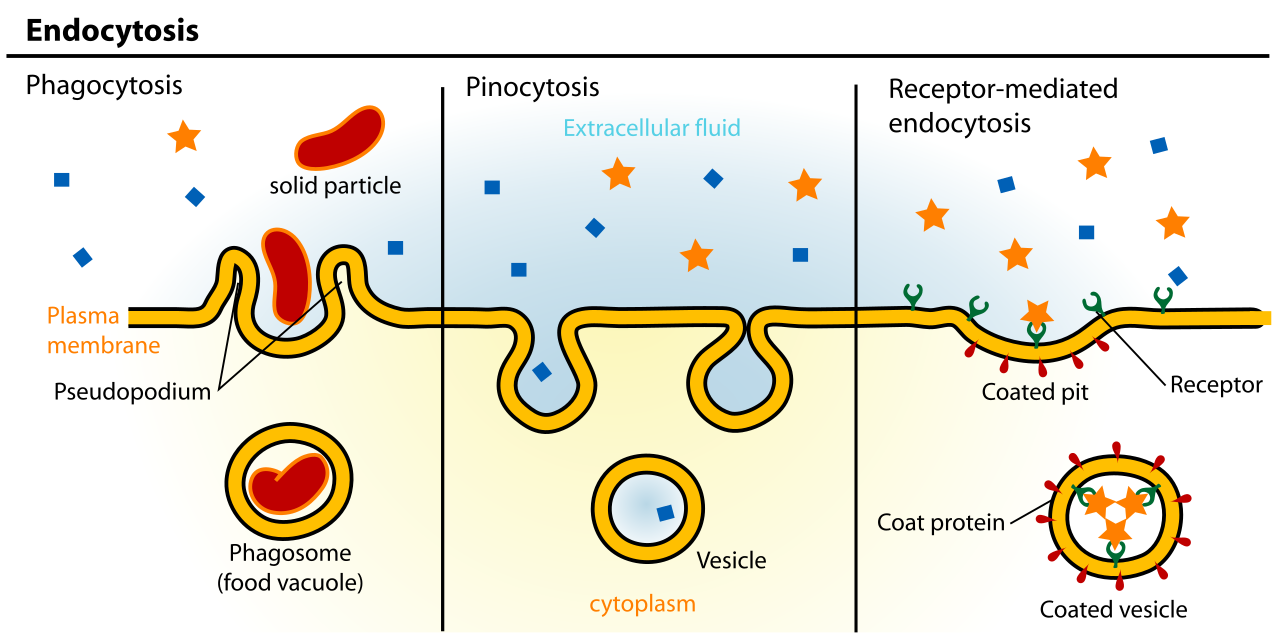
Endocytosis is the process of capturing a substance from outside the cell by engulfing the substance with the cell membrane. The membrane literally folds inward, creates a pocket around the target, pinches off, and forms a vesicle inside the cell.
There are three distinct variations of this process:
- Phagocytosis: Often called "cell eating." Phagocytosis is a type of endocytosis involving the ingestion of large particles or whole cells. When a patient has an infection, specialized white blood cells (macrophages) hunt down bacteria, engulf them via phagocytosis, and digest them.
- Pinocytosis: Often called "cell drinking." Pinocytosis is a type of endocytosis involving the ingestion of extracellular fluid and dissolved solutes. The cell takes a tiny gulp of the surrounding environment to sample the fluid and absorb nutrients.
- Receptor-mediated endocytosis: This is a highly targeted operation. Receptor-mediated endocytosis requires the binding of specific molecules to cell surface receptors before membrane invagination. The cell only swallows the cargo if the exact right "key" binds to the "lock" on the membrane. This is how cells absorb cholesterol from the blood.
Exocytosis (Moving Out)
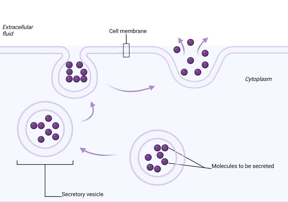
The exact reverse of endocytosis is used for cellular exporting. Exocytosis is the process of vesicles fusing with the plasma membrane to release the vesicle contents to the outside of the cell.
When a neuron needs to send a chemical message to the next neuron, it packages thousands of neurotransmitter molecules into a vesicle. That vesicle travels to the cell border, merges seamlessly with the phospholipid bilayer, and spills its chemical contents into the gap, triggering the next nerve to fire.
By mastering these physical mechanisms—from the passive drifting of simple diffusion to the energy-hungry mechanical work of active and bulk transport—you arm yourself with the foundational mechanics of human physiology. When you sit for the HESI A2, remember the underlying logic: the cell is a fortress, the membrane is its gate, and every molecule moving across it is strictly governed by the elegant, unyielding laws of physics and biology.