Cell Division and Classification
Not sure you’re ready?
Take the ~3-minute readiness diagnostic and see where you stand.
When a scalpel makes an incision, thousands of cells are destroyed. Yet, within days, the tissue knits itself back together perfectly, a flawless reconstruction of the microscopic architecture that was lost. This is not magic; it is the highly regulated mechanics of cell division at work. To understand human health—how a fetus develops, how a laceration heals, or how a localized tumor becomes a systemic crisis—we must understand the fundamental rules governing how cells replicate and how life is organized. In the clinical environment, you will encounter the outcomes of these microscopic processes every single day. From tracking the rapid division of epithelial cells in wound care to relying on the precise taxonomic classification of a bacterial pathogen to administer the correct antibiotic, the principles of cellular reproduction and biological organization form the bedrock of medical science.
The cell cycle is the series of events involving the growth and division of a cell. If you picture a cell as an intricate manufacturing plant, it cannot simply pinch itself in half whenever it feels like it. If a factory splits without first building a second set of machines and copying the master blueprints, both resulting halves will fail.
To prevent this catastrophic failure, the cell cycle consists of interphase and the mitotic phase. Interphase is the crucial preparatory period, accounting for the vast majority of a cell's life. Think of interphase as the time when the factory is running at full capacity, stockpiling resources, and copying its instruction manuals.
Interphase is divided into the G1 phase, the S phase, and the G2 phase. Let us look at what happens in each:
- During the G1 phase of the cell cycle, a cell grows in size and synthesizes cellular components. The "G" stands for Gap, but nothing is lacking here. The cell is busily accumulating the building blocks of chromosomal DNA and the associated proteins, alongside storing sufficient energy reserves.
- During the S phase of the cell cycle, a cell replicates its DNA. The "S" stands for Synthesis. Deep in the nucleus, the cell takes its genetic blueprint and makes a flawless, identical copy. This ensures that when the cell eventually divides, each new cell receives a complete manual for survival.
- During the G2 phase of the cell cycle, a cell prepares for division by producing necessary proteins. It replenishes energy stores and synthesizes the proteins necessary for manipulating chromosomes. The cell also undergoes a final check to ensure all DNA was copied without damage.
Only after these three preparatory steps are complete does the cell enter the mitotic phase, the brief window of actual, physical division.
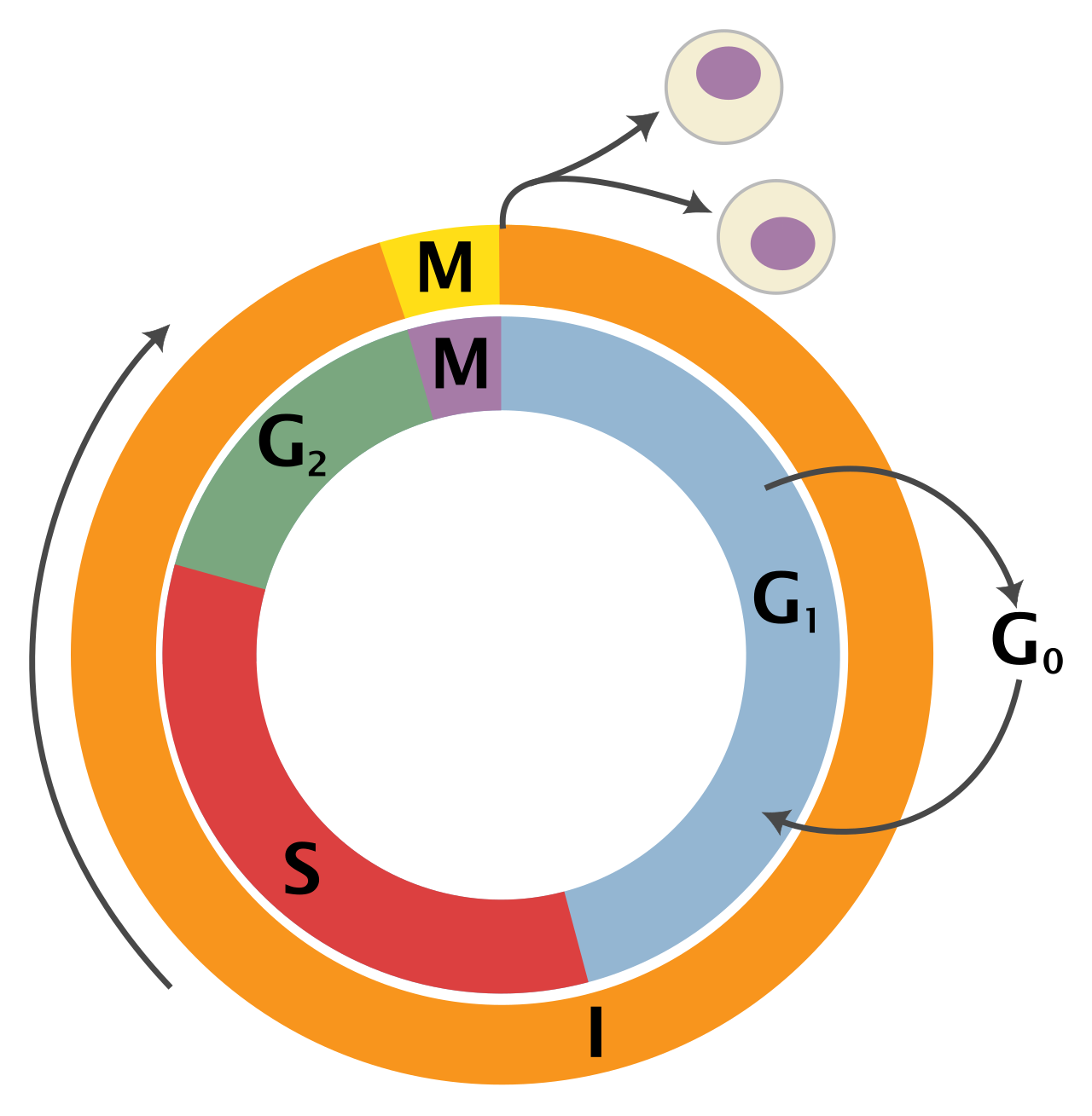
When we talk about the growth of a child, the regeneration of a liver, or the healing of a burn, we are talking about mitosis. Mitosis is a form of asexual cell division. It requires no partner. Somatic cells divide through the process of mitosis. Somatic cells are the standard "body" cells—everything from your neurons to your skin cells, excluding the reproductive sperm and egg cells.
The biological imperative of mitosis is perfect replication. Mitosis produces two genetically identical daughter cells. There is no room for creativity here; a skin cell must produce another skin cell. Because no genetic material is halved, the daughter cells produced by mitosis are diploid.
Diploid cells contain two complete sets of chromosomes—one set inherited from the mother, and one from the father. To be precise, human somatic cells contain 46 chromosomes (23 pairs).
The Stages of Mitosis
How does a cell mechanically ensure that 46 chromosomes are perfectly segregated into two new cells? It uses a beautifully choreographed, four-step dance. The stages of mitosis are prophase, metaphase, anaphase, and telophase. (You can remember this with the mnemonic PMAT).
- Prophase: During prophase of mitosis, chromatin condenses into visible chromosomes. The loose, stringy DNA packs tightly into X-shaped structures, making them easier to move without tangling. Simultaneously, during prophase of mitosis, the nuclear envelope breaks down. The protective vault holding the DNA dissolves so the cellular machinery can access the chromosomes.
- Metaphase: During metaphase of mitosis, chromosomes align along the center of the cell. Spindle fibers attach to the center of each chromosome and physically drag them to the cellular equator. This creates a neat, organized line, ensuring neither side gets shortchanged.
- Anaphase: During anaphase of mitosis, sister chromatids are pulled toward opposite poles of the cell. The X-shaped chromosomes are ripped cleanly down the middle. One half goes left; the other goes right.
- Telophase: The chromosomes reach their destinations. During telophase of mitosis, new nuclear envelopes form around each set of chromosomes. The DNA uncoils back into its relaxed state, and the cell now has two complete, enclosed nuclei.

Cytokinesis: The Final Split
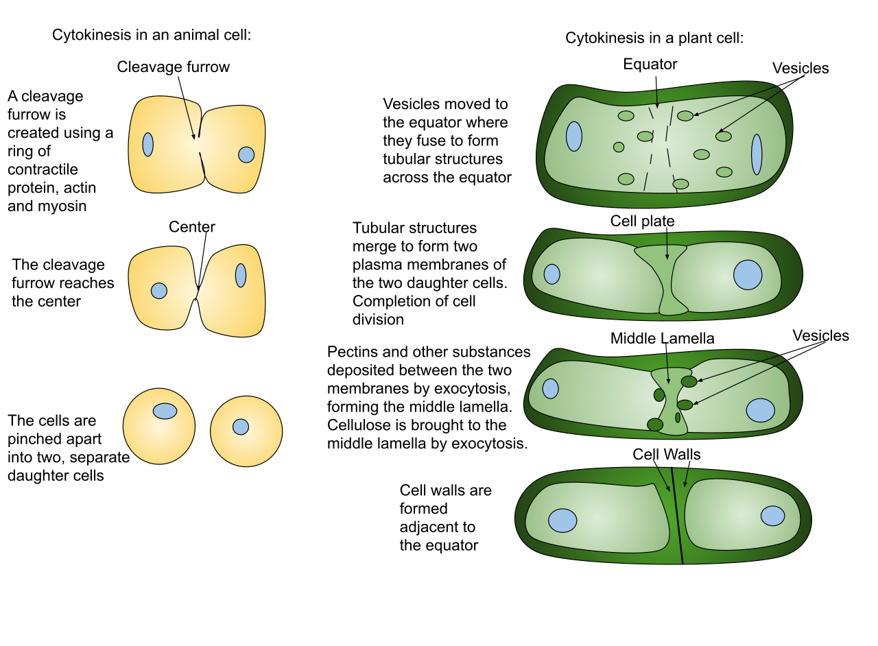
Mitosis strictly refers to the division of the nucleus. But the rest of the cell must divide too. Cytokinesis is the physical division of the cell cytoplasm.
Cytokinesis typically occurs simultaneously with the telophase stage of mitosis. However, the mechanism differs drastically depending on the organism's kingdom:
- During cytokinesis in animal cells, a cleavage furrow pinches the cell membrane in half. Imagine tightening a drawstring around the waist of a balloon until it pinches into two separate balloons.
- Because plants have rigid outer walls, they cannot be pinched. Instead, during cytokinesis in plant cells, a cell plate forms to divide the cytoplasm. A new wall is literally built down the middle of the cell, partitioning it into two walled rooms.
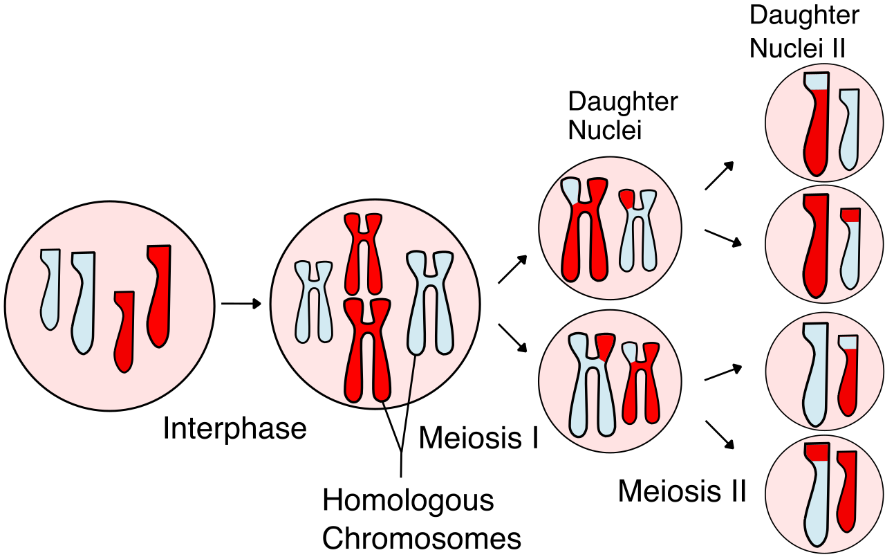
If mitosis is about perfect cloning, meiosis is about deliberate variation. Meiosis is a form of cell division required for sexual reproduction. Unlike somatic cells, gametes are formed through the process of meiosis. Gametes are the reproductive cells—sperm in males and ova (eggs) in females.
If two human cells, each with 46 chromosomes, fused together during reproduction, the resulting embryo would have 92 chromosomes. The generation after that would have 184. Life would collapse under the weight of its own DNA. To solve this, meiosis involves two consecutive rounds of cell division. Unsurprisingly, the two rounds of cell division in meiosis are known as meiosis I and meiosis II.
Because the parent cell divides twice but only replicates its DNA once, the math changes drastically. Meiosis produces four daughter cells. Crucially, the daughter cells produced by meiosis are haploid.
Haploid cells contain half the number of chromosomes as the parent cell. Therefore, human gametes contain 23 chromosomes. When a haploid sperm (23) fertilizes a haploid egg (23), the resulting human being is restored to the proper diploid number of 46.
The Magic of Crossing Over
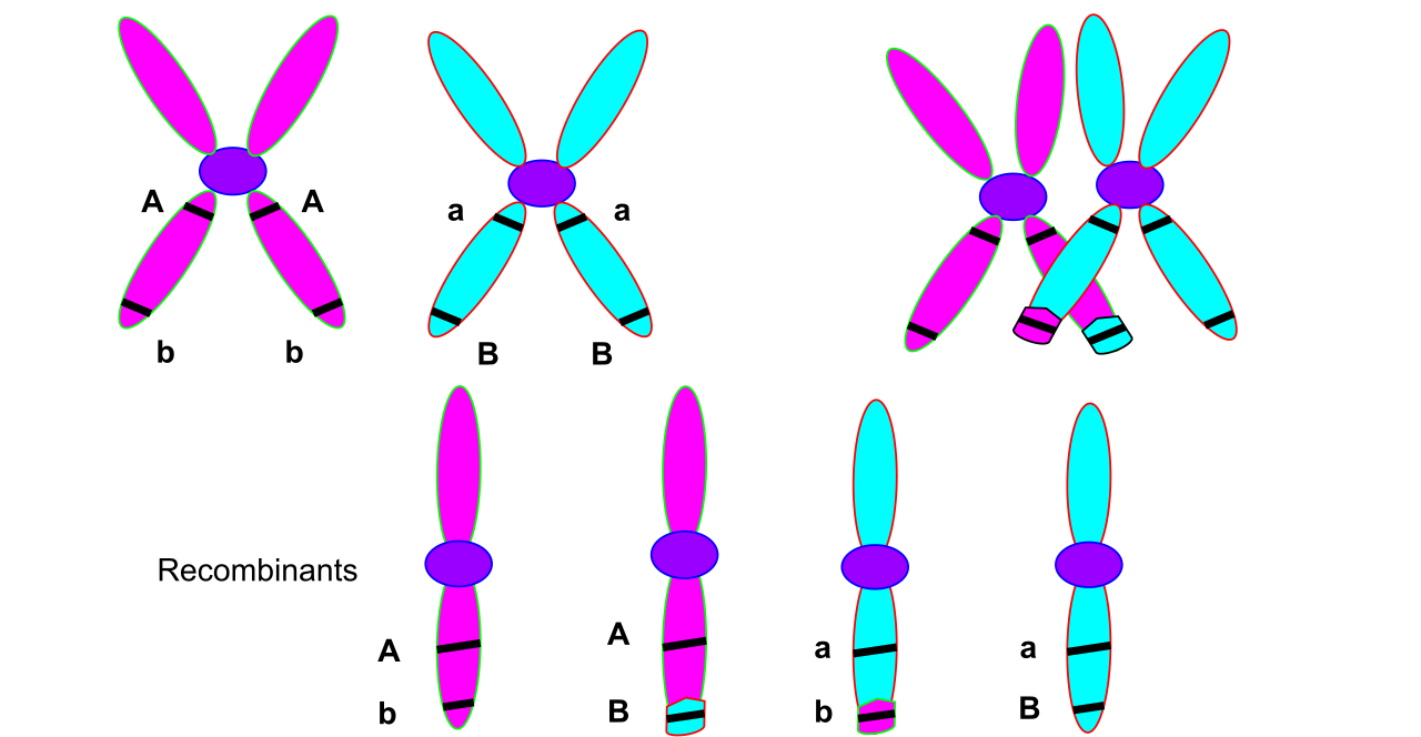
The daughter cells produced by meiosis are genetically distinct from the parent cell. This is the secret to human diversity—why you do not look exactly like your siblings, and why populations can adapt to diseases.
The heavy lifting of this variation happens at the very beginning of the process. During prophase I of meiosis, homologous chromosomes pair up and exchange genetic material. The chromosome you got from your father and the chromosome you got from your mother physically embrace and trade chunks of DNA. The exchange of genetic material between homologous chromosomes is called crossing over.
Because of crossing over, every single sperm or egg cell a human produces is completely unique. Crossing over increases genetic diversity in the resulting gametes, ensuring that every new generation carries novel combinations of traits.
High-Yield Comparison for the HESI A2
| Feature | Mitosis | Meiosis |
|---|---|---|
| Purpose | Growth, repair, asexual reproduction | Sexual reproduction |
| Cell Type | Somatic (body) cells | Gametes (sex cells) |
| Rounds of Division | 1 | 2 (Meiosis I and Meiosis II) |
| Daughter Cells Produced | 2 | 4 |
| Genetic Outcome | Genetically identical to parent | Genetically distinct from parent |
| Chromosome Count | Diploid (46 in humans) | Haploid (23 in humans) |
Understanding cellular biology is only half the battle. Once you comprehend the microscopic, you must zoom out to understand how organisms relate to one another. If a patient is infected with Escherichia coli, a nurse must know exactly what kind of organism that is to understand why a specific antibiotic is prescribed. This brings us to taxonomy.
Taxonomy is the scientific study of naming, defining, and classifying groups of biological organisms. It is the great filing system of the biological world. The modern taxonomic classification system was developed by Carl Linnaeus in the 18th century, bringing rigorous order to what was previously biological chaos.
The Taxonomic Hierarchy
Linnaeus realized that to categorize life, you need a nested hierarchy—like a set of matryoshka dolls. The standard biological classification hierarchy consists of eight primary taxonomic ranks.
From largest to smallest, the primary taxonomic ranks are domain, kingdom, phylum, class, order, family, genus, and species. (A common mnemonic used by medical students is: Dear King Philip Came Over For Good Soup).
- Domain is the broadest and most inclusive taxonomic rank. It separates all life into three massive groups: Bacteria, Archaea, and Eukarya (which includes humans, animals, plants, and fungi).
- On the opposite end, species is the most specific and least inclusive taxonomic rank.
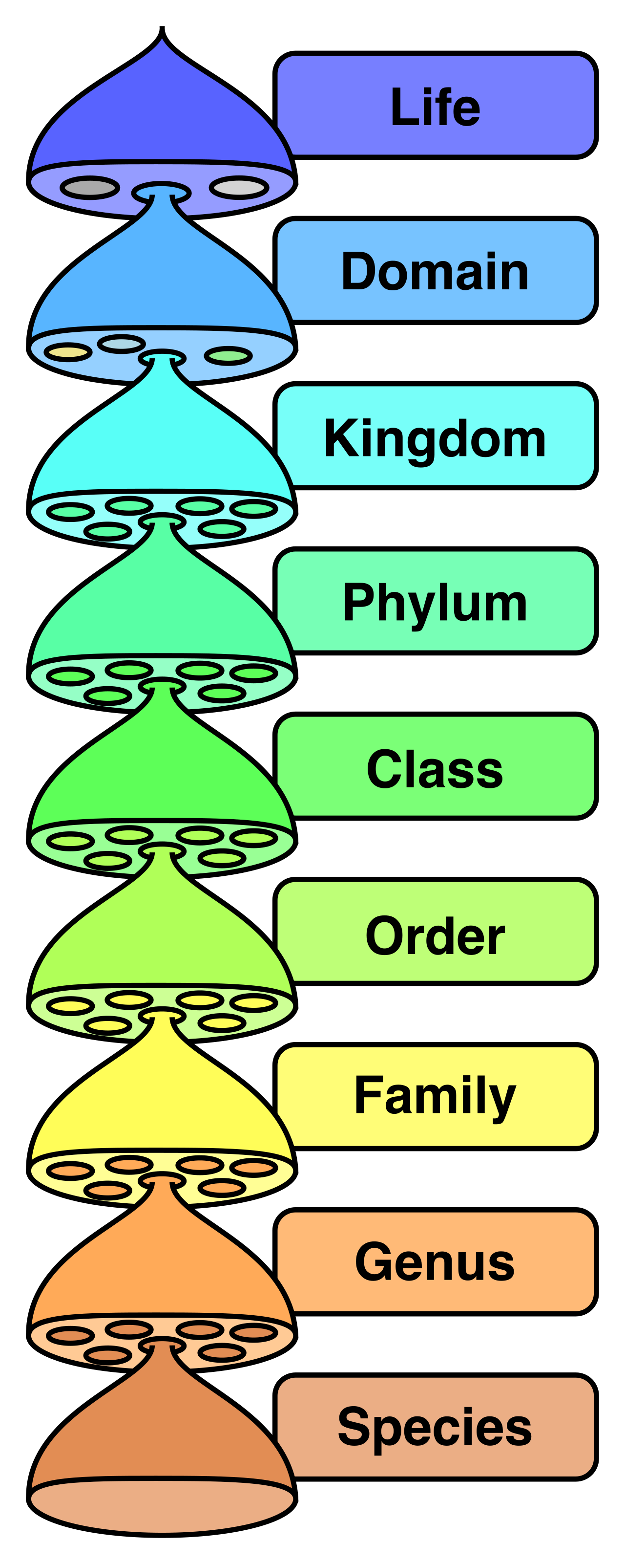
The evolutionary closeness of organisms is directly tied to this hierarchy. By definition, organisms placed in the same taxonomic family are more closely related than organisms placed in the same taxonomic order. If two animals are in the same family, they share a much more recent common ancestor than two animals that only share an order.
When we get down to the species level, the definition becomes functional. Organisms belonging to the same species can interbreed and produce fertile offspring. A horse and a donkey can interbreed to produce a mule, but because mules are largely sterile, horses and donkeys remain distinct species.
Binomial Nomenclature: Speaking the Language of Science
Because common names vary wildly across regions and languages (is it a cougar, a puma, or a mountain lion?), Linnaeus standardized how we talk about species. Binomial nomenclature is a formal scientific system of naming species.
As the prefix bi- suggests, a binomial nomenclature name consists of the genus name and the specific epithet.
There are strict, internationally recognized grammatical rules for writing these names—rules you will see constantly on patient charts, lab cultures, and pharmacology labels:
- In binomial nomenclature, the genus name is always capitalized.
- In binomial nomenclature, the specific epithet is always written in lowercase.
- Binomial nomenclature names are typically written in italics (or underlined if handwritten).
Consider the bacterium that causes standard staph infections: Staphylococcus aureus.
- Staphylococcus is the capitalized Genus.
- aureus is the lowercase specific epithet.
- The entire name is italicized.
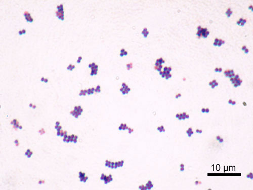
By understanding taxonomy and binomial nomenclature, you are not just memorizing trivia; you are learning the foundational language of global healthcare. When you look at a lab result for Streptococcus pneumoniae, you immediately know its genus, and you can leverage your knowledge of that broad biological family to anticipate the pathogen's behavior, its life cycle, and exactly how to destroy it.