DNA, RNA, and Genetics
Not sure you’re ready?
Take the ~3-minute readiness diagnostic and see where you stand.
Imagine a hospital administrator handing you a master blueprint for an entire medical facility. This blueprint is perfect, but it is locked in an underground vault, written in a four-letter code, and requires a team of specialized couriers and builders to translate it into functioning vital signs monitors, IV pumps, and isolation rooms.
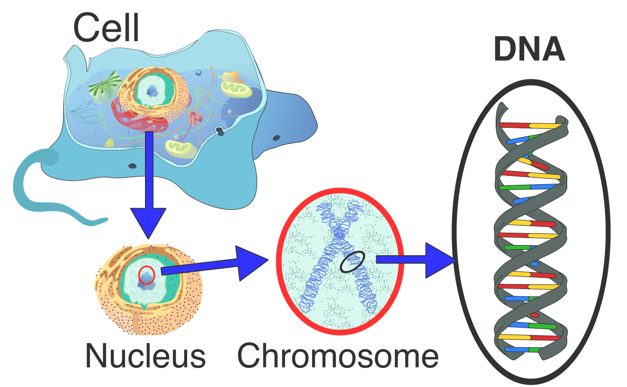
This is the operational reality of the human cell. The master blueprint is DNA, securely housed in the cellular vault—the nucleus. The couriers are RNA molecules, and the functional machinery they produce are the proteins that dictate every physiological process in a patient’s body. As a future healthcare professional, understanding this molecular supply chain is not a mere academic hurdle for the HESI A2; it is the fundamental basis for pharmacology, genetic counseling, oncology, and the mechanics of viral infections.
To understand how life stores information, we must look at the physical molecules that hold the code. Nucleic acids are polymers composed of monomer units called nucleotides. Just as a long train is made of individual boxcars linked together, a nucleic acid is a long chain of nucleotides.
A nucleotide consists of a three-part molecular anatomy:
When these nucleotides link together, they form a highly stable structure. The backbones of DNA and RNA molecules are formed by alternating sugar and phosphate groups. The nitrogenous bases project from this backbone like the teeth of a key, storing the actual genetic data.
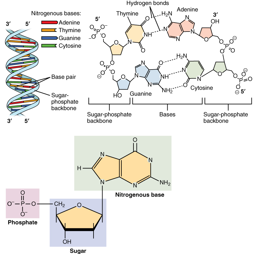
There are two primary types of nucleic acids, differing in their specific components and functions:
- DNA stands for deoxyribonucleic acid. As the name implies, the sugar in DNA is deoxyribose (meaning it is missing one oxygen atom compared to standard ribose). DNA is typically a double-stranded molecule forming a double helix structure. To maintain this structure, the two strands of a DNA molecule run in opposite directions, described as antiparallel.
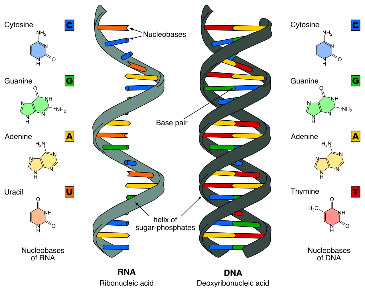
- RNA stands for ribonucleic acid. Accordingly, the sugar in RNA is ribose. Unlike DNA's massive double helix, RNA is typically a single-stranded molecule, making it smaller, more flexible, and capable of traveling out of the nucleus.
The Nitrogenous Bases: The Code Itself
The genetic code relies on a strict pairing system between nitrogenous bases.
- DNA contains the nitrogenous bases adenine, thymine, cytosine, and guanine.
- RNA contains the nitrogenous bases adenine, uracil, cytosine, and guanine. Crucially, RNA does not contain the nitrogenous base thymine.
Because of their chemical structures, these bases fit together in specific ways across the double helix.
Base Pairing Rules in DNA:
- In DNA, adenine always pairs with thymine via two hydrogen bonds.
- In DNA, cytosine always pairs with guanine via three hydrogen bonds.
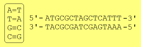
This specific pairing explains why the two strands are complementary; if you know the sequence of one strand, you instantly know the sequence of the other.
| Feature | DNA | RNA |
|---|---|---|
| Full Name | Deoxyribonucleic acid | Ribonucleic acid |
| Sugar | Deoxyribose | Ribose |
| Structure | Double-stranded (Double helix) | Single-stranded |
| Nitrogenous Bases | Adenine (A), Thymine (T), Cytosine (C), Guanine (G) | Adenine (A), Uracil (U), Cytosine (C), Guanine (G) |
The overarching principle of genetics is a directional flow of instructions. The central dogma of molecular biology states that genetic information flows from DNA to RNA to protein. The cell protects its master DNA, makes temporary RNA copies of specific sections, and uses those copies to build functional proteins.
DNA Replication: Preparing for Cell Division
Before a skin cell can divide to heal a patient's wound, it must provide a complete set of genetic instructions to the new cell. DNA replication is the process by which a cell copies its entire genome before cell division.
This process requires molecular machinery to open the blueprint and copy it:
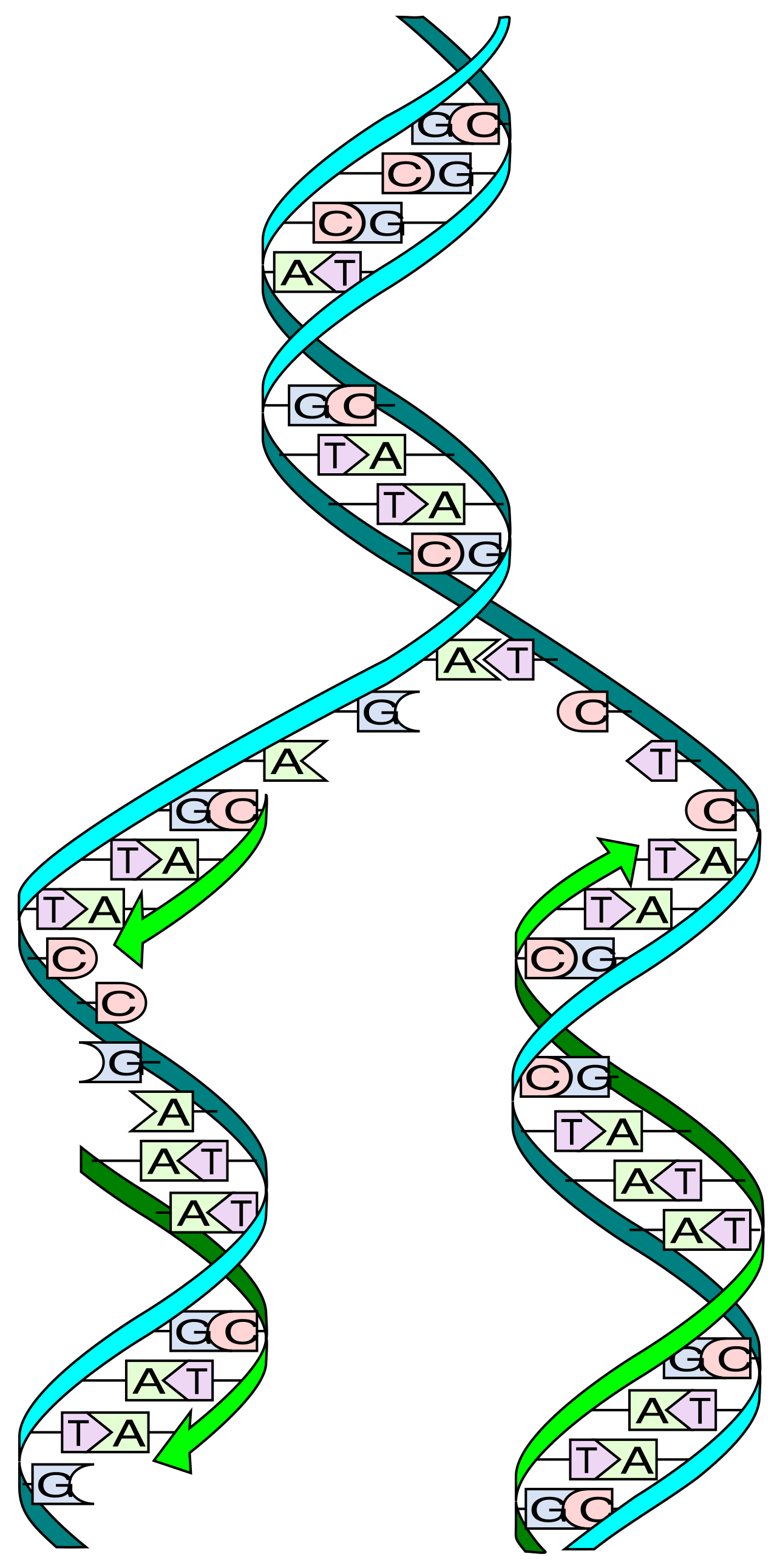
- The enzyme DNA helicase unwinds the DNA double helix during DNA replication. Think of helicase as a molecular zipper sliding down the DNA, separating the two intertwined, antiparallel strands.
- Once separated, the enzyme DNA polymerase adds complementary nucleotides to the growing DNA strand during replication. It reads the exposed bases and brings in the matching partners (A to T, C to G).
Because of this mechanism, DNA replication is described as semi-conservative. When the process finishes, you do not have one completely old double helix and one completely new double helix. Instead, in semi-conservative DNA replication, each new DNA double helix contains one original parent strand and one newly synthesized strand. The old strand acts as an uncorrupted template, minimizing genetic mutations.
Transcription: Drafting the Messenger
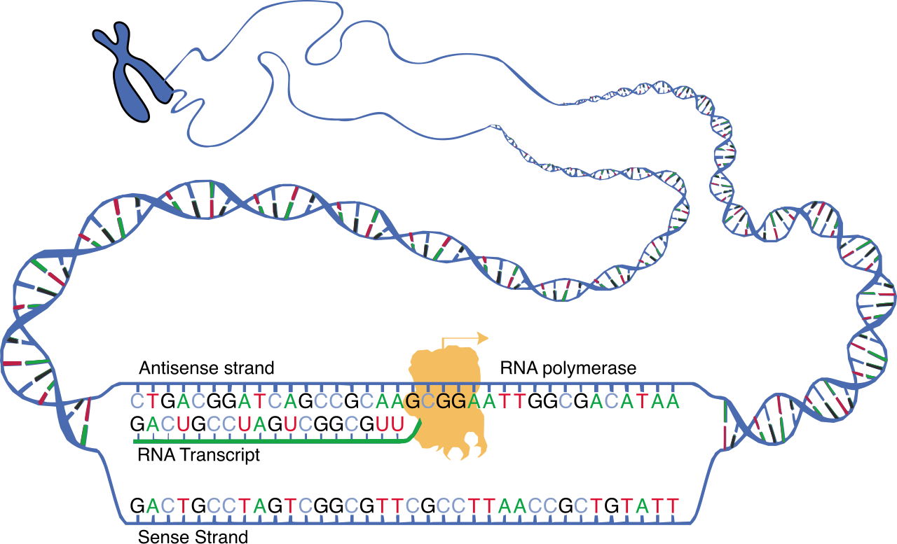
When a cell needs to manufacture a protein (like insulin), it does not send its master DNA out into the chaotic cellular environment. Instead, it makes a disposable photocopy of the specific instructions. Transcription is the process of synthesizing a messenger RNA molecule from a DNA template.
- In eukaryotic cells, transcription occurs in the nucleus. The DNA never leaves this protected vault.
- Instead of DNA polymerase, the enzyme RNA polymerase builds the messenger RNA strand during transcription.
- Because RNA lacks thymine, the pairing rules slightly shift. During transcription, the DNA base adenine pairs with the RNA base uracil. (If the DNA template reads T-A-C, the newly transcribed RNA will read A-U-G).
Translation: Manufacturing the Protein
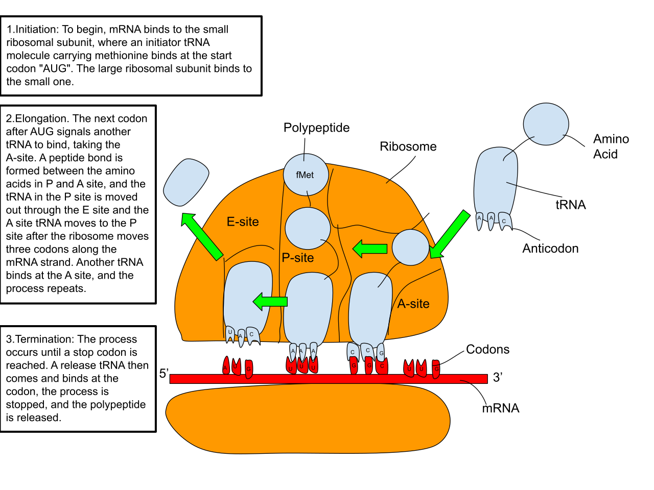
Once the RNA photocopy is complete, it must be read by cellular factories to build a physical product. Translation is the process of synthesizing a protein using the information carried by a messenger RNA molecule.
Translation occurs at the ribosomes in the cytoplasm or on the rough endoplasmic reticulum. This complex process requires the collaboration of three different types of RNA:
- Messenger RNA (mRNA) carries genetic instructions from the nucleus to the ribosomes. It is the blueprint photocopy.
- Ribosomal RNA (rRNA) is a structural component of ribosomes. It makes up the actual factory workbench where the protein is assembled.
- Transfer RNA (tRNA) carries specific amino acids to the ribosome during translation. Think of tRNA as the supply trucks delivering raw building materials.
The ribosome reads the mRNA in discrete three-letter "words." A codon is a sequence of three consecutive nucleotides on a messenger RNA molecule. The genetic code is a dictionary where each messenger RNA codon corresponds to a specific amino acid or a stop signal.
To ensure the correct amino acid is delivered, the tRNA features a matching lock-and-key mechanism. An anticodon is a sequence of three nucleotides on a transfer RNA molecule. During assembly, the transfer RNA anticodon binds to a complementary messenger RNA codon during translation.
As the ribosome moves down the mRNA, tRNA molecules continue dropping off their amino acids in the exact order specified by the codons. These amino acids are linked together by peptide bonds during translation to form a polypeptide chain, which then folds into a functional, three-dimensional protein.
Understanding molecular biology explains how traits are built, but Mendelian genetics explains how traits are passed from parents to offspring. When you take a patient's family medical history, you are looking for patterns of genetic inheritance.
A gene is a distinct sequence of nucleotides forming part of a chromosome. Because genes contain the instructions for synthesizing specific proteins, they are the fundamental units of heredity. However, not all copies of a gene are identical. Alleles are alternative versions of a specific gene. For example, the gene for blood type has different alleles that result in Type A, Type B, or Type O blood.
Genotype versus Phenotype
In genetics, we must strictly separate the underlying code from the physical reality.
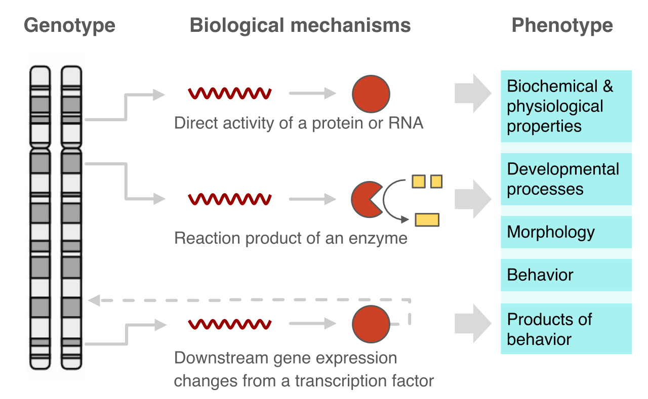
- Genotype refers to the specific combination of alleles an organism possesses for a given trait. This is the hidden genetic code.
- Phenotype refers to the observable physical or biochemical characteristics of an organism. This is the manifested trait, such as eye color, height, or the presence of a genetic disorder like sickle cell anemia.
Importantly, genetics is rarely destiny on its own. An organism's phenotype is determined by the interaction between the organism's genotype and environmental factors. A patient may have a genotype that predisposes them to type 2 diabetes, but environmental factors like diet and exercise will heavily influence their actual phenotype.
Dominant and Recessive Alleles
Because humans inherit one set of chromosomes from each parent, we carry two alleles for every trait.
- A homozygous organism has two identical alleles for a particular genetic trait.
- A heterozygous organism has two different alleles for a particular genetic trait.
When an individual is heterozygous, the two differing alleles must compete. A dominant allele masks the expression of a recessive allele in a heterozygous organism. For notation purposes in clinical genetics and biology:
- Dominant alleles are conventionally represented by uppercase letters (e.g., 'A').
- Recessive alleles are conventionally represented by lowercase letters (e.g., 'a').
Therefore, an individual with the genotype 'Aa' is heterozygous, but their physical phenotype will only display the dominant 'A' trait, completely masking the recessive 'a' trait.
To calculate the statistical likelihood of an offspring inheriting a genetic condition, we use a specific mathematical tool. A Punnett square is a diagram used to predict the probability of an offspring inheriting particular genotypes.
For the HESI A2, you will most commonly encounter a monohybrid cross, which examines the inheritance of a single genetic trait.
Setting up a Punnett square is a standardized process:
- In a Punnett square, the possible alleles from one parent are aligned along the top margin.
- In a Punnett square, the possible alleles from the second parent are aligned along the left margin.
You then fill in the interior boxes by combining the row and column alleles, simulating fertilization.
The Classic Heterozygous Cross
Consider the most frequently tested scenario: crossing two heterozygous parents (Aa x Aa) for a trait exhibiting complete dominance.
Statistical Outcomes of a Heterozygous Cross (Aa x Aa):
- A cross between two heterozygous individuals yields a 1:2:1 genotypic ratio. This breaks down as 1 homozygous dominant (AA), 2 heterozygous (Aa), and 1 homozygous recessive (aa).
- Because the dominant allele masks the recessive one, the AA and Aa offspring will look physically identical. Therefore, a cross between two heterozygous individuals for a completely dominant trait yields a 3:1 phenotypic ratio. (Three offspring display the dominant trait; one displays the recessive trait).
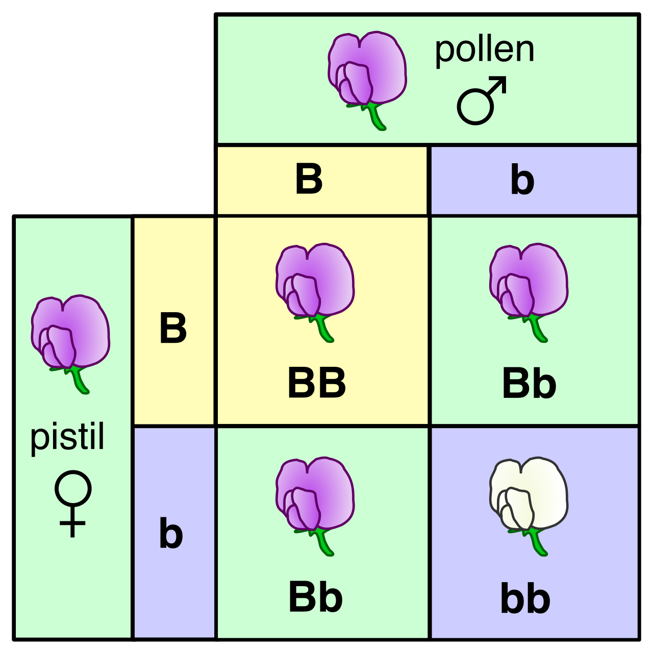
Understanding the distinction between that 1:2:1 genotypic ratio and the 3:1 phenotypic ratio is paramount. It bridges the gap between invisible molecular sequences and the living, breathing patients you will one day treat. Every physical symptom, metabolic pathway, and inherited risk factor relies on the elegant translation of DNA to RNA, RNA to protein, and protein to human life.