Cells and Organelles
Not sure you’re ready?
Take the ~3-minute readiness diagnostic and see where you stand.
Imagine the human body not as a single solid entity, but as a vast, bustling metropolis of over 30 trillion autonomous micro-cities. Each of these cities is a cell, possessing its own power grid, manufacturing plants, security forces, and command center. For a future nurse or allied health professional, understanding human health begins with mastering the architecture of these microscopic cities. When a patient is given a dose of antibiotics to clear a bacterial infection, or when a failing liver struggles to clear a toxic medication from the bloodstream, the drama plays out entirely on the cellular level. To intervene in human disease, one must first understand the fundamental unit of life.
Before we can look inside the cell, we must establish the boundaries of what constitutes life. In the 19th century, biologists formalized a unifying concept that remains the bedrock of modern biology and clinical medicine.
Cell Theory states:
- All living organisms are composed of one or more cells.
- The cell is the basic structural unit of all living organisms.
- The cell is the basic functional unit of all living organisms.
- All cells arise from pre-existing cells through cell division.
Why does this matter in clinical practice? When a surgical wound heals, it is because pre-existing skin cells are dividing to create new tissue. When a tumor grows, it is because the regulation of that cellular division has failed. Life does not spontaneously generate; it propagates from cell to cell.
If you look at all the cells on Earth, they fall into two distinct architectural categories. The presence or absence of a nucleus dictates the fundamental division between eukaryotic and prokaryotic cells.
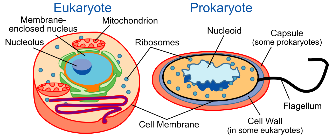
Prokaryotic Cells
Prokaryotes are the minimalist survivors of the biological world. Bacteria and Archaea are examples of prokaryotic organisms. In a clinical setting, every time you swab a throat for strep or administer penicillin, you are dealing with prokaryotes.
- Prokaryotic cells lack a membrane-bound nucleus. Their genetic material floats freely within the cell.
- Prokaryotic cells lack membrane-bound organelles. They do not have specialized "rooms" for different metabolic tasks.
- Size: They are generally smaller than eukaryotic cells, allowing them to reproduce rapidly.
Eukaryotic Cells
Eukaryotes are complex, highly compartmentalized structures. Animals, plants, fungi, and protists are composed of eukaryotic cells. As a healthcare provider, your patients—and the fungal or protist pathogens that occasionally infect them—are eukaryotic.
- Eukaryotic cells contain a membrane-bound nucleus to carefully protect their genetic blueprints.
- Eukaryotic cells possess membrane-bound organelles, allowing them to perform complex metabolic functions simultaneously in isolated compartments.
| Feature | Prokaryotic Cells | Eukaryotic Cells |
|---|---|---|
| Organisms | Bacteria, Archaea | Animals, Plants, Fungi, Protists |
| Nucleus | Absent | Present (Membrane-bound) |
| Organelles | Lacks membrane-bound organelles | Possesses membrane-bound organelles |
| Size | Generally smaller | Generally larger |
To be a discrete city, a cell must have a wall or a border to separate the inside from the outside.
The Cell Membrane
The boundary of the cell is the cell membrane, which is primarily composed of a phospholipid bilayer. This membrane is selectively permeable to control the internal cellular environment, acting as a highly regulated border checkpoint.
The structure of this bilayer is brilliantly adapted to the watery environment of the body. It features hydrophilic (water-loving) phosphate heads facing the aqueous environment on both the outside and inside of the cell. Conversely, the hydrophobic (water-fearing) lipid tails point inward away from water, hiding in the middle of the membrane sandwich. Because this lipid core repels water and charged particles, embedded membrane proteins serve as transport channels across the cell membrane, allowing specific nutrients in and pumping waste out.
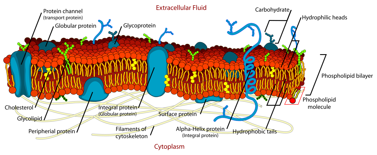
(Clinical translation: When you administer a medication, its chemical structure—whether it is lipid-soluble or water-soluble—determines how easily it can slip past this phospholipid bilayer to do its job.)
The Cytoplasm
Once inside the membrane, you enter the cytoplasm, which is the jelly-like intracellular fluid filling the cell. The cytoplasm is not just empty space; it acts as a medium for cellular metabolic reactions, allowing molecules to dissolve, collide, and interact.
The Nucleus
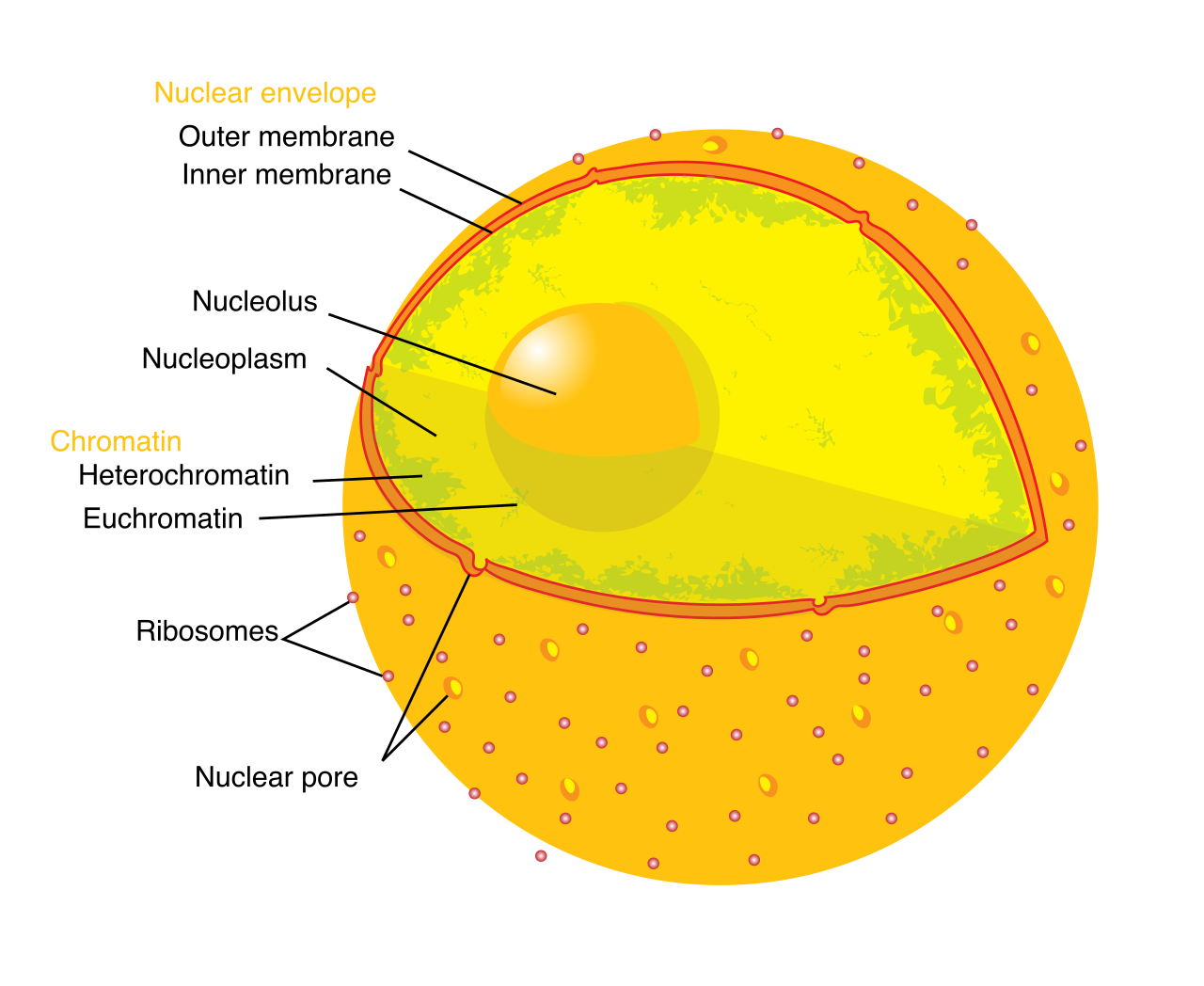
In a eukaryotic cell, the nucleus houses the chromosomal DNA, the master blueprint for every protein the body will ever need to build. Because this DNA is incredibly precious, it is protected by the nuclear envelope, a highly selective double membrane that surrounds the cell nucleus.
However, blueprints are useless if they cannot be sent to the factory floor. Therefore, the nuclear envelope contains nuclear pores, which regulate the transport of molecules between the nucleus and the cytoplasm.
Deep inside the nucleus sits a dense structure called the nucleolus. The nucleolus is the primary site of ribosome synthesis.
Ribosomes
If the nucleus is the command center, ribosomes are the cellular sites of protein synthesis—the robotic workers that read genetic blueprints and assemble proteins.
- Structurally, ribosomes are cellular structures composed of RNA and proteins.
- Location-wise, they operate in two distinct areas: Ribosomes exist freely suspended within the cytoplasm (building proteins that will remain in the cytoplasm) and are found attached to the rough endoplasmic reticulum (building proteins destined for the cell membrane or for export).
Proteins and lipids must be manufactured, folded, and shipped. This is handled by a continuous membrane system radiating outward from the nucleus.
The Endoplasmic Reticulum (ER)
The ER comes in two distinct varieties, rough and smooth, defined by their appearance under a microscope.
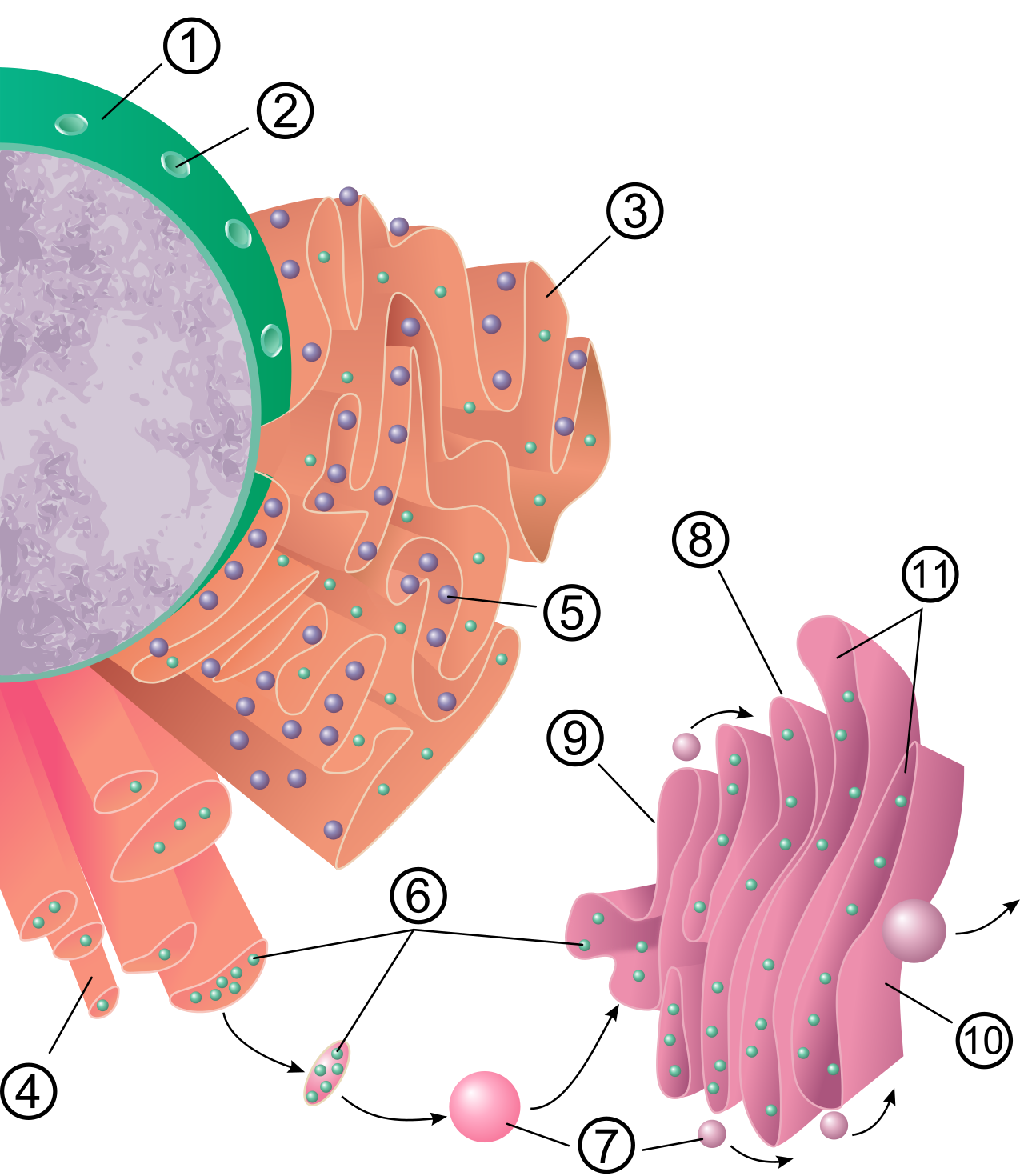
1. The Rough Endoplasmic Reticulum (RER) The rough ER features ribosomes on its outer surface, giving it a studded, "rough" texture. Because of these ribosomes, the rough endoplasmic reticulum facilitates the synthesis of proteins, specifically those destined to be embedded in membranes or secreted from the cell. Once the raw protein chain is created, the rough endoplasmic reticulum facilitates the folding of proteins into their functional, three-dimensional shapes.
2. The Smooth Endoplasmic Reticulum (SER) The smooth ER lacks surface ribosomes, so it has nothing to do with protein synthesis. Instead, the smooth endoplasmic reticulum synthesizes cellular lipids, including the phospholipids needed to repair the cell membrane.
Crucially for pharmacology and medicine, the smooth ER acts as the cell's detox center. The smooth endoplasmic reticulum detoxifies metabolic byproducts and detoxifies drugs. (Clinical translation: If a patient has a history of heavy alcohol or barbiturate use, the liver cells will respond by drastically expanding their smooth ER to handle the toxic load, leading to drug tolerance—meaning you will require higher doses of anesthesia to sedate them.)
The Golgi Apparatus
Once proteins and lipids are built in the ER, they are shipped to the Golgi apparatus, which looks like a series of flattened membrane-bound sacs. Think of the Golgi as the cellular post office.
- The Golgi apparatus modifies proteins received from the endoplasmic reticulum (for example, by adding sugar tags to them).
- The Golgi apparatus sorts proteins into vesicles.
- The Golgi apparatus packages proteins into vesicles for targeted cellular delivery, ensuring a digestive enzyme goes to a lysosome, while a hormone goes to the cell membrane for export into the bloodstream.
Mitochondria
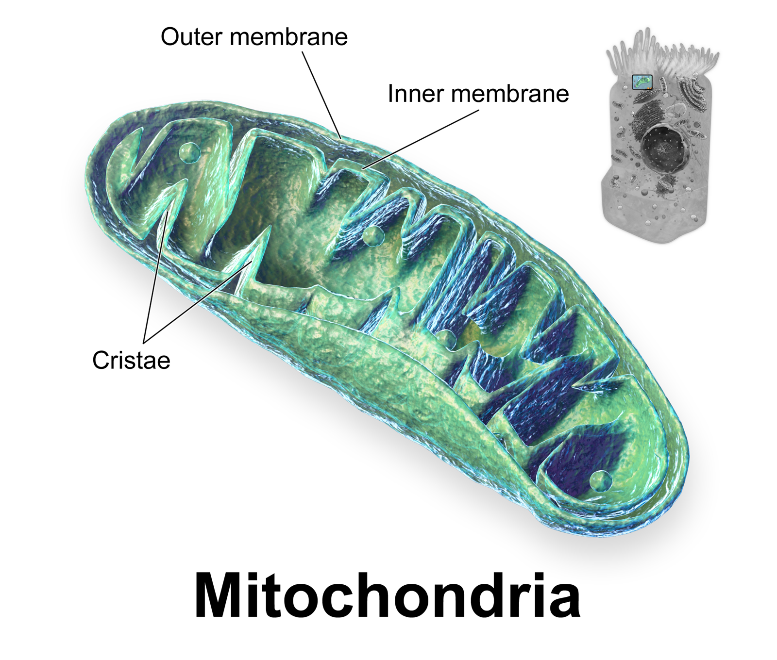
No city runs without a power plant. The mitochondria perform cellular respiration, taking the glucose from the food we eat and the oxygen from the air we breathe to generate adenosine triphosphate (ATP) to provide energy for cellular activities. ATP is the universal energy currency of life.
Mitochondria are structurally unique. Mitochondria possess a double-membrane structure, and the inner membrane of a mitochondrion contains folds called cristae, which maximize the surface area for energy production. Fascinatingly, mitochondria contain their own circular DNA, distinct from the DNA in the nucleus, a remnant of an ancient evolutionary event where a primitive eukaryotic cell swallowed a bacterium.
Lysosomes
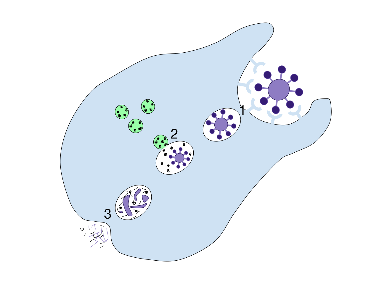
Metabolism creates waste, and cellular structures eventually break down. Lysosomes are membrane-bound organelles containing hydrolytic enzymes—powerful acidic chemicals capable of dissolving biological molecules.
- Lysosomes digest worn-out cellular organelles, recycling their parts for future use.
- Lysosomes break down engulfed foreign particles and pathogens. (Clinical translation: When a patient's white blood cells (macrophages) engulf an invading streptococcus bacterium, they trap it in a vesicle and merge it with a lysosome. The lysosome's hydrolytic enzymes literally digest the pathogen alive, curing the infection.)
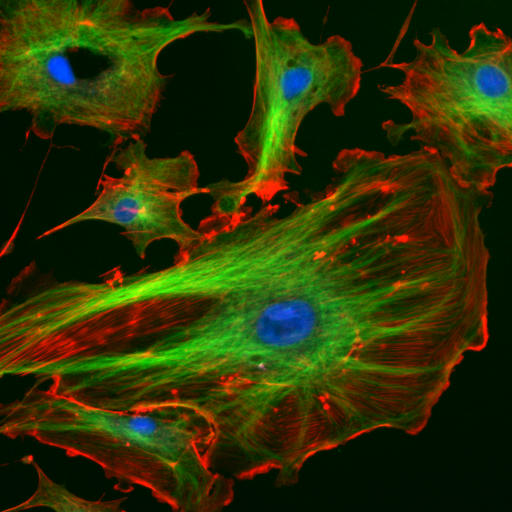
To maintain its shape and move internal cargo, the cell relies on an internal scaffolding system. The cytoskeleton is a network of protein filaments located throughout the cytoplasm.
- The cytoskeleton provides structural support to maintain cell shape.
- The cytoskeleton facilitates the movement of organelles within the cell, acting as a microscopic railway system along which vesicles are hauled.
During cell division (mitosis), animal cells rely on centrioles, which are cylindrical structures found in animal cells. Their specific job is to organize the mitotic spindle during animal cell division, ensuring that when the cell splits, each new daughter cell receives an exact copy of the DNA.
While nursing focuses heavily on human (animal) cells, the HESI A2 exam evaluates foundational biological literacy, meaning you must be able to contrast animal cells with plant cells. Plant cells face different environmental challenges than animal cells—they cannot run away from predators, and they cannot hunt for food—and their organelle structure reflects this.
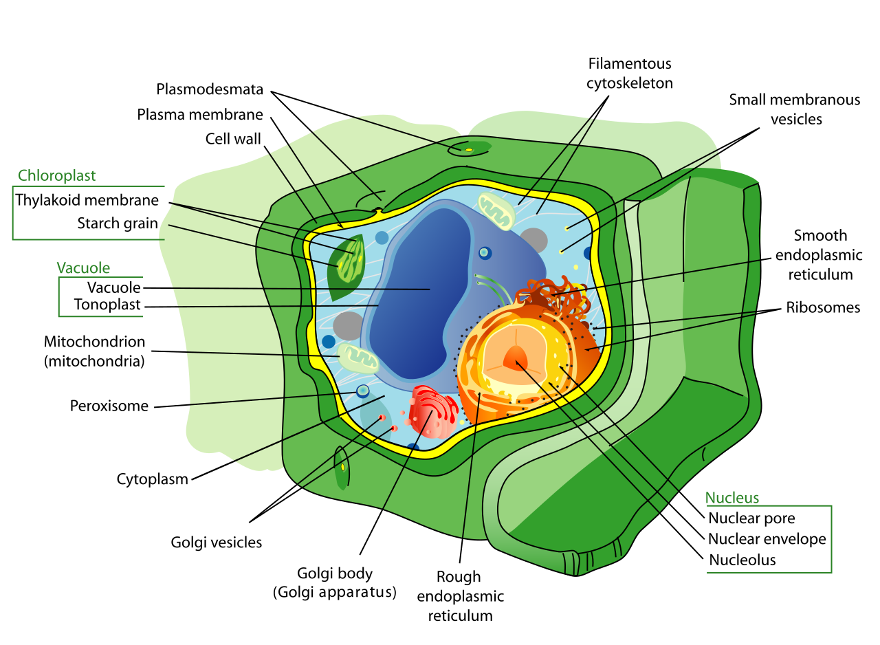
1. Cell Walls Unlike animal cells, which are squishy and surrounded only by a fluid membrane, plants require rigid structural support. Cell walls are rigid outer layers found in plant cells (sitting outside the cell membrane). The plant cell wall is primarily composed of cellulose. Its primary function is simple: the cell wall provides mechanical protection to the plant cell and allows trees to grow hundreds of feet tall without collapsing.
2. Chloroplasts Plants do not eat; they manufacture their own food. Chloroplasts are organelles found in plant cells and certain algae. They contain the green pigment chlorophyll and are uniquely designed to capture light energy to perform photosynthesis, converting solar energy into usable glucose.
3. Vacuoles While animal cells may have tiny, temporary vesicles, plants require massive storage tanks to survive droughts. Vacuoles are membrane-bound sacs utilized for cellular storage. More specifically, plant cells typically feature a single large central vacuole. Beyond just holding water and nutrients, the central vacuole regulates turgor pressure within a plant cell. When you forget to water a houseplant and it wilts, it is because the central vacuoles in its cells have lost their water, causing a drop in turgor pressure that collapses the rigid architecture of the tissue.