Cells and Tissues
Not sure you’re ready?
Take the ~3-minute readiness diagnostic and see where you stand.
When you examine a surgical incision, you are not merely looking at a disruption of flesh; you are observing a microscopic breach in a highly precise, highly organized biological architecture. To understand human healing, disease, and intervention, we must drop down to the cellular level and examine how life organizes itself into functional materials. Histology is the microscopic study of tissues, and in biological terms, a tissue is a group of similar cells working together to perform a specific function. Despite the vast complexity of human anatomy—from the pumping chambers of the heart to the filtering tubules of the kidney—the human body contains exactly four primary tissue types. Every organ you will ever assess, palpate, or treat is constructed entirely from variations of these four fundamental building blocks: epithelial, connective, muscle, and nervous tissue.
To master histology for the HESI A2, you must approach tissues not as a list of anatomical trivia, but as biological engineering. Form always follows function. How a tissue looks under a microscope tells you exactly what it does in the body.
1. Epithelial Tissue: The Barriers and Glands
Think of epithelial tissue as the body's boundary layer. Epithelial tissue covers external body surfaces (like the outer layer of your skin) and lines internal hollow body cavities (like the inside of your respiratory tract and digestive system). Furthermore, when epithelial cells fold inward and specialize in secretion, epithelial tissue forms the secretory portions of human glands, such as your sweat, salivary, and thyroid glands.
Because epithelial tissue forms boundaries, its physical structure must be impenetrable. Therefore, epithelial cells are tightly packed together, and the tissue itself contains very little extracellular matrix between its cells. They are biological brick walls.
However, this tight packing comes with a strict anatomical rule: epithelial tissue lacks a direct blood supply.
Crucial Concept: Because they are strictly avascular, avascular epithelial tissues receive nutrients through diffusion from underlying vascular connective tissue. The oxygen and glucose must literally seep upward from the blood vessels in the tissue directly beneath them.
Because epithelial tissues are constantly exposed to friction, harsh environments, and physical trauma, cells are continually damaged and shed. To compensate, epithelial tissue undergoes rapid cell division to enable continuous renewal and regeneration. When you assess a healing superficial wound, you are observing this rapid epithelial mitosis in real time.
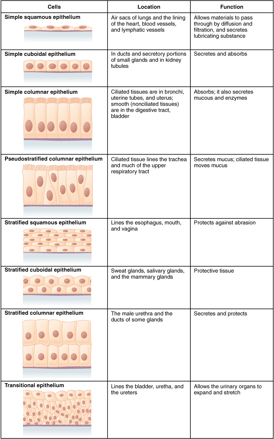
Classification of Epithelial Tissue Epithelial tissues are named using a two-word system based on their layers and their cellular shape.
- By Layers:
- Simple epithelial tissue consists of a single layer of cells. (Ideal for areas where diffusion or filtration is needed, like the lungs).
- Stratified epithelial tissue consists of two or more layers of cells. (Ideal for areas requiring protection against friction, like the skin).
- By Shape:
2. Connective Tissue: The Scaffolding and Glue
If epithelium forms the borders, connective tissue fills the spaces. Connective tissue is the most abundant primary tissue type in the human body. Its roles are highly diverse: connective tissue provides structural support to the body, protects other internal body structures, and physically binds different body structures together.
While epithelial cells are tightly packed, connective tissue is the exact opposite. Connective tissue is characterized by large amounts of nonliving extracellular matrix. The cells are widely spaced, floating or embedded within this matrix like fruit suspended in gelatin.
The Matrix Composition: The extracellular matrix in connective tissue contains ground substance (a fluid, gel, or solid background material) and contains various protein fibers (such as strong collagen or stretchy elastin).
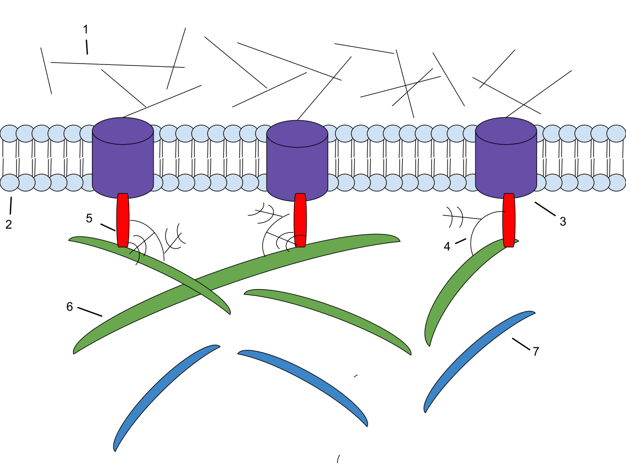
Unlike the avascular epithelium, most connective tissues possess a rich blood supply. However, there is one critical exception you must remember: cartilage is a specialized connective tissue that lacks a direct blood supply. Because cartilage is avascular, it heals notoriously slowly when injured—a vital clinical fact for patients with joint trauma.
Connective tissue takes many surprisingly distinct forms in the body. Examples include:
- Bone is classified as a connective tissue, where the extracellular matrix is hardened by calcium salts.
- Blood is a specialized liquid connective tissue, where the "matrix" is the fluid plasma.
- Adipose tissue is a loose connective tissue specialized for lipid storage, functioning as cellular bubble wrap and energy reserves.
3. Muscle Tissue: The Engines of Movement
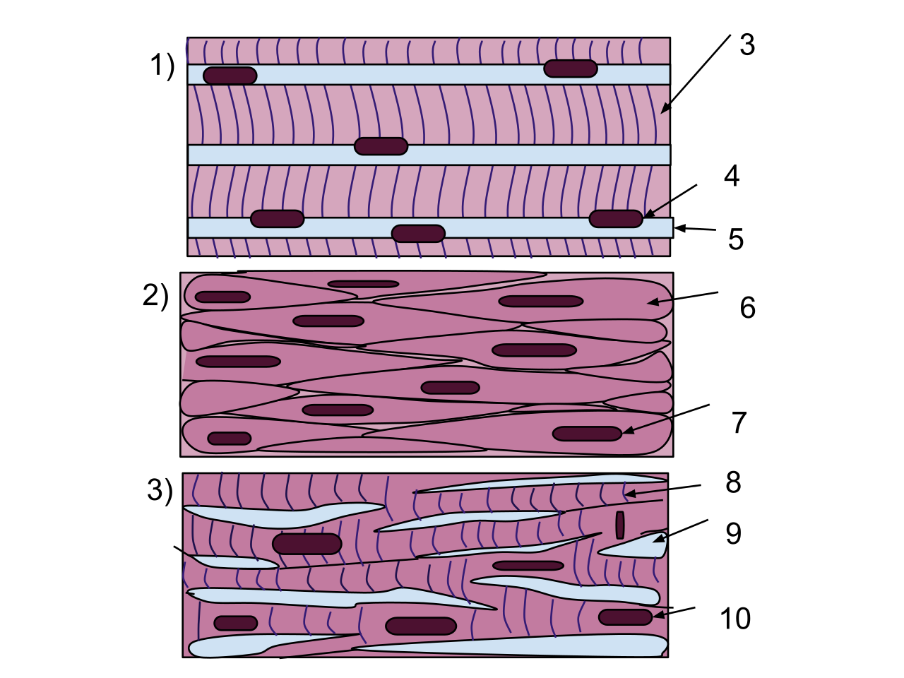
Muscle tissue specializes in cellular contraction to produce physical force. It is the only tissue capable of actively shortening to create movement. The human body contains skeletal, cardiac, and smooth muscle tissue types.
| Tissue Type | Microscopic Appearance | Location | Control | Key Features |
|---|---|---|---|---|
| Skeletal | Skeletal muscle cells exhibit a visible striated appearance under a microscope. | Skeletal muscle tissue attaches to bones to facilitate voluntary body movement. | Voluntary | Skeletal muscle cells have a long, cylindrical shape and, remarkably, are multinucleated (containing multiple nuclei per cell). |
| Cardiac | Cardiac muscle cells exhibit a visible striated appearance under a microscope. | Cardiac muscle tissue is found exclusively in the walls of the heart. | Involuntary | Cardiac muscle contraction operates under involuntary control. Cardiac muscle cells typically contain a single central nucleus. |
| Smooth | Smooth muscle cells completely lack visible striations under a microscope. | Smooth muscle tissue is located in the walls of hollow internal organs. | Involuntary | Smooth muscle contraction operates under involuntary control. |
Special Features of Cardiac and Smooth Muscle: In cardiac tissue, the cells must beat in perfect, synchronized rhythm. To achieve this, cardiac cells are linked by intercalated discs, which are specialized cell junctions that structurally and functionally connect cardiac muscle cells, allowing electrical signals to flash across the heart wall instantly.
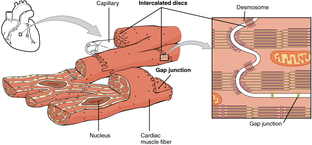
Smooth muscle, lacking the organized striping (striations) of the other two types, functions primarily to squeeze and propel substances through tubes. For instance, the stomach wall contains smooth muscle tissue to churn food, and blood vessel walls contain smooth muscle tissue to regulate blood pressure via vasodilation and vasoconstriction.
4. Nervous Tissue: The Command Network
If muscle provides the engine, nervous tissue acts as the wiring. Nervous tissue specializes in receiving and transmitting electrochemical impulses. It serves as the body's rapid communication system. Anatomically, nervous tissue forms the structural basis of the brain, forms the structural basis of the spinal cord, and comprises the peripheral nerves that branch out to your fingertips and toes.
Under the microscope, nervous tissue contains two distinct categories of cells:
- Neurons: These are highly specialized nerve cells that generate and conduct electrochemical nerve impulses. They are the communicators.
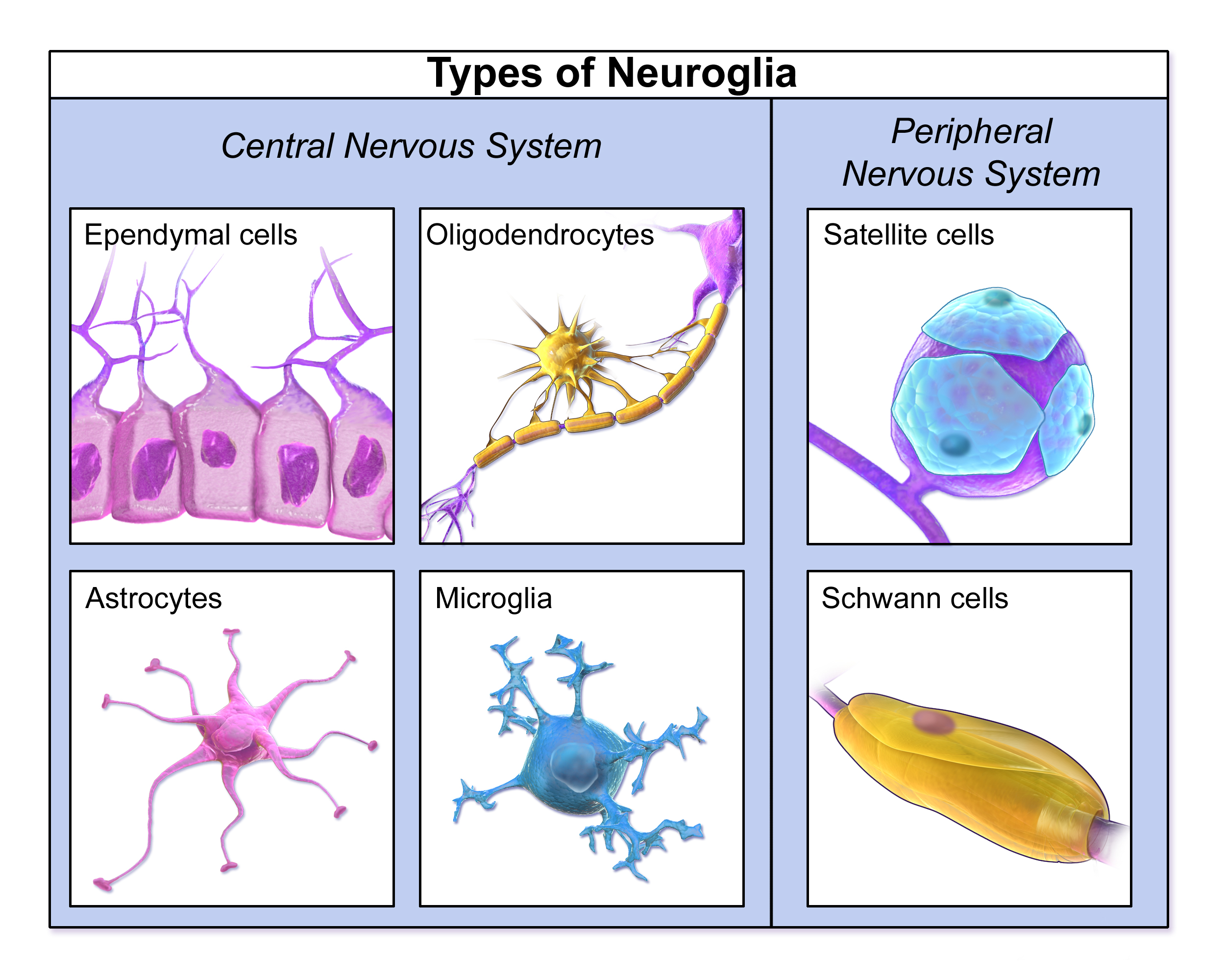
- Neuroglia: Neurons are fragile and demanding; they cannot survive alone. Neuroglia are specialized supporting cells in the nervous system. They act as the biochemical caretakers for the neurons. Specifically, neuroglia provide electrical insulation to neurons (ensuring signals travel fast), provide physical protection to neurons, and deliver essential nourishment to neurons.
Tissues are not static. To grow, heal, and reproduce, the cells within these tissues must divide. The body utilizes two fundamentally different processes for cell division, depending on the goal.
Mitosis: The Engine of Growth and Repair
When you scrape your knee, how does the stratified squamous epithelium regenerate? Through mitosis. Mitosis is the specific process of cell division in human somatic cells.
Definition: Somatic cells are all the cells in the body except for the reproductive gametes. Your skin, bone, liver, and lung cells are all somatic cells.
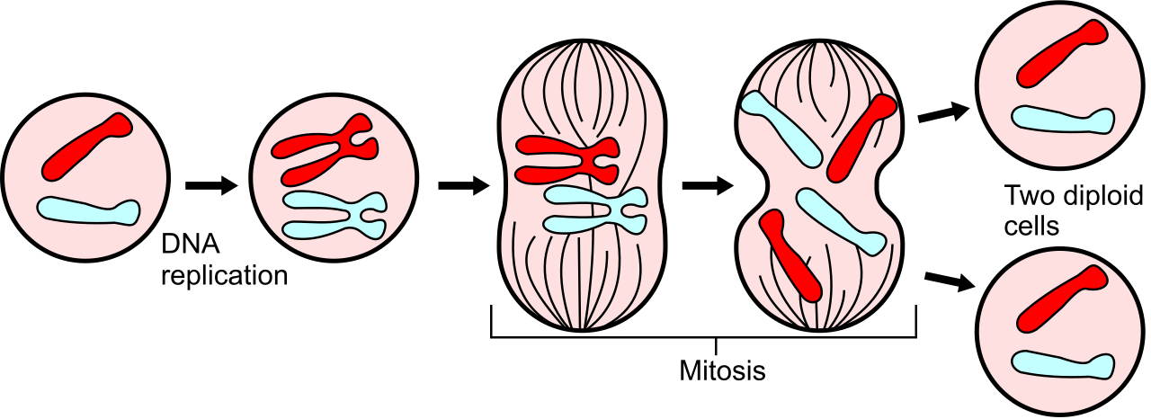
The goal of mitosis is creating an exact replica. Mitosis enables tissue growth in the human body, as well as tissue repair and cellular replacement in the human body.
During this process:
- A single round of mitosis produces exactly two daughter cells.
- The daughter cells produced by mitosis are genetically identical to the original parent cell.
- Mitosis produces diploid daughter cells.
- Diploid cells contain two complete sets of chromosomes (46 total in humans, with 23 inherited from each parent).
To divide the cellular blueprints flawlessly, mitosis follows four strictly choreographed sequential phases:
- Prophase is the initial phase of mitosis, where chromosomes condense and become visible.
- Metaphase is the second sequential phase of mitosis, where chromosomes align perfectly along the center (equator) of the cell.
- Anaphase is the third sequential phase of mitosis, where the sister chromatids are violently pulled apart toward opposite poles.
- Telophase is the final phase of mitosis, where two new nuclear envelopes form around the separated genetic material.
Meiosis: The Architecture of Inheritance
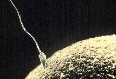
While mitosis builds and repairs the individual, meiosis ensures the continuation of the species. Meiosis is a highly specialized type of cell division dedicated to producing human gametes. In human biology, human sperm cells are reproductive gametes, and human egg cells are reproductive gametes.
Unlike somatic cells, a sperm or an egg cannot be diploid. If a sperm with 46 chromosomes fertilized an egg with 46 chromosomes, the resulting embryo would have 92 chromosomes—a fatal genetic overload.
Therefore, meiosis halves the total chromosome number in gametes to ensure the resulting zygote has the correct diploid number upon fertilization.
To achieve this mathematical reduction and introduce genetic diversity, meiosis operates differently than mitosis:
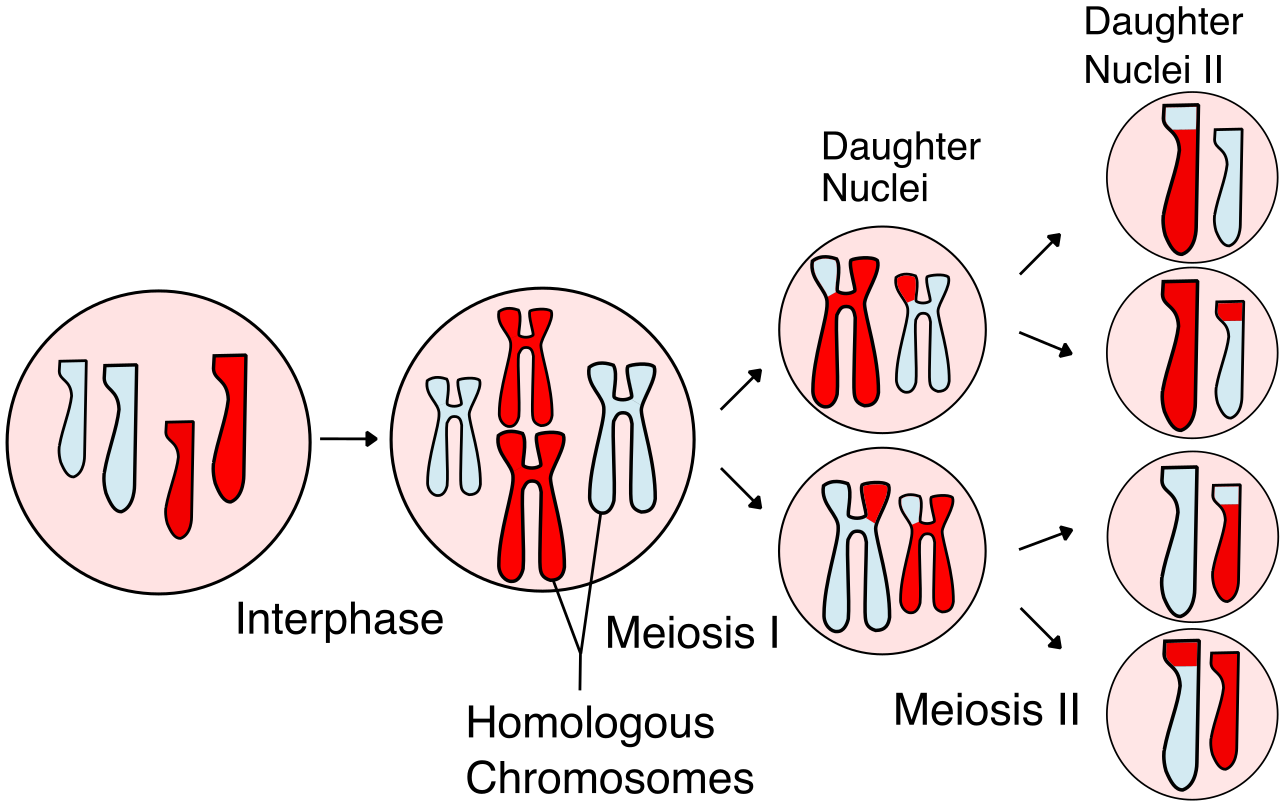
- Instead of one round, meiosis requires two consecutive rounds of cellular division (Meiosis I and Meiosis II).
- Because of these two rounds, a complete meiotic cycle produces exactly four daughter cells from a single parent cell.
- Through a process called genetic recombination (or "crossing over"), the daughter cells produced by meiosis are genetically distinct from one another.
- Most importantly, meiosis produces haploid daughter cells.
- Haploid cells contain exactly one single set of chromosomes (just 23 total).
When a haploid sperm (23 chromosomes) meets a haploid egg (23 chromosomes), they fuse to form a beautifully complete, genetically unique, diploid human cell (46 chromosomes). From that single cell, millions of rounds of mitosis will eventually construct the intricately woven tissues—epithelial, connective, muscle, and nervous—that make up the human body.