Organization and Terminology
Not sure you’re ready?
Take the ~3-minute readiness diagnostic and see where you stand.
A map is only useful if everyone agrees on which way is north. When a trauma patient arrives in the emergency department with a laceration to the forearm, a miscommunication about its exact location could mean the difference between a simple suture and a severed nerve. Medicine is fundamentally a collaborative discipline, and precise communication requires an absolute, universally understood reference frame. Before we can discuss the pathology of a disease or the mechanics of a treatment, we must master the architecture of the human form. We need a shared language to describe how the body is built, how it is divided, and how it actively fights to maintain its own survival.
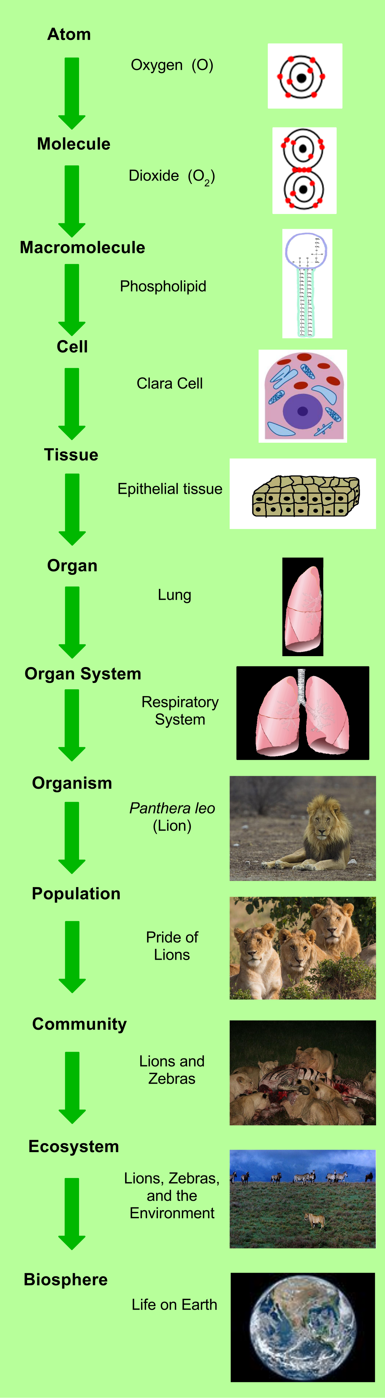
To understand a complex machine, you do not start by staring at the entire engine; you start by looking at the gears. The human body is organized into multiple structural levels of increasing complexity, with each tier building sequentially upon the last.
- The Chemical Level: This is the simplest level of human organization. Before we even reach the threshold of life, we are simply physics and chemistry. The chemical level includes atoms (the fundamental building blocks of matter) combining to form molecules (like water, proteins, and DNA).
- The Cellular Level: This level is composed of cells, which are the basic structural units and the basic functional units of a living organism. A muscle cell and a nerve cell look vastly different because their forms dictate their specific functions.
- The Tissue Level: A single cell rarely works alone. The tissue level of human organization consists of groups of similar cells working together to perform a specific function. For example, thousands of cardiac muscle cells contract in unison to form cardiac tissue.
- The Organ Level: The organ level of human organization consists of structures composed of two or more different types of tissues. The heart is an organ because it contains cardiac muscle tissue, nervous tissue to pace it, and connective tissue to hold its valves together.
- The Organ System Level: The organ system level of human organization consists of related organs working together to accomplish a common physiological purpose. The cardiovascular system unites the heart, blood, and blood vessels to transport nutrients. The human body contains eleven major organ systems, from the integumentary to the reproductive.
- The Organismal Level: The organismal level is the highest level of human structural organization. It represents the sum total of all eleven organ systems working in harmony to keep the human alive.
If a patient is lying face down on an operating table and the surgeon asks for an incision on the "front" of the arm, do they mean the part of the arm currently facing the ceiling, or the anatomical front? To eliminate this ambiguity, every anatomical direction assumes the body is in standard anatomical position.
Standard anatomical position requires the human body to be configured exactly as follows:
- Standing upright
- Feet to be parallel and flat on the floor
- Arms to be at the sides of the body
- Palms of the hands to face forward
- Head to face forward
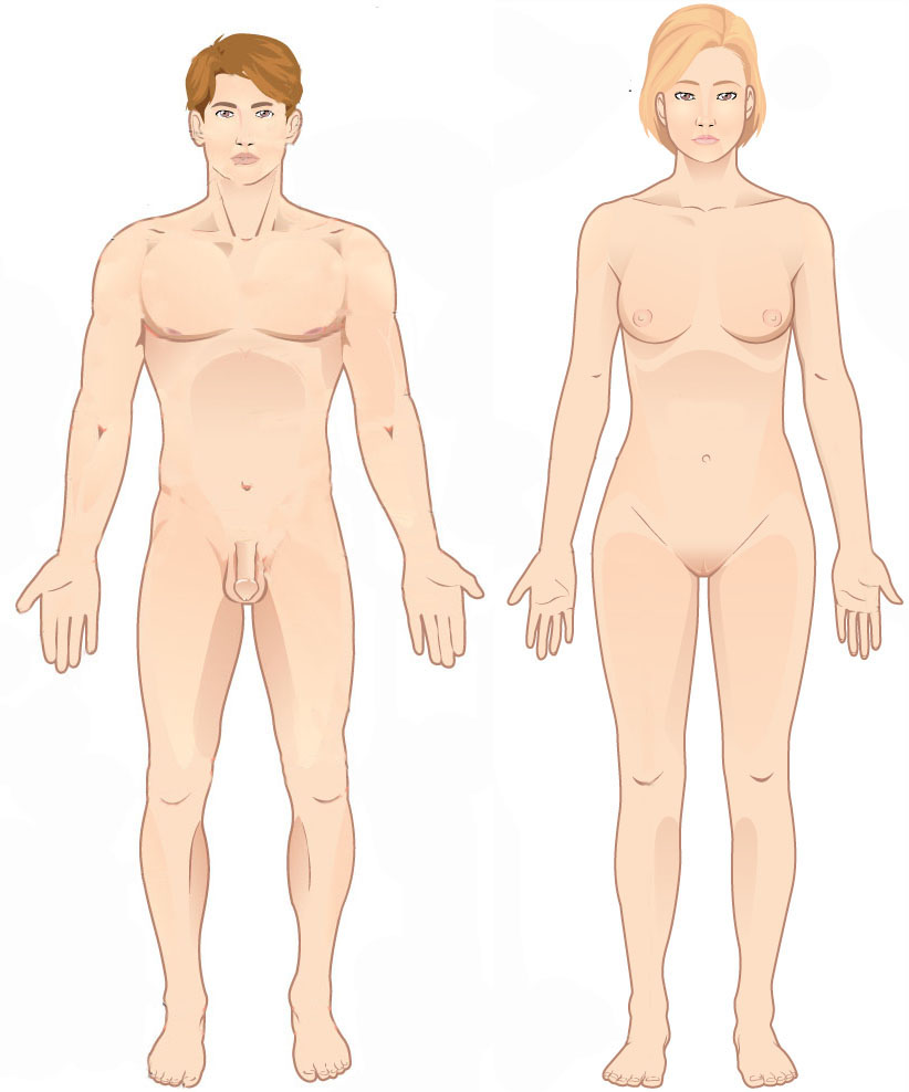
Clinical Insight: Even if a patient is curled up in a hospital bed or inverted in an MRI machine, all directional terminology describes them as if they were standing in standard anatomical position.
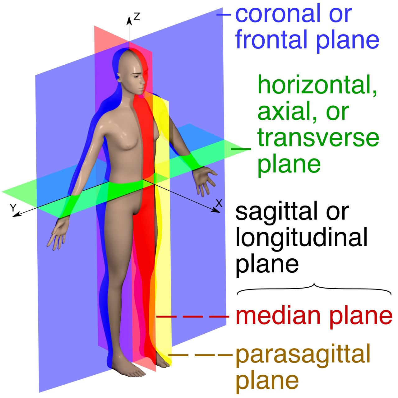
Because we inhabit a three-dimensional world, we must often visualize the internal structures of the body by "slicing" it. Body planes are imaginary flat surfaces used to divide the body into specific sections. These planes dictate how CT scans and MRIs are presented on a monitor.
| Body Plane | Definition |
|---|---|
| Sagittal Plane | Divides the body into right and left portions. |
| Midsagittal Plane | A specific sagittal plane that runs perfectly down the midline, dividing the body into equal right and left halves. |
| Parasagittal Plane | A sagittal plane offset from the midline, dividing the body into unequal right and left portions. |
| Frontal Plane | Also known as the coronal plane. It divides the body into anterior (front) and posterior (back) portions. |
| Transverse Plane | Also known as the horizontal plane. It divides the body into superior (upper) and inferior (lower) portions. |
| Oblique Plane | Divides the body at an angle other than 90 degrees. |
Directional terms describe the position of one body structure relative to another body structure. They always come in opposing pairs.
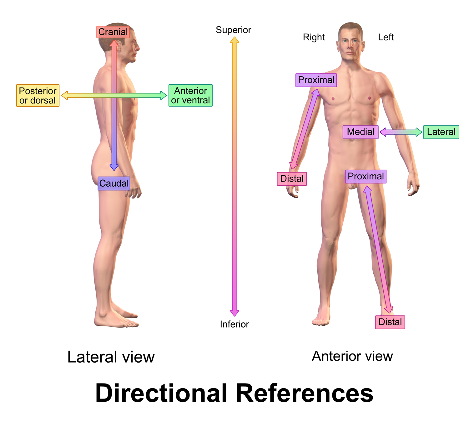
Up and Down, Front and Back
- Superior indicates a structure is closer to the head. (The nose is superior to the mouth).
- Inferior indicates a structure is closer to the feet. (The chin is inferior to the nose).
- Anterior indicates a structure is toward the front of the body. The directional term ventral is often used interchangeably with anterior in human anatomy.
- Posterior indicates a structure is toward the back of the body. The directional term dorsal is often used interchangeably with posterior in human anatomy.
Midline and Limbs
- Medial indicates a structure is closer to the midline of the body. (The heart is medial to the lungs).
- Lateral indicates a structure is farther from the midline of the body. (The ears are lateral to the eyes).
- Proximal indicates a structure is closer to the point of attachment to the trunk. We use this primarily for limbs. (The elbow is proximal to the wrist).
- Distal indicates a structure is farther from the point of attachment to the trunk. (The fingers are distal to the wrist).
Depth
- Superficial indicates a structure is closer to the surface of the body. (The skin is superficial to the muscles).
- Deep indicates a structure is farther from the surface of the body. (The bones are deep to the muscles).
The body is not a solid block of tissue; it is hollowed out into distinct, fluid-filled compartments called cavities that house and protect our vital organs.
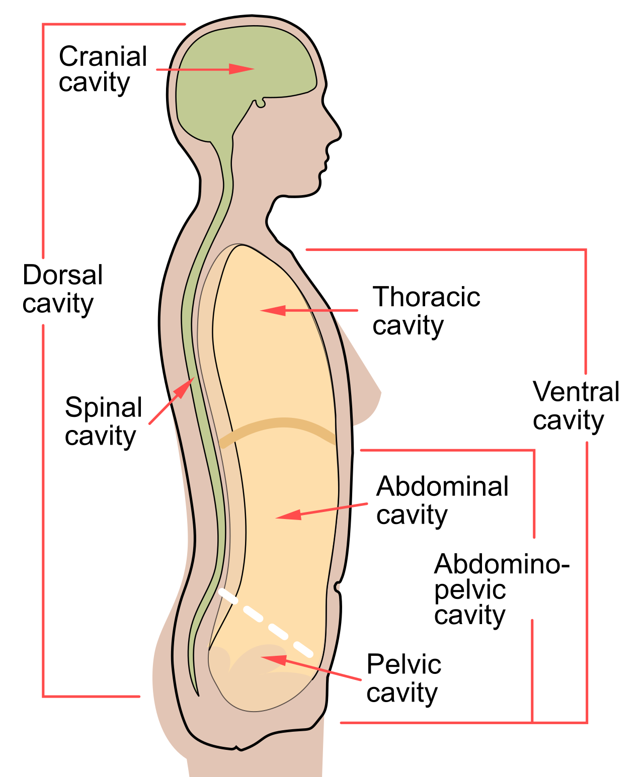
The Dorsal Body Cavity
Located along the posterior (back) surface of the body, the dorsal body cavity is encased in heavy bone to protect the delicate tissues of the central nervous system. It contains two connected sub-cavities:
- The Cranial Cavity: Houses the brain.
- The Spinal Cavity: Houses the spinal cord. This is also known as the vertebral cavity.
The Ventral Body Cavity
Located along the anterior (front) surface of the body, the ventral body cavity is much larger and houses our visceral organs. It is partitioned into two main sections by the diaphragm, a dome-shaped muscle vital for breathing that physically separates the thoracic cavity from the abdominopelvic cavity below it.
1. The Thoracic Cavity (Superior to the diaphragm)
- Pleural cavities: Two separate compartments that surround the lungs.
- Pericardial cavity: A compartment that surrounds the heart.
- Mediastinum: The central region of the thoracic cavity located between the lungs. It contains the pericardial cavity, the esophagus, and the trachea.
2. The Abdominopelvic Cavity (Inferior to the diaphragm) As the name suggests, the abdominopelvic cavity consists of two contiguous regions:
- The Abdominal cavity: Houses the digestive organs, meaning the stomach is located within the abdominal cavity, as well as the liver.
- The Pelvic cavity: Safely tucked inside the bony pelvis, it houses the reproductive organs, the rectum, and the urinary bladder.
Mapping the Abdomen
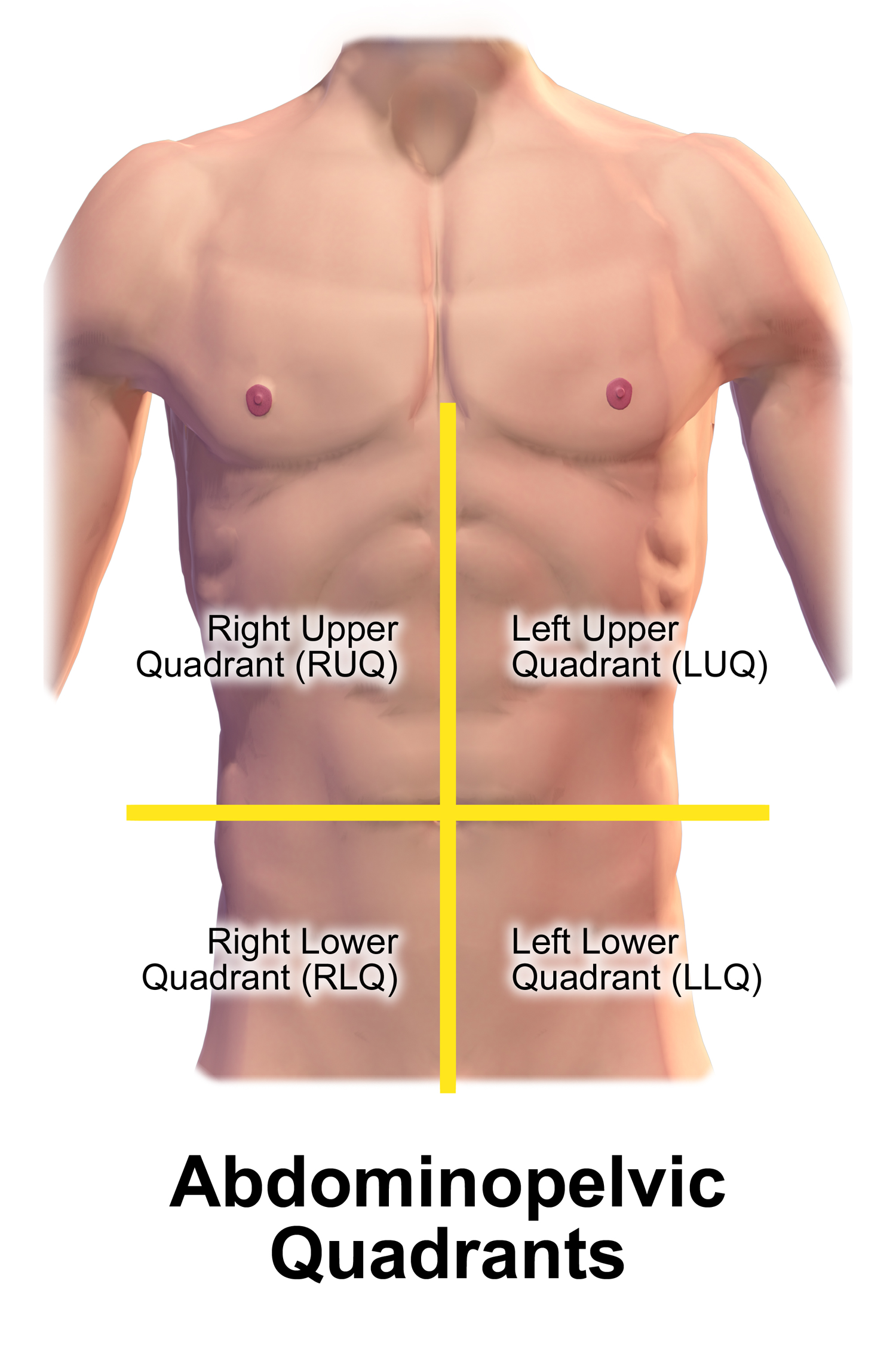
Because the abdominopelvic cavity contains so many organs, healthcare professionals divide it into smaller zones to localize pain and pathology.
For clinical use, the abdominopelvic cavity is divided into four quadrants by drawing a vertical and horizontal line right through the navel (Right Upper, Left Upper, Right Lower, Left Lower). If a patient complains of sharp pain in the right lower quadrant, a nurse immediately suspects appendicitis.
For anatomical precision, the abdominopelvic cavity is divided into nine anatomical regions, visualized like a tic-tac-toe board over the abdomen:
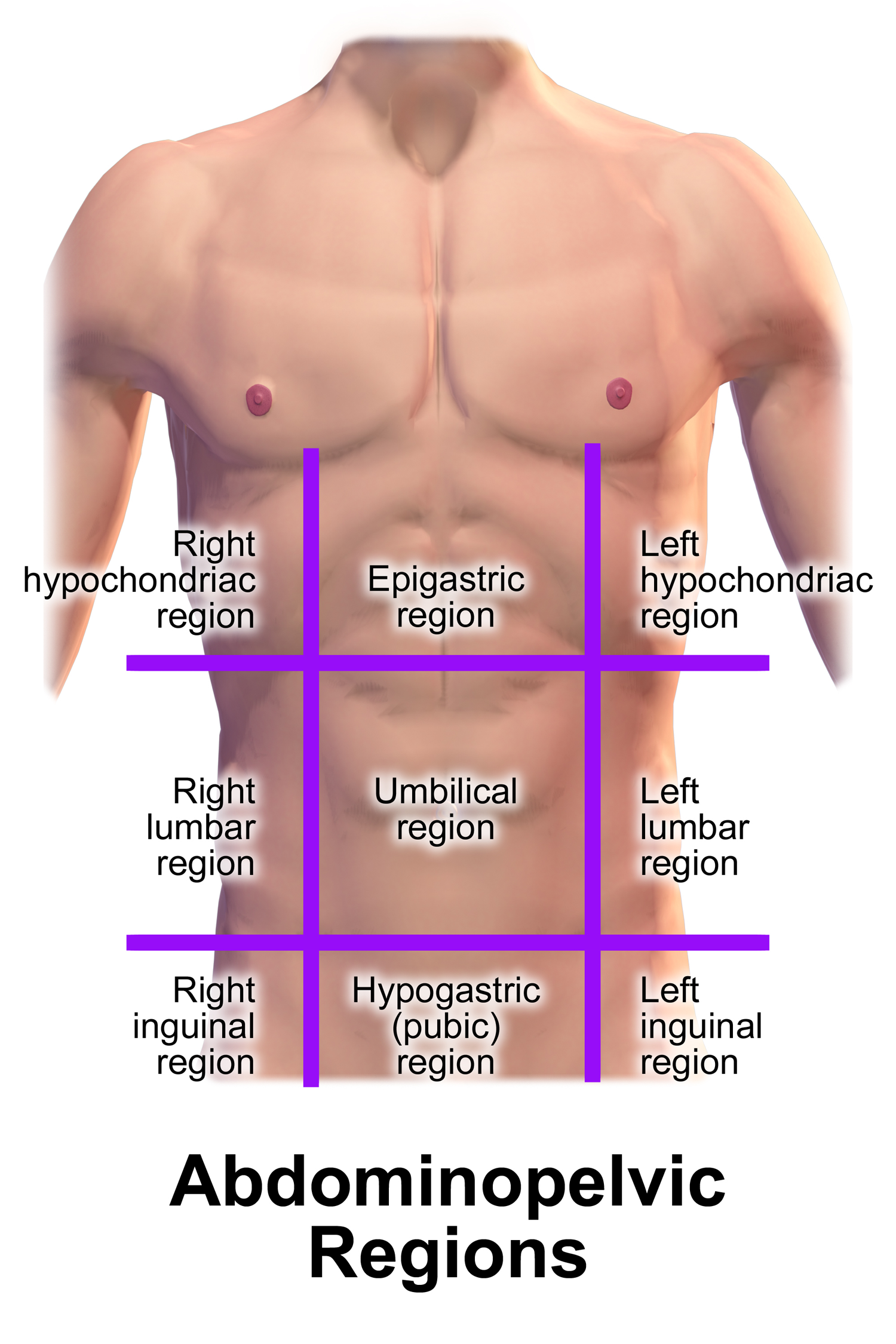
| Region | Anatomical Location |
|---|---|
| Umbilical Region | The central anatomical region surrounding the navel. |
| Epigastric Region | Located superior to the umbilical region (over the stomach). |
| Hypogastric Region | Located inferior to the umbilical region (the pubic area). |
| Right & Left Hypochondriac Regions | Located lateral to the epigastric region (tucked under the ribs). |
| Right & Left Lumbar Regions | Located lateral to the umbilical region. |
| Right & Left Iliac Regions | Located lateral to the hypogastric region. These are also known as the inguinal regions. |
All of this structural organization—from the smallest atom to the complexity of the nine abdominal regions—exists to serve one ultimate purpose: keeping the organism alive in a chaotic universe.
Homeostasis is the maintenance of a relatively stable internal environment despite external changes. Whether you are running a marathon in the summer heat or sleeping in a freezing cabin, your blood pH, core temperature, and hydration levels must remain remarkably constant. If homeostasis fails, disease or death swiftly follows.
The Machinery of Homeostasis
Homeostatic control mechanisms require three interconnected components to function:
- A Receptor: This is a sensor that monitors the internal environment. A homeostatic receptor responds to environmental changes known as stimuli.
- A Control Center: The brain or endocrine gland receives the signal from the receptor and determines the set point at which a physiological variable (like temperature or blood pressure) is to be maintained.
- An Effector: A homeostatic effector provides the means for the control center's response to the stimulus.
Negative Feedback Loops
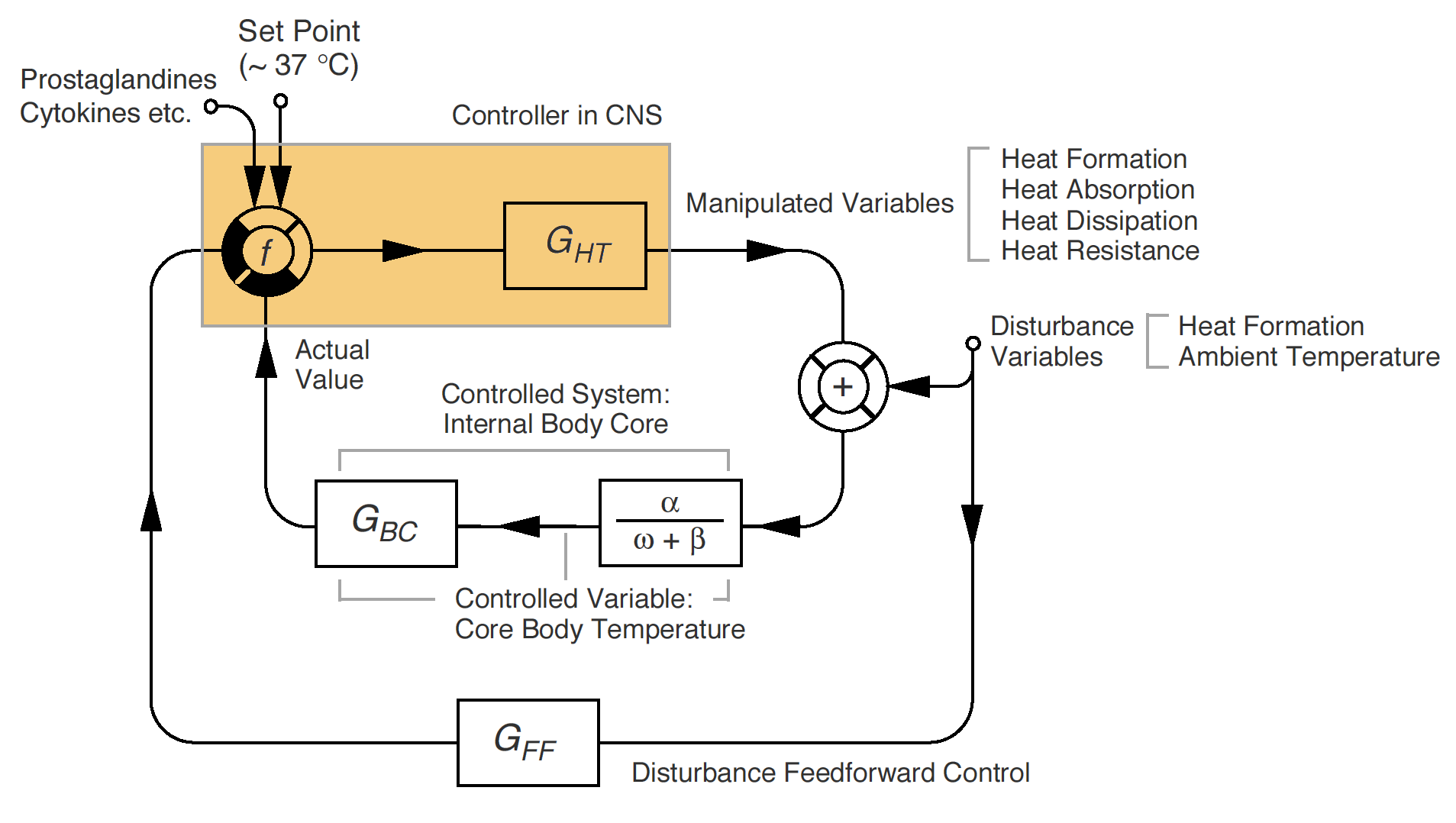
Negative feedback is the most common homeostatic control mechanism in the human body. By definition, negative feedback mechanisms reduce the intensity of the original homeostatic stimulus, and they can shut off the original homeostatic stimulus entirely once balance is restored.
Think of it like a thermostat. If the room gets too cold (stimulus), the thermostat (control center) turns on the heater (effector). Once the room warms up, the heater turns off.
- Human body temperature regulation is an example of a negative feedback mechanism. If you overheat, you sweat to cool down, negating the original heat stimulus.
- Human blood glucose regulation is an example of a negative feedback mechanism. If blood sugar spikes after a meal, insulin is released to lower it, negating the original spike.
Positive Feedback Loops
While rare, positive feedback mechanisms are highly powerful. Instead of negating the stimulus, positive feedback mechanisms enhance and exaggerate the original homeostatic stimulus. They create a snowball effect that pushes a physiological process to a rapid completion.
- Blood clotting is an example of a positive feedback mechanism in the human body. When a vessel tears, platelets rush to the site and release chemicals that attract even more platelets, creating an escalating cascade until the clot is fully formed and the bleeding stops.
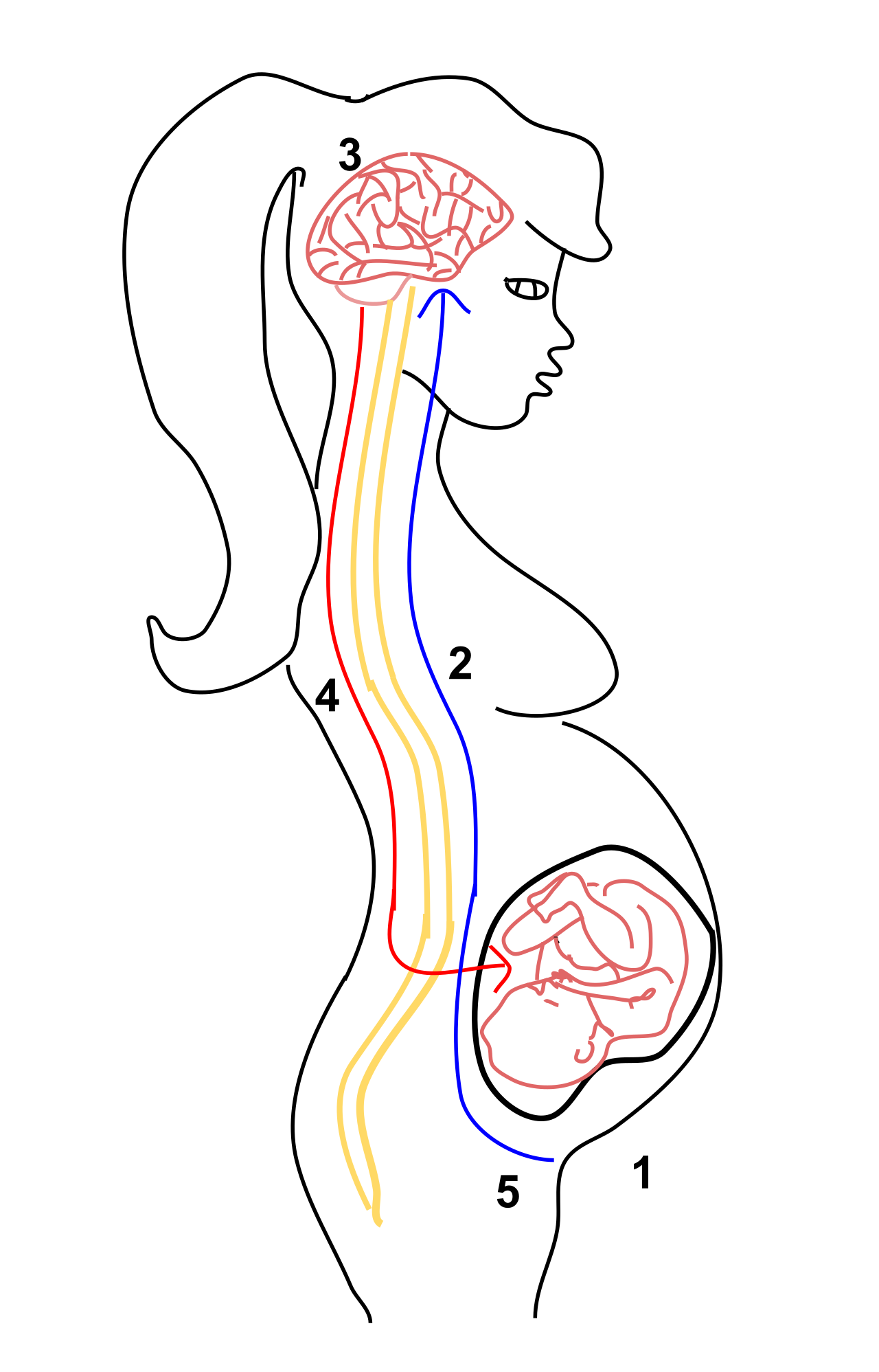
- Uterine contractions during childbirth are an example of a positive feedback mechanism in the human body. The baby's head pushes against the cervix (stimulus), triggering the release of oxytocin, which causes a contraction. This pushes the baby harder against the cervix, releasing more oxytocin, causing stronger contractions until the child is born.