Alterations in Body Systems and Pathophysiology
Not sure you’re ready?
Take the ~3-minute readiness diagnostic and see where you stand.
The Master Mechanic of the Human Body: Alterations in Systems and Pathophysiology
Welcome! Pull up a chair. If you are going to be a brilliant practical nurse, you need to stop memorizing disconnected lists of symptoms and start looking at the human body for what it really is: the most magnificent, intricate, interconnected machine in the universe.
When you look at a patient, you are looking at a living ecosystem of plumbing, electrical wiring, and pressure dynamics. When illness strikes, this machine doesn't just fail randomly; it fights back, it compensates, and it leaves clues. Your job is to be the detective that reads those clues.
Let’s take a walk through the wards and decode the pathophysiology behind the signs and symptoms you will face on the NCLEX-PN.
When a pathogen invades the body, nature doesn't just sit back. It mounts an organized, energetic defense.
Think about what happens when you cut your finger and it gets infected. Purulent drainage from a wound indicates a localized tissue infection. The pus you see is literally a graveyard of dead bacteria and white blood cells—evidence of a localized battle.
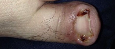
But what if the invader gets into the blood? The battle goes system-wide. Fever is a systemic clinical sign of an active infection. The body intentionally turns up the core thermostat because bacteria hate the heat. But a hotter, working body demands more oxygen and nutrients. So, what does the heart do? It speeds up! Therefore, tachycardia is a physiological compensatory response to systemic infection.
To fight this war, the body needs soldiers.
The Normal Fleet: A normal white blood cell count ranges from 5,000 to 10,000 cells per microliter of blood.
If you draw blood and see a number jumping up to 15,000 or 18,000, you know the factories in the bone marrow are working overtime. An elevated white blood cell count indicates an active infectious process in the body.
The Stealth Invader: Urinary Tract Infections
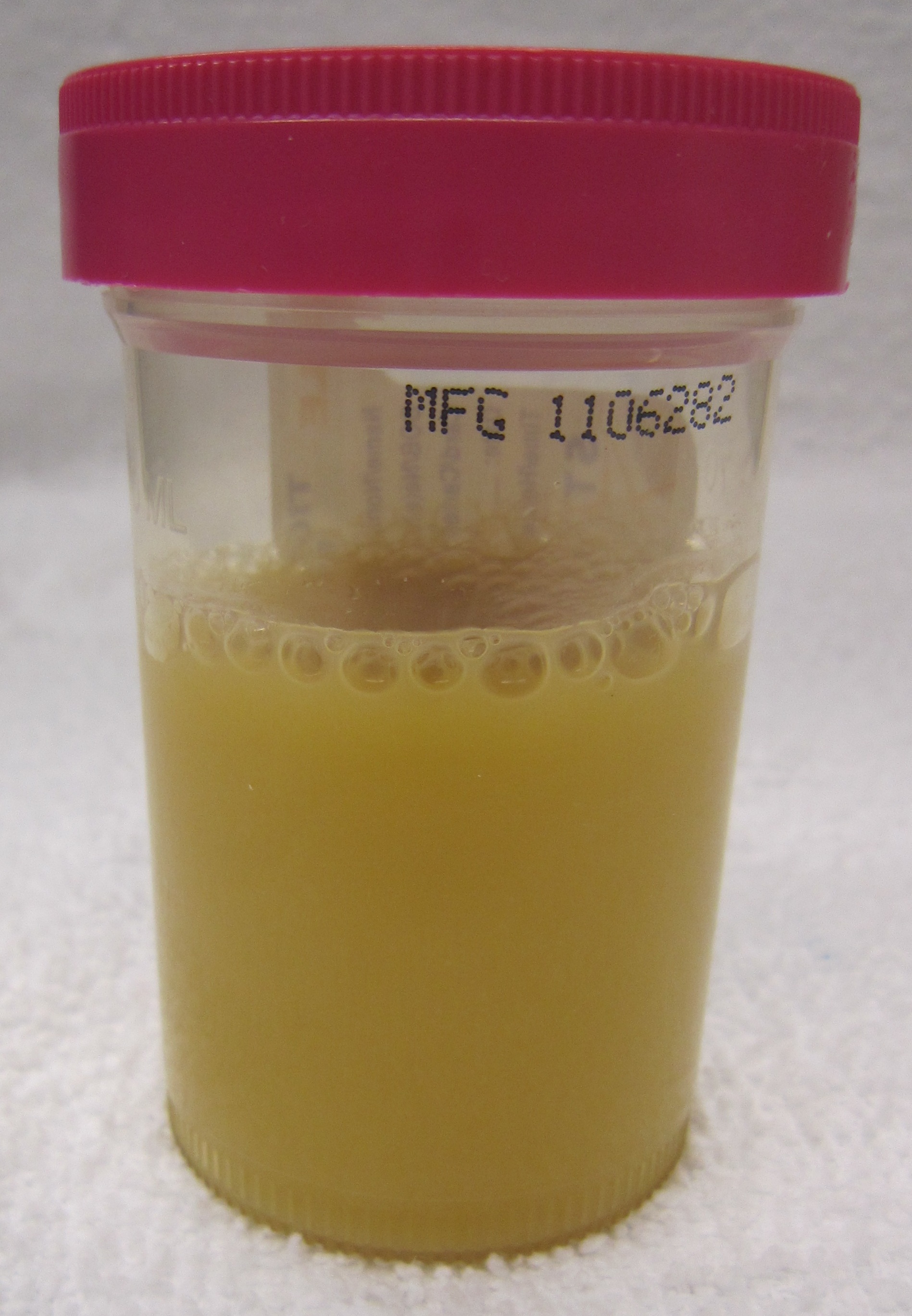
Not all infections are obvious. If the kidneys and bladder are under attack, the waste disposal system shows the evidence. Cloudy urine with a foul odor is a primary clinical indicator of a urinary tract infection.
But here is a fascinating quirk of human biology: the brains of older adults are highly sensitive to systemic inflammation. Long before an elderly patient complains of burning urination, their brain feels the stress. Sudden confusion is often the first clinical sign of a urinary tract infection in older adult clients. Remember this! If sweet Grandma suddenly doesn't know what year it is, check the urine.
The heart is a mechanical pump, but it runs entirely on an electrical grid. If you understand the wiring, reading an ECG monitor strip is like reading a book.
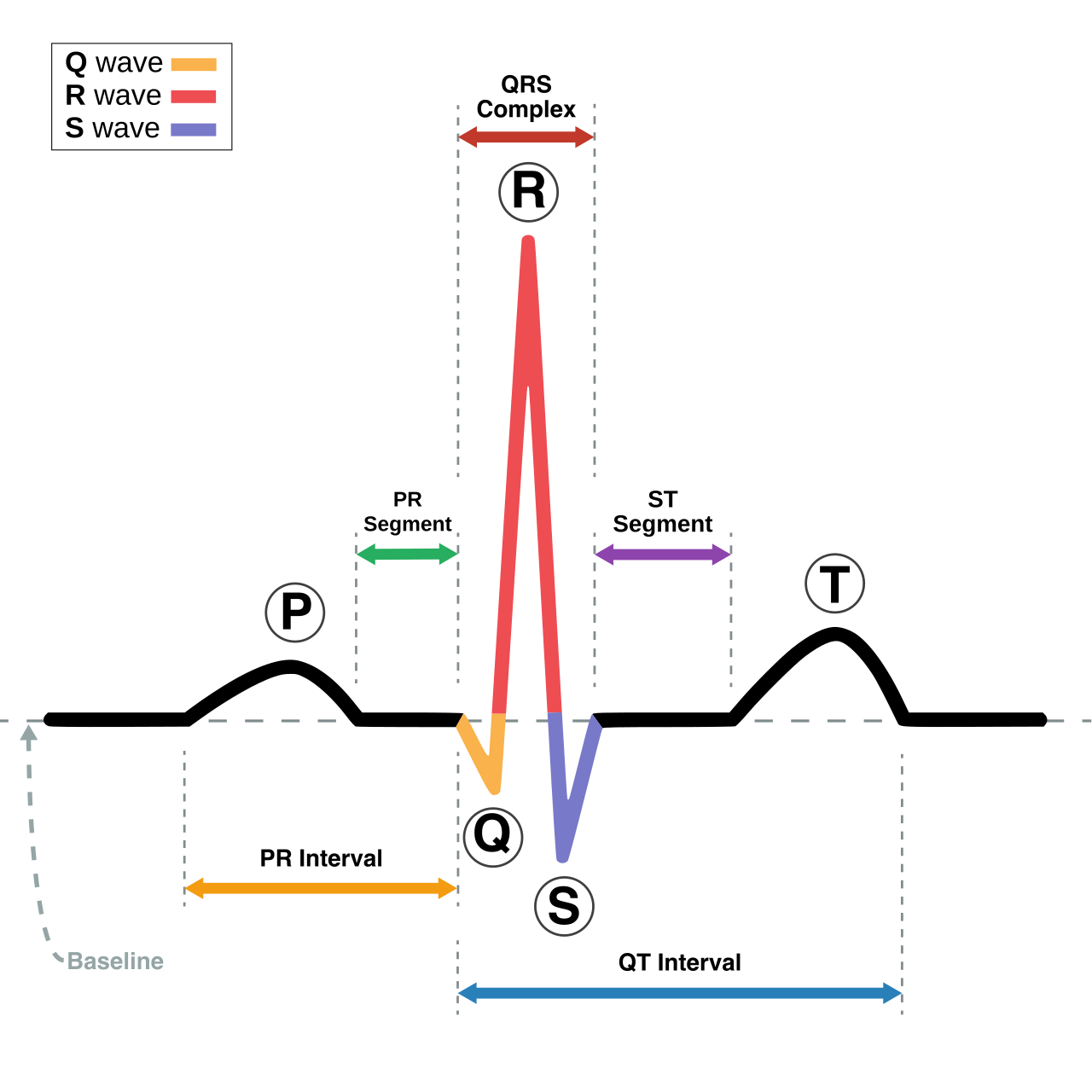
The main power switch of the heart is the sinoatrial (SA) node. A normal sinus rhythm originates from the sinoatrial node of the heart. When this pacemaker fires beautifully, normal sinus rhythm has a regular heart rate between 60 and 100 beats per minute.
If the rhythm is regular but the rate drops below 60, we call it sinus bradycardia (defined as a regular heart rhythm with a rate below 60 beats per minute). If the regular rhythm speeds up past 100, that is sinus tachycardia (a regular heart rhythm with a rate above 100 beats per minute).
Electrical Misfires & Chaos
What happens when the electrical signal gets delayed? As the signal travels from the top of the heart (atria) to the middle (AV node), it should be quick. A prolonged PR interval indicates a delay in electrical conduction through the atrioventricular node. It’s like a traffic jam on the electrical highway.
Now, let's look at when the electricity goes completely haywire:
| Rhythm | Pathophysiology & ECG Appearance | Clinical Consequence |
|---|---|---|
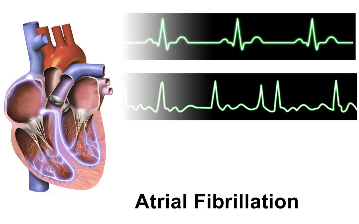
| Atrial Fibrillation (A-Fib) | The atria quiver instead of squeezing. Atrial fibrillation presents on an electrocardiogram as an irregular heart rhythm with no identifiable P waves. | Loss of atrial "kick," risk of blood clots. |
| Ventricular Fibrillation (V-Fib) | The main pumping chambers just twitch chaotically. Ventricular fibrillation produces a chaotic waveform with no discernible QRS complexes on an electrocardiogram. | Ventricular fibrillation results in a complete absence of cardiac output. The patient is clinically dead. Start CPR! |
| Asystole | Total electrical failure. Asystole appears as a flat baseline line on an electrocardiogram monitor strip. | No pulse. CPR and Epinephrine required. |
Fluid dynamics in the body are governed by a simple rule: If a pump fails, fluid backs up into whatever feeds it.
Left vs. Right Heart Failure
The left side of the heart receives blood from the lungs. If the left ventricle fails, the blood has nowhere to go but backwards into the pulmonary system. Left-sided heart failure causes pulmonary venous congestion. The fluid leaks into the lung tissue, which is why auscultated crackles in the lung bases are a primary clinical sign of left-sided heart failure.
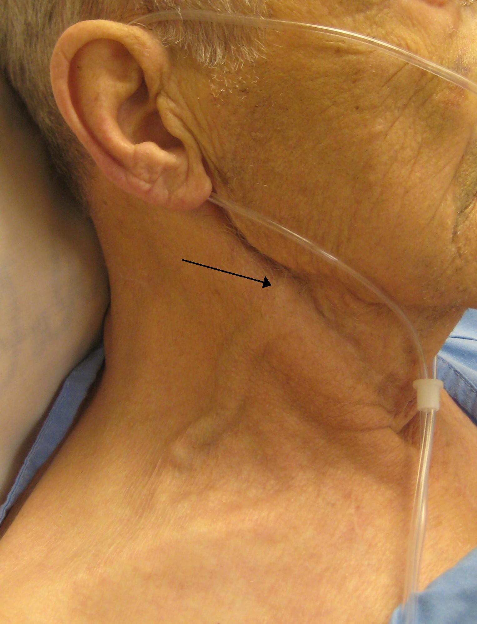
The right side of the heart receives blood from the rest of the body. Right-sided heart failure causes systemic venous congestion. The fluid backs up into the veins. You can literally see the backup in the neck—jugular vein distention is a primary clinical sign of right-sided heart failure.
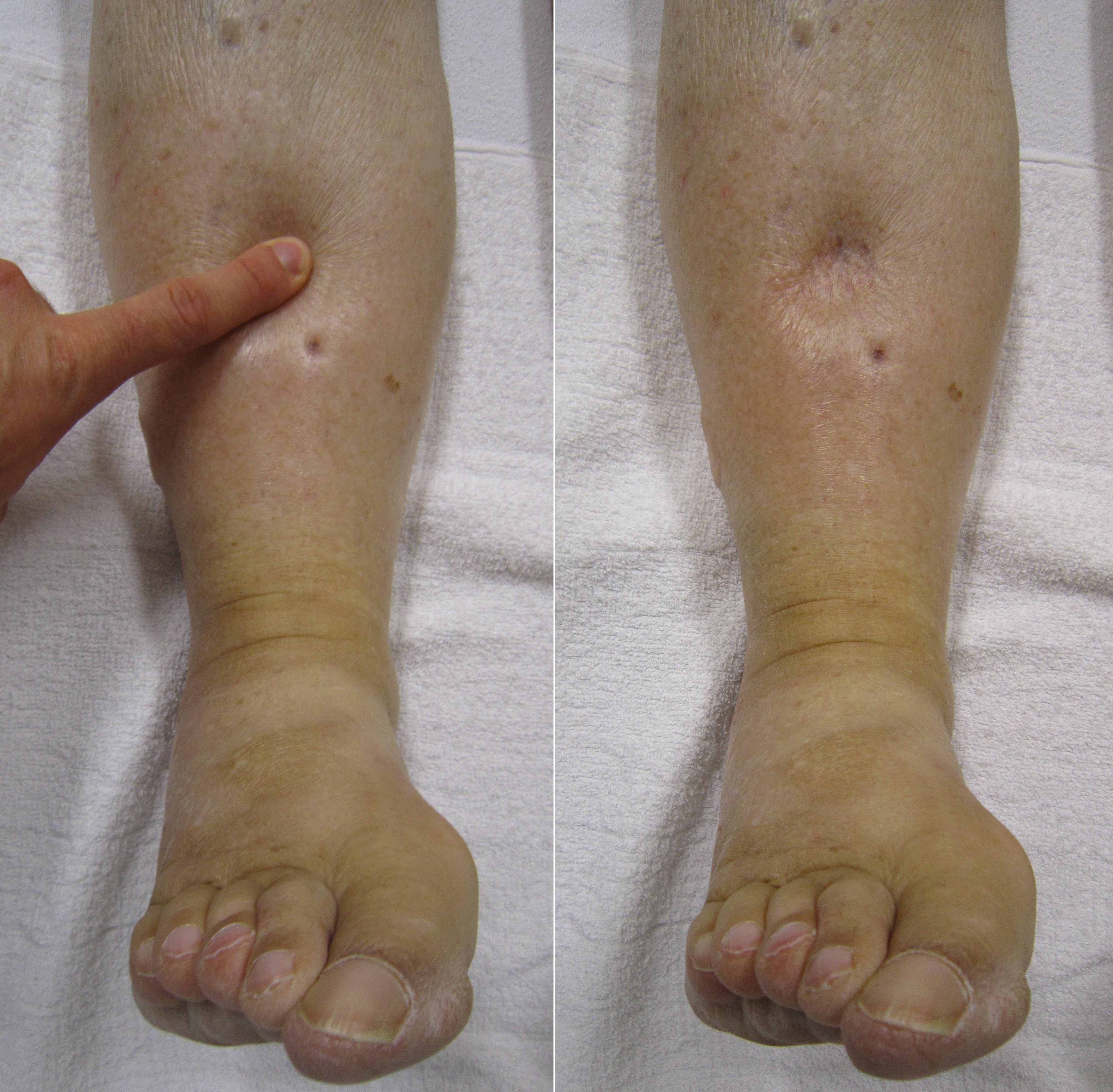
Gravity then pulls this backed-up fluid down to the lowest points, meaning dependent peripheral edema is a common clinical manifestation of right-sided heart failure.
Hypovolemia
What if there just isn't enough fluid in the pipes? Hypovolemia decreases total circulating blood volume. Because there is less liquid to push against the vessel walls, severe hypovolemia presents with a weak and thready peripheral pulse.
When the natural airway fails, we build a new one. A tracheostomy provides a secure artificial airway through an incision in the anterior neck.
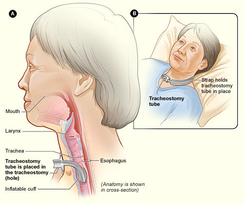
The Rules of Tracheostomy Care
Because we have bypassed the body's natural air filters (the nose and mouth), the lungs are highly vulnerable. Therefore, tracheostomy suctioning requires strict sterile technique to prevent lower respiratory tract infections.
Suctioning removes mucus, but it also steals the patient's oxygen. You must prepare them! The nurse must pre-oxygenate the client with 100 percent oxygen before initiating tracheostomy suctioning, and to prevent suffocation, each individual tracheostomy suctioning pass must not exceed 10 seconds in duration.
For maintenance, routine tracheostomy care requires cleaning the inner cannula with sterile normal saline.
The Golden Safety Rule: What happens if the patient coughs so hard the whole tube flies out? You need to get a new one in immediately. An obturator must be kept at the bedside of a client with a new tracheostomy at all times. Why? Because the obturator is used to safely guide the reinsertion of an accidentally dislodged tracheostomy tube.
Mechanical Ventilators
Think of a ventilator as a smart bicycle pump. Mechanical ventilators provide positive pressure to inflate the lungs of a compromised client.
The vent is always measuring how hard it has to push (resistance). Understanding ventilator alarms is pure physics:
- High-Pressure Alarms: A high-pressure ventilator alarm indicates increased airflow resistance in the airway circuit. The machine is trying to push air in, but something is blocking the path. What causes blocks?
- Excessive airway secretions trigger a high-pressure ventilator alarm.
- A kinked ventilator tube triggers a high-pressure ventilator alarm.
- A client biting the endotracheal tube triggers a high-pressure ventilator alarm.
- Low-Pressure Alarms: A low-pressure ventilator alarm indicates a sudden decrease in airway resistance. The machine pushes air, and it meets zero resistance. The air is escaping!
- A disconnected ventilator tubing circuit triggers a low-pressure ventilator alarm.
- An endotracheal tube cuff leak triggers a low-pressure ventilator alarm.
Bonus Vent Safety: Having a tube in the throat allows bacteria to slide right down into the lungs. Frequent oral care with chlorhexidine reduces the risk of ventilator-associated pneumonia.
When the bowel is diseased, surgeons bring a piece of it through the abdominal wall, creating a stoma.
Reading the Stoma
A stoma is inside-out intestine. It should look like the inside of your cheek. A healthy ostomy stoma appears beefy red and moist.
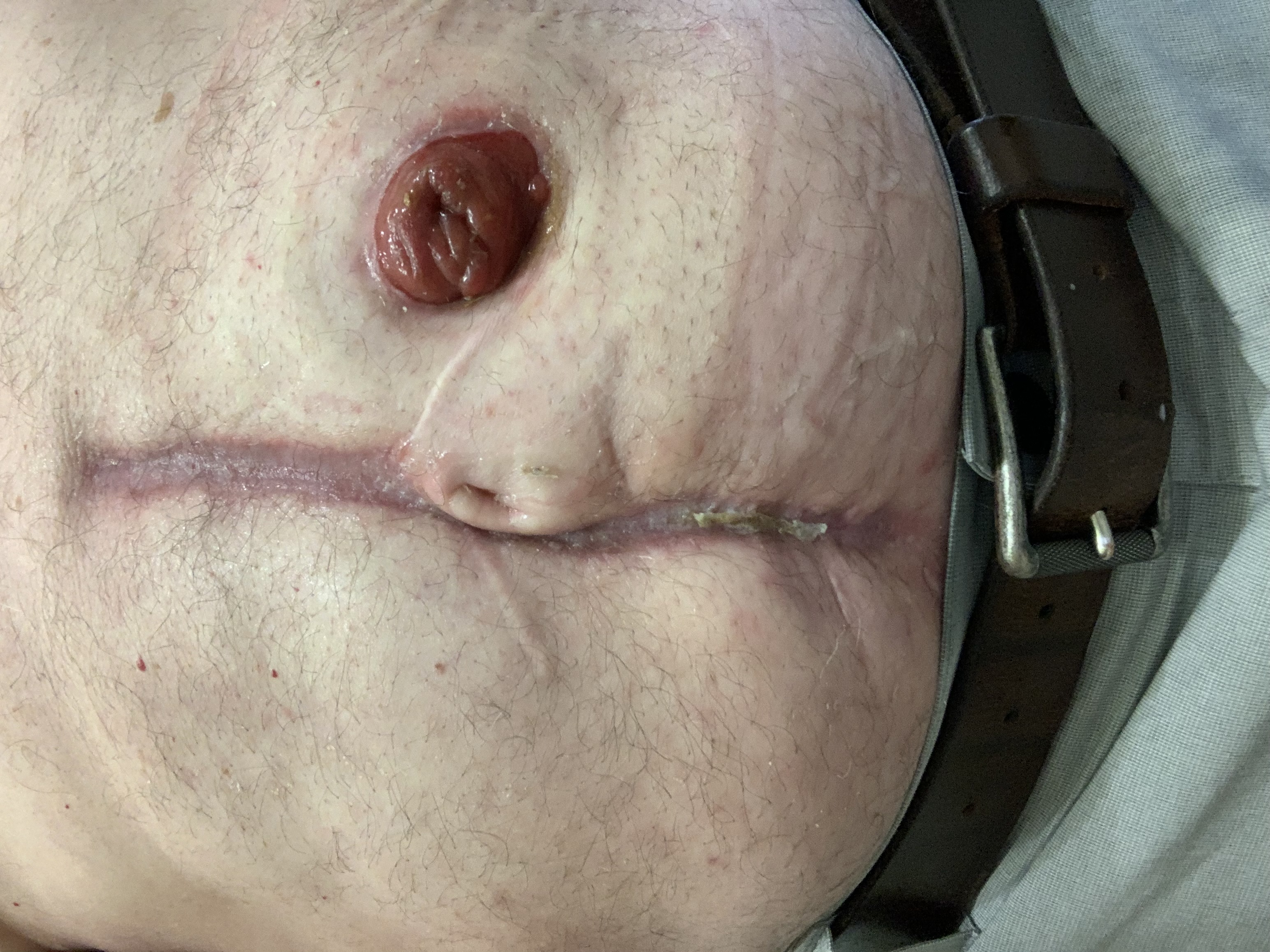
If the color changes, you have a blood flow emergency.
- A pale ostomy stoma indicates inadequate tissue perfusion (think low hemoglobin or poor flow).
- A dusky blue or black ostomy stoma indicates severe stomal ischemia. This is dying tissue. Alert the provider instantly!
Pouch Mechanics & Output
Gravity is your enemy with ostomy bags. An ostomy appliance should be emptied when the pouch is one-third to one-half full. Why so early? Because emptying the ostomy pouch before the pouch becomes completely full prevents weight-induced appliance leakage. A heavy bag pulls the adhesive right off the skin.
What comes out of the bag depends on where the ostomy was placed:
- Ileostomy (Small Intestine): This bypassed the entire large intestine (where water is normally absorbed). Thus, ileostomy drainage is typically liquid and continuous. Because they are losing so much water, clients with a new ileostomy must increase daily fluid intake to prevent dehydration.
- Colostomy (Large Intestine): If the stoma is way down the line, water has already been absorbed. Colostomy drainage from the descending colon is typically solid and formed.
Finally, let's look at how the entire body behaves when the chemistry or pressure is thrown off balance.
Oxygen Deprivation: The brain is a greedy organ. It uses 20% of the body's oxygen. If oxygen drops, the brain panics long before the patient turns blue. Cerebral hypoxia causes early behavioral changes such as restlessness and agitation. If your previously calm patient suddenly won't stay in bed, check their oxygen!
Blood Sugar Battles:
- Hypoglycemia (Low Sugar): The body dumps adrenaline to try and free up stored glucose. This adrenaline rush is why hypoglycemia frequently presents with profuse diaphoresis and muscle tremors.
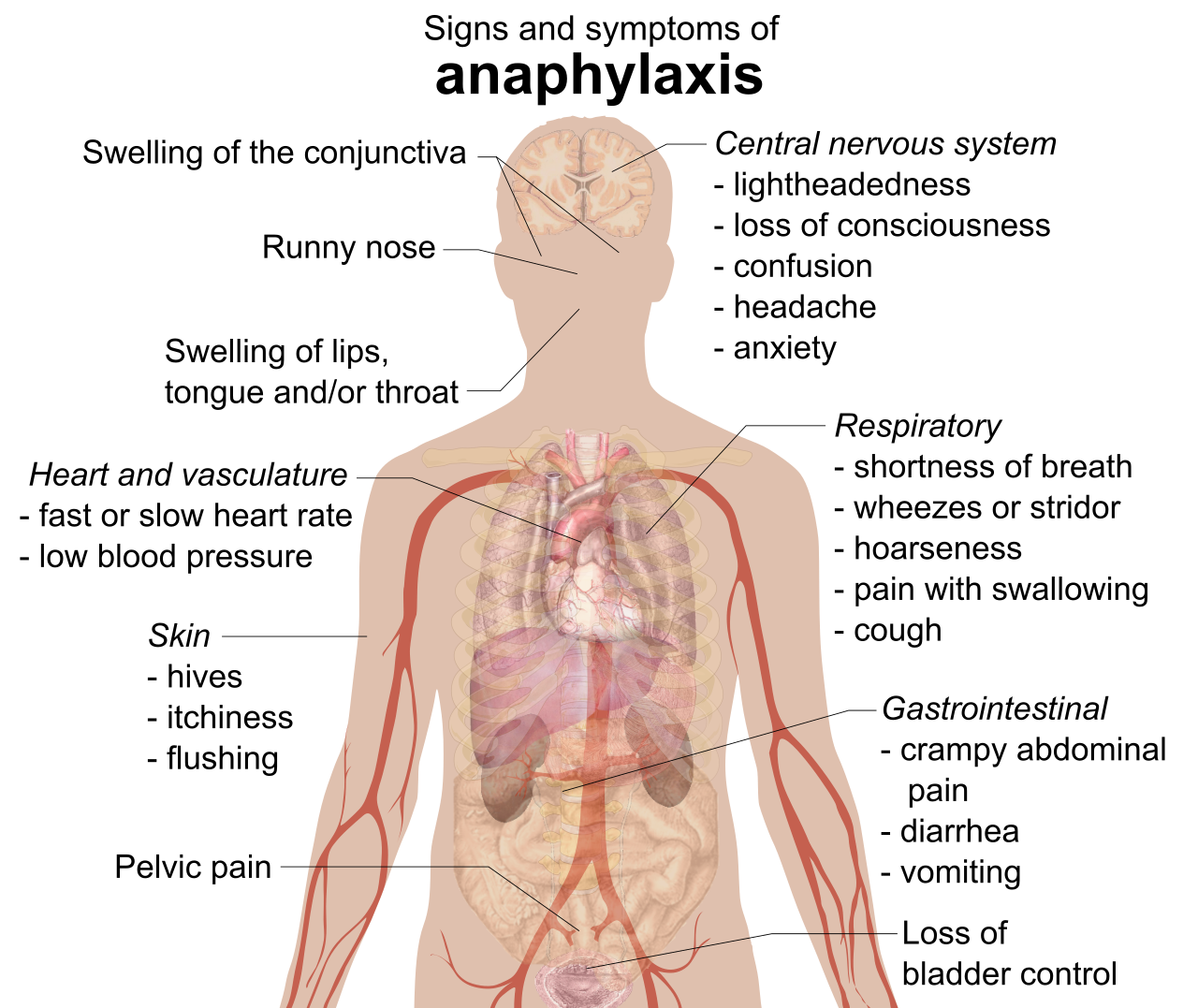
- Diabetic Ketoacidosis (High Sugar/No Insulin): When cells starve without insulin, they burn fat, creating acid (ketones). The body tries to fix this extreme acidity by blowing off carbon dioxide (an acid) through the lungs. This is why diabetic ketoacidosis causes deep and rapid Kussmaul respirations. Those broken-down ketones also off-gas through the lungs, meaning diabetic ketoacidosis produces a distinct fruity breath odor.
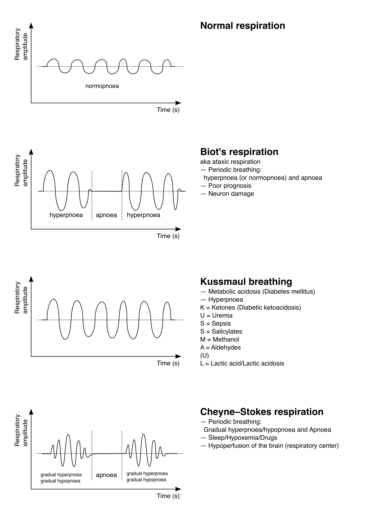
Anaphylaxis: When an allergic reaction goes nuclear, mast cells release massive amounts of histamine. Histamine does two terrible things simultaneously: Anaphylaxis causes severe systemic vasodilation (crashing blood pressure) AND anaphylaxis causes acute bronchoconstriction (closing the airways).
Increased Intracranial Pressure (ICP): The skull is a closed, rigid box. If the brain swells, pressure builds. This pressure crushes the brainstem, which controls vital signs, resulting in a terrifying phenomenon called Cushing's Triad:
- The heart tries to force blood into the highly pressurized skull, so systolic blood pressure shoots up while diastolic stays low. Increased intracranial pressure causes a widened pulse pressure.
- The crushed vagus nerve drastically slows the heart. Increased intracranial pressure causes severe bradycardia.
- The respiratory center is compressed. Increased intracranial pressure causes an irregular respiratory pattern.
Final Thoughts
Look at the logic here! Whether it's the heart pumping faster to deliver white blood cells, the lungs breathing deeply to blow off acid in DKA, or a ventilator screaming because of a kinked hose—none of this is random. By understanding why the body systems alter, you stop being just a nurse who follows orders, and you become a critical thinker who saves lives.
Now, go out there, look at your patients, and read the machine.