Parenteral and Intravenous Therapies
Not sure you’re ready?
Take the ~3-minute readiness diagnostic and see where you stand.
Mastering Parenteral and Intravenous Therapies: The River of Life
Welcome! Let’s talk about one of the most remarkable, powerful, and frankly, perilous things you will do as a nurse: intravenous (IV) therapy.
Think about it. The human body has spent millions of years evolving brilliant defenses—the skin, the acidic stomach, the complex mucosal linings—just to keep the outside world out. And what do we do? We take a sharp needle, bypass all of that magnificent evolutionary architecture, and establish a direct pipeline into the central vascular highway.
It is a profound intervention. You are introducing fluids and medications that act instantly. Because of this, the margin for error is razor-thin. If you understand the physics, the biology, and the mechanics of what you are actually doing, the NCLEX questions won't just be things you memorize—they will be things you understand intuitively. Let’s dive in.
Before you can deliver a payload, you need the right delivery route. You are looking for a vessel that is accessible, durable, and safe.
In adults, the cephalic vein, the basilic vein, and the median cubital vein are all entirely appropriate sites for peripheral intravenous access. These veins are the sturdy, reliable highways of the arm.
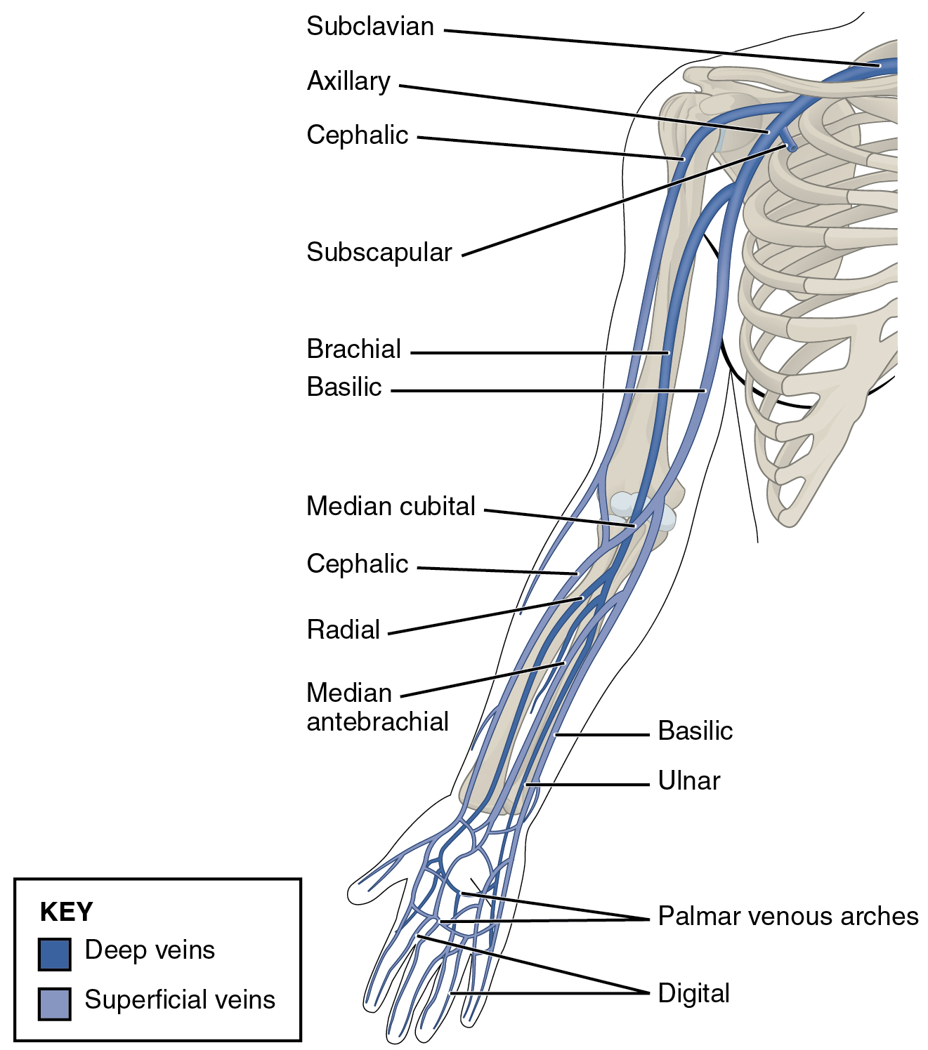
But choosing a vein isn't just about finding a blue line under the skin; it’s about anticipating the future.
- Avoid the joints: Veins located in areas of joint flexion carry a profoundly high risk of peripheral catheter dislodgement. Every time the patient bends their wrist or elbow, the catheter acts like a microscopic saw. Consequently, veins located in areas of joint flexion carry a high risk of mechanical phlebitis (physical irritation of the vein).
- Respect the restricted zones: Some highways are strictly off-limits due to altered hemodynamics or lymphatics. The presence of an arteriovenous fistula (used for dialysis) absolutely contraindicates the placement of a peripheral intravenous line in that extremity. You do not mess with a patient's lifeline. Similarly, a history of mastectomy or a history of lymph node dissection contraindicates the placement of a peripheral intravenous line on the affected side, as the compromised lymphatic drainage could lead to severe lymphedema or infection.
The Physics of the Venipuncture
Now, how do we make the stick successful? We manipulate pressure and temperature.
- Pressure: A tourniquet acts as a dam, allowing arterial blood in but stopping venous blood from getting out. This tourniquet is typically applied four to six inches above the intended venipuncture site to create the perfect hydraulic back-pressure to engorge the vein.
- Temperature: If the veins are hiding, physics is your friend. Applying a warm compress to an extremity promotes vasodilation for easier venipuncture. Heat expands; it’s that simple.
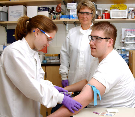
Preparation and The "Air-Dry" Rule
When you breach the skin, you must obliterate the local microbial life. Chlorhexidine gluconate is the preferred antiseptic solution for peripheral intravenous site preparation because it has incredible residual antimicrobial activity.
But here is where nurses make a critical physics error: they get impatient and wave their hands over the wet site. Do not do this! Fanning the chlorhexidine skin preparation area introduces airborne pathogens to the insertion site. You are literally creating miniature wind currents that drag bacteria off your scrubs and onto the patient's sterilized skin. Chlorhexidine gluconate skin preparation must air dry completely prior to venipuncture for the chemical lysis of the bacterial cell walls to finish.
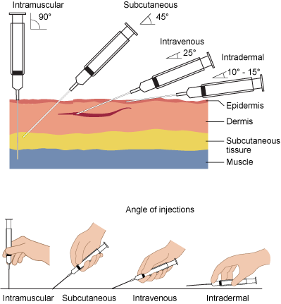
When you insert the needle, the mechanics matter. An intravenous needle is inserted with the bevel facing upward. Why? Because the sharp point must pierce the skin and vein wall first to create a clean slice rather than a jagged tear. To slide smoothly into the vessel lumen without going out the other side, the typical angle of insertion for a peripheral intravenous catheter is between 10 and 30 degrees.
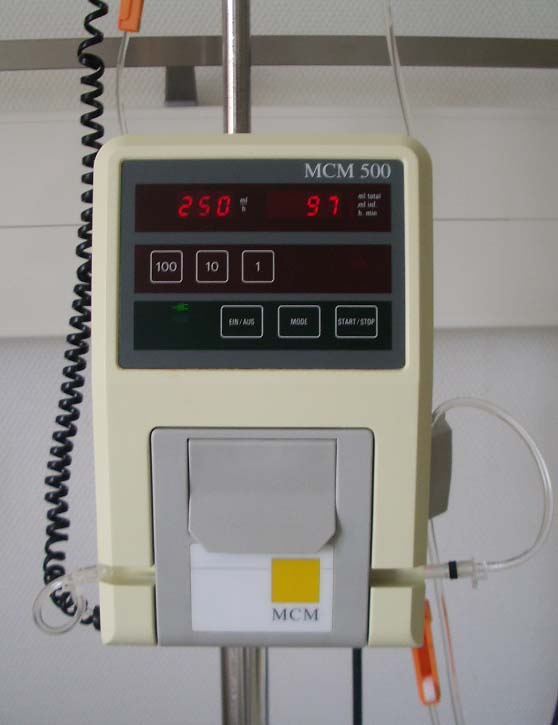
Once you are in the vein, you often hook the patient up to a robot. Electronic infusion pumps deliver intravenous fluids at a precisely regulated rate measured in milliliters per hour.
Because these machines do the math and the heavy lifting, they operate fundamentally differently than a gravity drip. Electronic infusion pumps deliver intravenous fluids under positive pressure. They actively force fluid into the patient. This positive pressure means you cannot blindly trust the machine; nurses must independently verify that electronic infusion pump settings match the provider prescription. The machine doesn't know what medication is in the bag; it only knows how hard and fast to push.
Listening to the Alarms
When the pump screams, it's telling you a physics problem:
- Occlusion alarm: This indicates a physical blockage in the intravenous line. The positive pressure has hit a wall. For example, kinked intravenous tubing triggers a high-pressure occlusion alarm on an electronic infusion pump. The pump tries to push, the resistance spikes, and the alarm sounds.
- Air-in-line alarm: This on an electronic infusion pump indicates the presence of gas bubbles within the tubing. The pump uses ultrasonic sensors; when the sound waves hit the vastly different density of a gas bubble compared to fluid, the machine stops to save the patient's life.
You’ve established access. Now, you must defend it.
We cover the site with a window. Transparent semipermeable dressings allow continuous visual inspection of the intravenous insertion site while keeping water and dirt out. But this barrier is fragile. Peripheral intravenous dressings require immediate changing upon becoming damp, visibly soiled, or loose. A compromised dressing is a red carpet for bacteria.
To ensure your access is still good, you flush it. Flushing an intravenous catheter with normal saline confirms the patency of the vascular access device (proving the highway is open) and removes residual medication from the internal lumen (preventing dangerous drug precipitates from forming in the plastic tubing).
The Microbial Clocks
Bacteria love sugar and water, which is exactly what IV tubing is full of. To prevent microbial growth, we have strict, evidence-based expiration timers:
- Continuous tubing: The Centers for Disease Control and Prevention recommends changing continuous intravenous tubing no more frequently than every 96 hours. Constant flow keeps bacterial colonization down.
- Intermittent tubing: Because the fluid stops and starts, the risk of colonization spikes. Thus, intermittent intravenous infusion tubing must be changed every 24 hours.
- The Fluid Bags: Primary intravenous fluid bags must be replaced every 24 hours to prevent microbial growth. Even if the bag is half full, throw it away. Stagnant fluid at room temperature is a petri dish.
The body will tell you when IV therapy is failing. You must know how to translate its distress signals.
1. The Local Disasters: Infiltration, Extravasation, and Phlebitis
| Complication | What is happening? | Clinical Signs | Nursing Action |
|---|---|---|---|
| Phlebitis | Phlebitis is the inflammation of the inner layer of a vein. The vessel is physically or chemically irritated. | Signs include localized erythema along the venous pathway and a palpable venous cord (the vein feels hard and ropey like a cooked noodle). | The immediate nursing intervention for phlebitis is the removal of the peripheral intravenous catheter. Do not wait. |
| Infiltration | Infiltration occurs when non-vesicant intravenous fluid leaks into the surrounding subcutaneous tissue. The catheter popped out of the vein, and now you are watering the tissues. | Because the fluid is usually cool room temperature and dilutes local blood flow, signs include localized skin pallor at the insertion site, coolness of the skin, and tissue edema around the insertion site. | Stop infusion, remove IV, elevate extremity. |
| Extravasation | Extravasation occurs when vesicant intravenous fluid leaks into the surrounding subcutaneous tissue. This is an emergency. | Vesicant medications cause severe tissue necrosis during an extravasation event. They literally burn the tissue black. | The initial nursing action for an extravasation is to immediately stop the intravenous infusion. Crucially, DO NOT immediately pull the IV out! A syringe is used to aspirate residual vesicant medication from the intravenous catheter before removal to prevent pushing more poison into the tissues. |
2. The Systemic Catastrophe: Air Embolism
If a large amount of air enters the line, it travels to the heart. Air embolism is a potentially fatal complication resulting from the entry of air into the venous system.
Feynman's Physics Check: What happens to air in water? It rises! So, if air enters the right side of the heart, we must prevent it from being pumped into the pulmonary artery, where it will cause an instant pulmonary infarction.
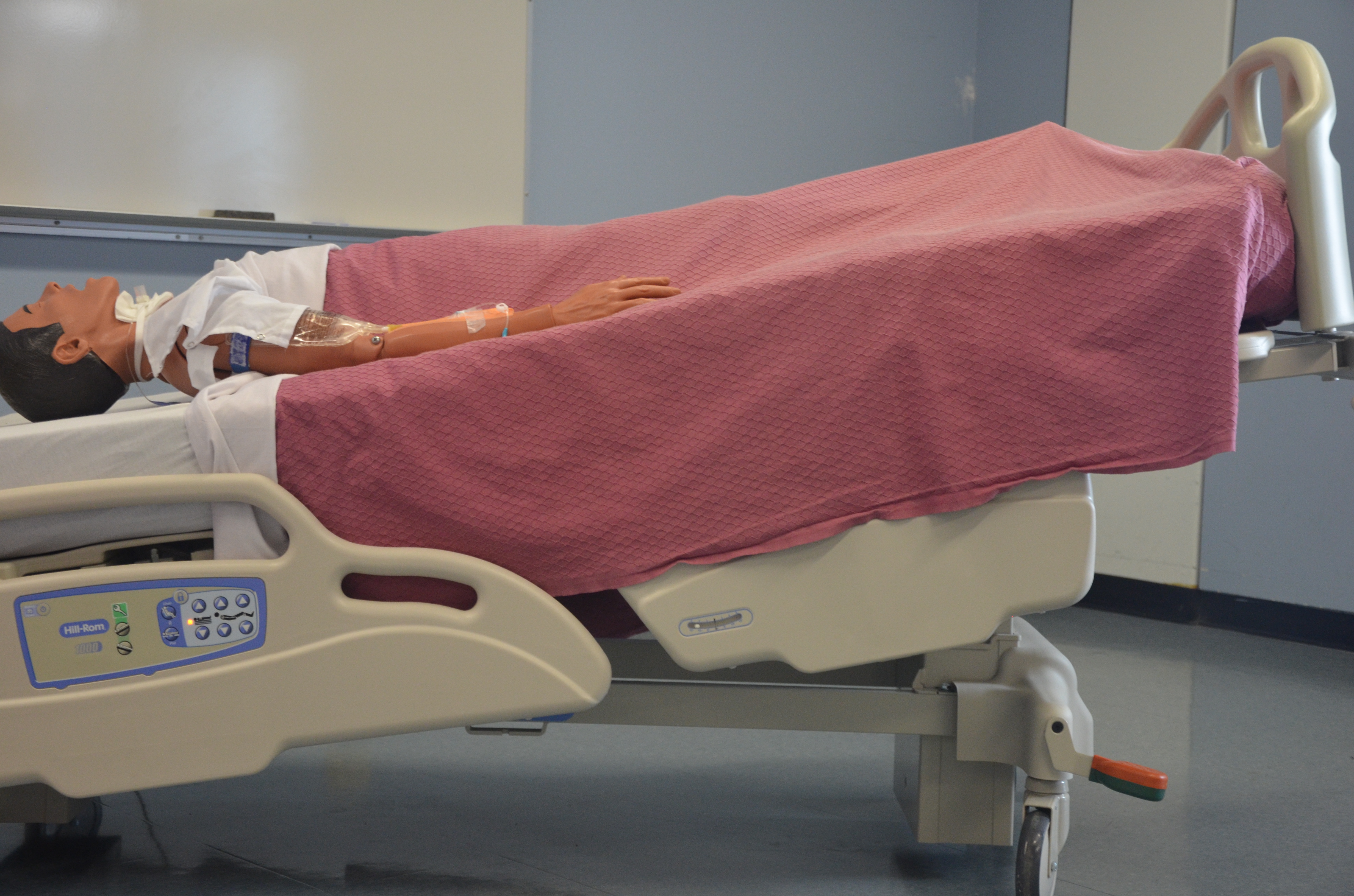
The solution? Placing a patient in the left lateral Trendelenburg position traps a venous air embolism in the right apex of the heart. By turning them on their left side and tilting their head down, you use gravity to make the right ventricle the highest point. The air bubble floats to the top of the right ventricle, trapping it safely out of the way of the pulmonary valve while the blood flows underneath it. It is pure, elegant physics saving a life!
Often, we don't run fluids continuously. Intermittent parenteral fluid therapy delivers specific volumes of fluids at scheduled intervals. For instance, intermittent parenteral fluid therapy is commonly utilized for the administration of intravenous antibiotics.
Whenever you are administering intermittent medications, you are delivering sudden boluses of foreign substances. Therefore, nurses must continuously monitor patients for signs of hypersensitivity reactions during intermittent intravenous medication administration.
Evaluating the Body's Hydraulic Balance
When we give IV fluids, we are manipulating the patient's total blood volume. You must monitor if they have too little (deficit) or too much (overload).
Systemic Fluid Volume Deficit (Dehydration/Hypovolemia)
Imagine a car engine running low on oil. The pump has to work frantically to maintain pressure.
- Tachycardia is an early physiological indicator of systemic fluid volume deficit. The heart beats faster to try and circulate the diminished fluid volume.
- Hypotension is a late physiological indicator of systemic fluid volume deficit. When the volume gets too low, the pump simply cannot maintain the pressure anymore, and the blood pressure crashes.
Fluid Volume Overload
What happens if you open the floodgates too wide? Fluid volume overload is a potential physiological complication of rapid intravenous fluid administration.
The vascular system gets over-pressurized, and fluid starts leaking out into places it shouldn't belong:
- Into the lungs: Bilateral pulmonary crackles are a clinical manifestation of intravenous fluid volume overload. (You are literally hearing air bubbling through fluid-filled alveoli).
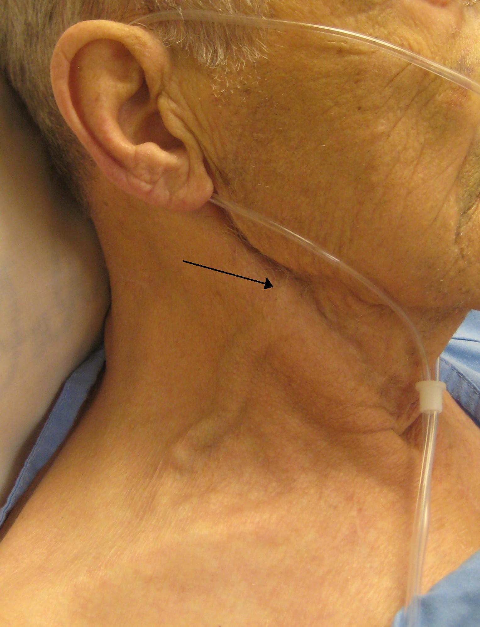
- Into the major vessels: Jugular vein distention (JVD) is a clinical manifestation of intravenous fluid volume overload. The heart can't process the volume fast enough, so it backs up into the neck veins.
- Into the tissues: Peripheral edema is a clinical manifestation of intravenous fluid volume overload. The hydrostatic pressure forces fluid down into the legs and feet.
How Do We Measure Success?
How do we know our hydration therapy is working? We use data.
- The Scale Never Lies: Daily weight measurement is the most sensitive indicator of changes in systemic fluid volume status. One liter of fluid weighs exactly one kilogram (2.2 lbs). If a patient gains 2 kg overnight, they didn't gain fat; they retained 2 liters of fluid!
- Kidney Output: The kidneys only make urine when they have enough fluid flow. Therefore, a therapeutic response to intermittent intravenous hydration is evidenced by an increase in hourly urine output. Remember this benchmark: Normal minimum hourly urine output for an adult is 30 milliliters per hour.
- Lab Values (Dilution is the Solution):
- Blood Urea Nitrogen (BUN) measures concentrated waste. When you hydrate a dry patient, you dilute the blood. Thus, a progressive decrease in blood urea nitrogen levels indicates an improvement in patient hydration status.
- Specific gravity measures how dark and concentrated the urine is. Well-hydrated kidneys make clear, watery urine. Therefore, a decrease in urine specific gravity indicates an improvement in patient hydration status.
Final Thoughts for the NCLEX
Whenever you look at an IV therapy question, ask yourself: Where is the fluid going? What forces are acting upon it? What is the body's natural physiological response?
Respect the catheter. Guard the insertion site from bacteria. Watch your infusion rates, and always, always listen to what the patient's hemodynamics—their heart rate, lung sounds, and urine output—are telling you. You aren't just passing meds; you are managing the river of life.