Potential for Alterations in Body Systems
Not sure you’re ready?
Take the ~3-minute readiness diagnostic and see where you stand.
Applied Physics and Physiology: The Nurse’s Guide to Defending the Body’s Systems
Welcome to the bedside! Let’s get something straight right from the beginning: nursing is not just about compassion. Compassion is your engine, but your daily practice? That is applied physics, fluid dynamics, and biological engineering.
When you stand in front of a client, you are looking at a brilliantly complex, highly pressurized system of pipes, pumps, and protective envelopes. When this system breaks down, we face three massive enemies: fluid going down the wrong pipe (aspiration), the breakdown of the protective envelope (tissue ischemia), and the failure of the delivery pipes (poor perfusion).
Let’s pull these concepts apart, see exactly how they work, and look at how we, as clinicians, manipulate physics and biology to keep our clients safe.
The human throat is a remarkably flawed design. We use the same central tube for eating a sandwich and breathing oxygen, separated only by a tiny flap of tissue called the epiglottis. When the swallowing mechanism loses its coordination—a condition known as dysphagia—it increases the risk of aspiration during oral intake.
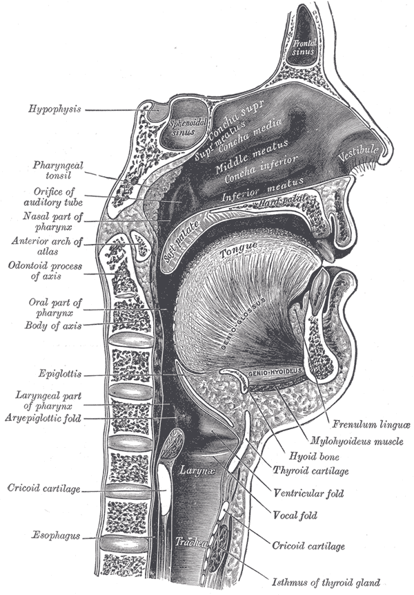
How do we know the system is vulnerable? First, check the reflexes. An impaired gag reflex indicates a high risk for pulmonary aspiration. If the body cannot automatically eject foreign material, the lungs are wide open to invasion. Furthermore, coughing during or immediately after swallowing is a clinical indicator of potential aspiration. It’s the body’s alarm bell ringing, telling you fluid is knocking on the vocal cords.
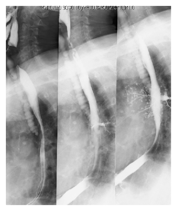
Engineering the Airway: Angles, Viscosity, and Geometry
If a client is at risk, we don't just hope for the best; we change the physical parameters of how they eat and drink.
-
Leveraging Gravity (Head of Bed): If you pour water on a flat table, it spreads everywhere. If you tilt the table, it flows exactly where you want it. Elevating the head of the bed utilizes gravity to reduce aspiration risk during enteral feeding. The golden rule? The recommended head of bed elevation to prevent aspiration during tube feeding is 30 to 45 degrees.
Crucial Warning: If you need to reposition the client, lay them flat, or change their linens, enteral feeding must be paused prior to lowering the head of the bed to prevent aspiration. Never flatten a client while the pump is running!
-
Altering Fluid Dynamics (Thickened Liquids): Water is fast. It splashes. For a poorly coordinated throat, fast is dangerous. Thickened liquids flow more slowly than thin liquids to allow better control during swallowing. By increasing the viscosity, we give the brain and muscles a few extra milliseconds to close the airway.
-
Changing the Anatomical Geometry: If a client has had a stroke and suffers from unilateral weakness, placing food on the unaffected side of the mouth decreases aspiration risk because that side still has the muscular control to manipulate the bolus. Additionally, we teach a mechanical trick: tucking the chin to the chest during swallowing narrows the airway opening to prevent aspiration. It forces the epiglottis downward, shielding the trachea.
-
Post-Meal Protocols: Gravity’s job doesn't end when the chewing stops. Educating clients with dysphagia to remain upright for at least 30 minutes after meals reduces postprandial aspiration risk by keeping residual food in the stomach where it belongs.
We also bring in the experts. A speech-language pathologist performs clinical swallowing evaluations to determine aspiration risk. They are the biomechanical engineers of the throat.
But what if, despite all our physics tricks, a micro-aspiration happens? We alter the biological environment. Providing thorough oral care reduces the bacterial load available for aspiration into the lungs. If they aspirate clean saliva, they get a cough. If they aspirate bacteria-laden plaque, they get fatal pneumonia.
Your skin is a tough, elastic envelope, but it has a fundamental weakness: it requires a constant flow of microscopic blood to stay alive.
When a client lies in bed, gravity traps their tissue between a heavy skeleton and a hard mattress. Prolonged mechanical pressure over bony prominences causes localized tissue ischemia and skin breakdown. No blood flow means no oxygen, which means cell death.
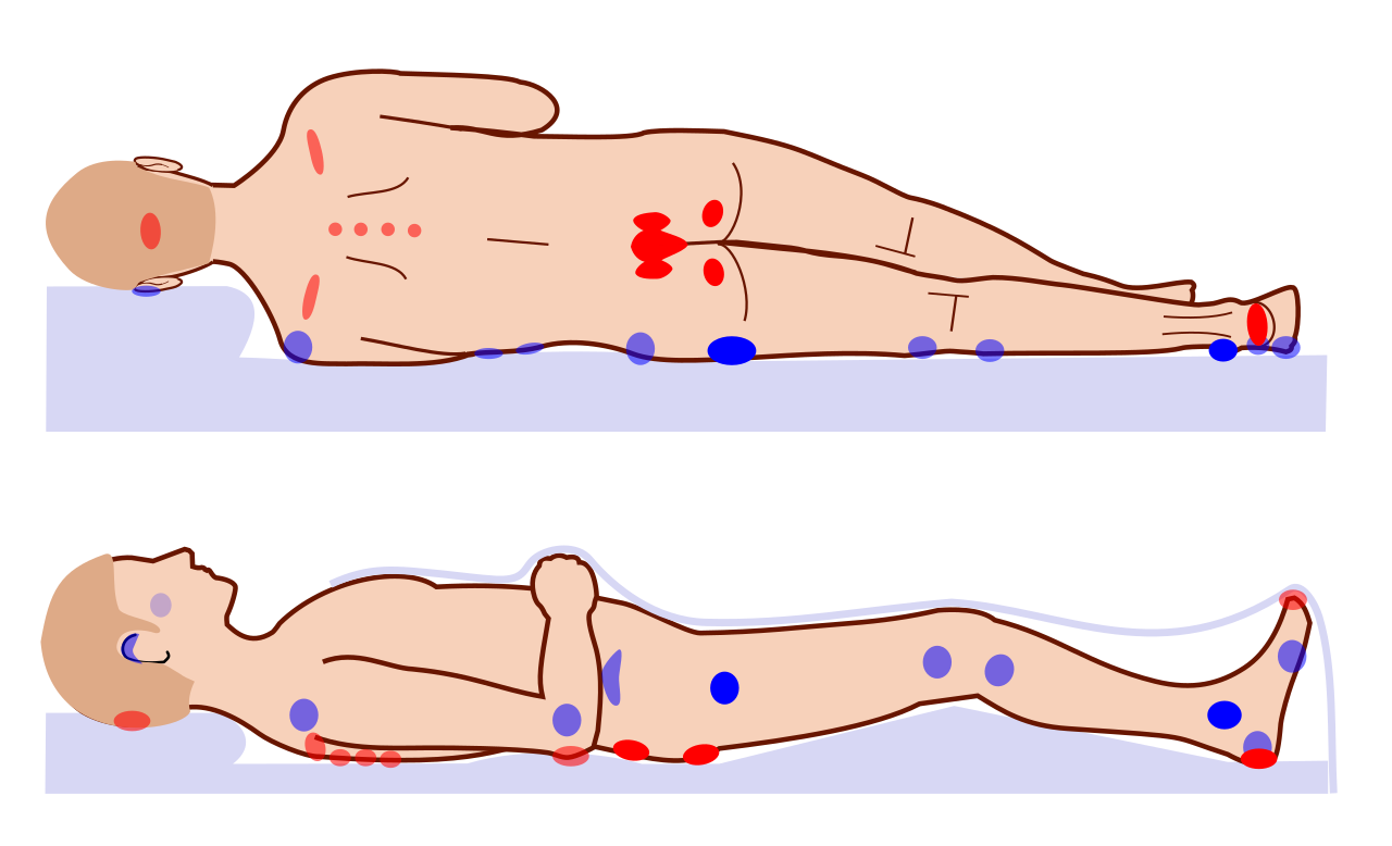
To predict this, we use the Braden Scale, which acts as our early warning radar. The Braden Scale assesses sensory perception, moisture, activity, mobility, nutrition, and friction and shear.
The Magic Number: A Braden Scale score of 18 or less indicates a risk for developing pressure injuries. If the score dips to 18 or below, your nursing interventions must immediately kick into high gear.
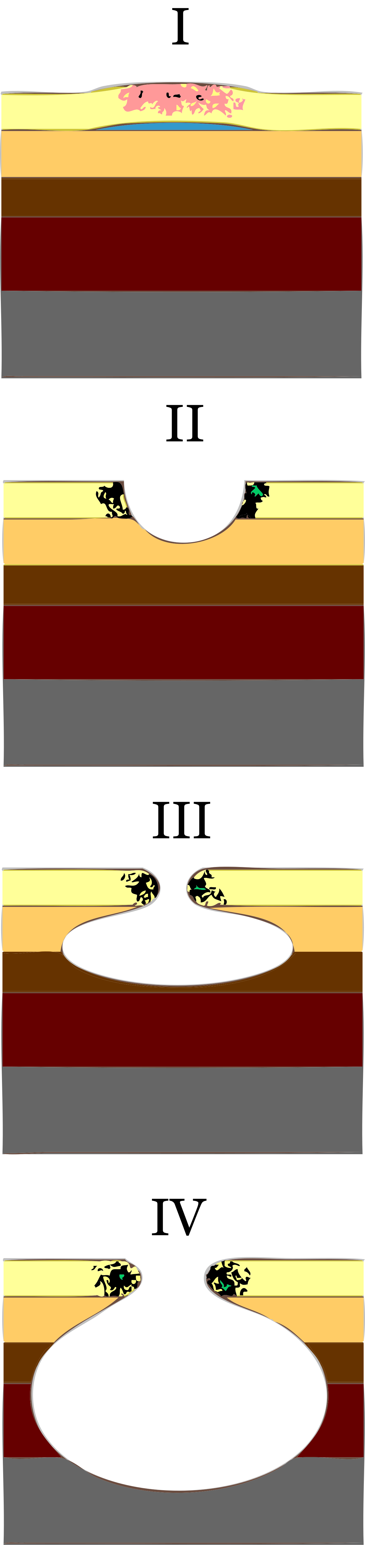
The Physics of Tissue Damage: Pressure, Friction, and Shear
Skin damage isn't just about downward pressure. It's about lateral forces.
- Friction damages the epidermal layer when skin rubs against support surfaces. Think of a rug burn. It’s a surface-level scrape.
- Shear force distorts deep tissue layers when the skeleton moves while the overlying skin remains stationary. Imagine sliding down in bed; your tailbone drags downward, but your skin sticks to the sheets. The blood vessels in between get stretched and snapped.
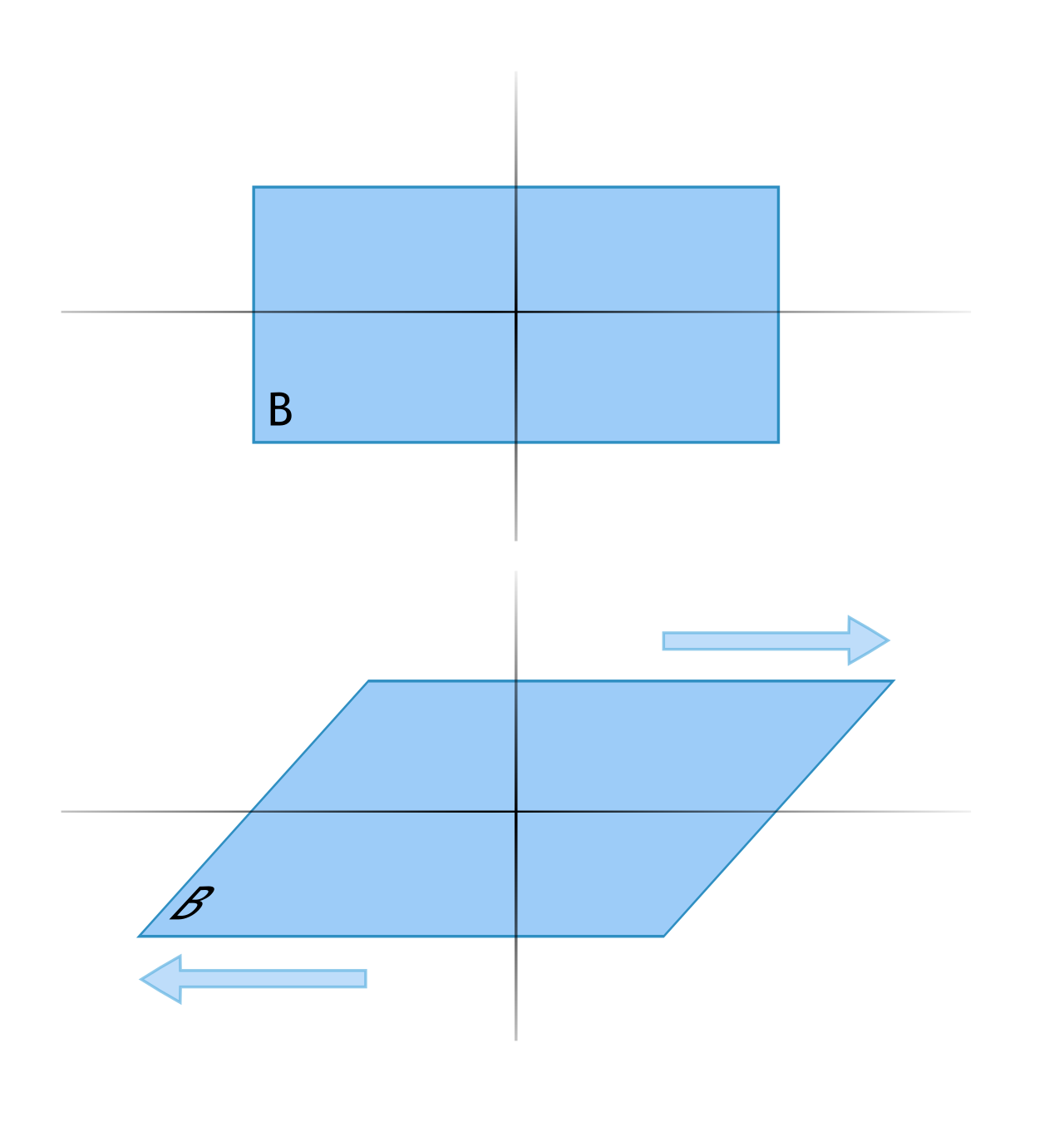
To combat shear, we face a fascinating nursing paradox. While we want the head of the bed high for breathing, maintaining the head of the bed at or below 30 degrees reduces shear forces on the sacrum. You must constantly balance the risk of aspiration against the risk of shear!
Defending the Skin: Mechanical and Chemical Strategies
- Offloading Pressure: Repositioning immobile clients every two hours relieves continuous pressure on vulnerable skin areas. For the most vulnerable bones, like the heel, we go further. Floating the heels off the bed completely eliminates pressure on the vulnerable calcaneus bone. We also use foam dressings [which] provide localized cushioning to reduce pressure over bony prominences.
- Managing Moisture: Wet skin is weak skin. Incontinence-associated dermatitis compromises skin integrity due to prolonged exposure to urine or feces. The fix? We make the skin waterproof. Applying a moisture barrier ointment protects perineal skin from breakdown related to incontinence.
- The Raw Materials of Repair: You can't rebuild a wall without bricks. Adequate dietary protein intake is necessary for maintaining tissue integrity and preventing skin breakdown. How do we measure this? We look at the blood. Serum albumin levels below 3.5 grams per deciliter indicate poor nutritional status and increased risk for skin breakdown. Finally, educating clients to consume a diet high in vitamin C supports collagen synthesis for skin maintenance.
The cardiovascular system is a closed loop of plumbing. You have supply lines (arteries) pushing oxygenated blood out, and return lines (veins) pulling deoxygenated blood back. When these fail, tissues starve.
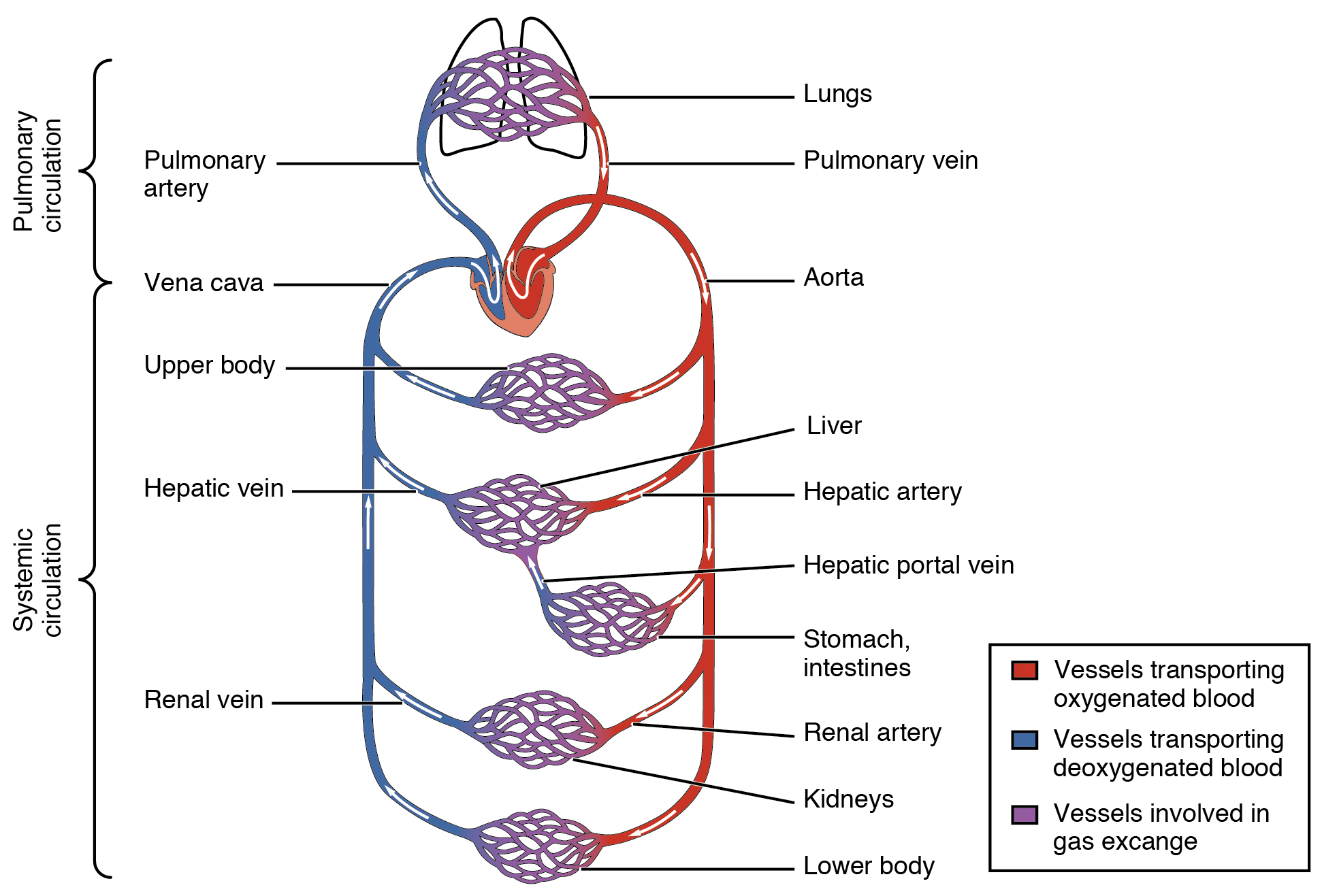
The Supply Lines: Arterial Perfusion
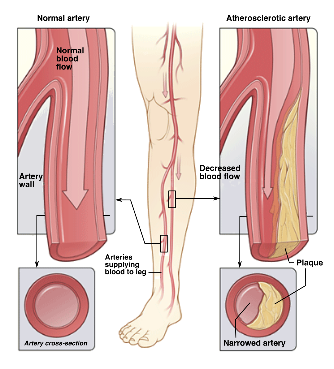
Arterial occlusion reduces oxygen delivery to distal tissues. If the pipe gets blocked, the tissue downstream immediately starts suffocating. We recognize this emergency by memorizing the six manifestations of acute arterial occlusion: pain, pallor, pulselessness, paresthesia, paralysis, and poikilothermia (inability to regulate temperature, meaning the limb turns cold).
A quick bedside test? Squeeze the fingernail. Capillary refill time greater than three seconds indicates delayed peripheral vascular perfusion.
To help our clients with Peripheral Arterial Disease (PAD), we educate them on lifestyle factors. Smoking causes vasoconstriction and significantly exacerbates peripheral arterial disease—it literally shrinks the pipes. We also empower the client: instructing clients to inspect their feet daily facilitates early detection of impaired tissue integrity, since they may lack the sensation to feel a wound forming.
The Return Lines: Venous Perfusion
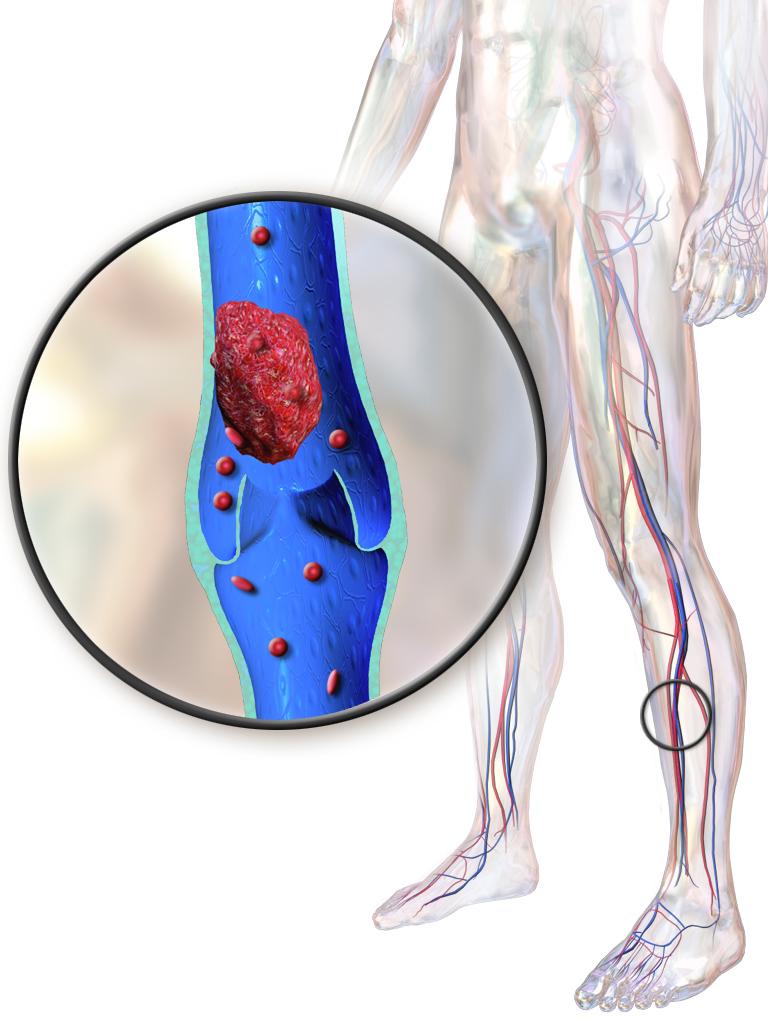
Veins are lazy. They operate at low pressure and rely entirely on skeletal muscle to squeeze blood back up to the heart. When a client lies in bed, the blood pools. Venous stasis increases the risk of deep vein thrombosis (DVT) formation.
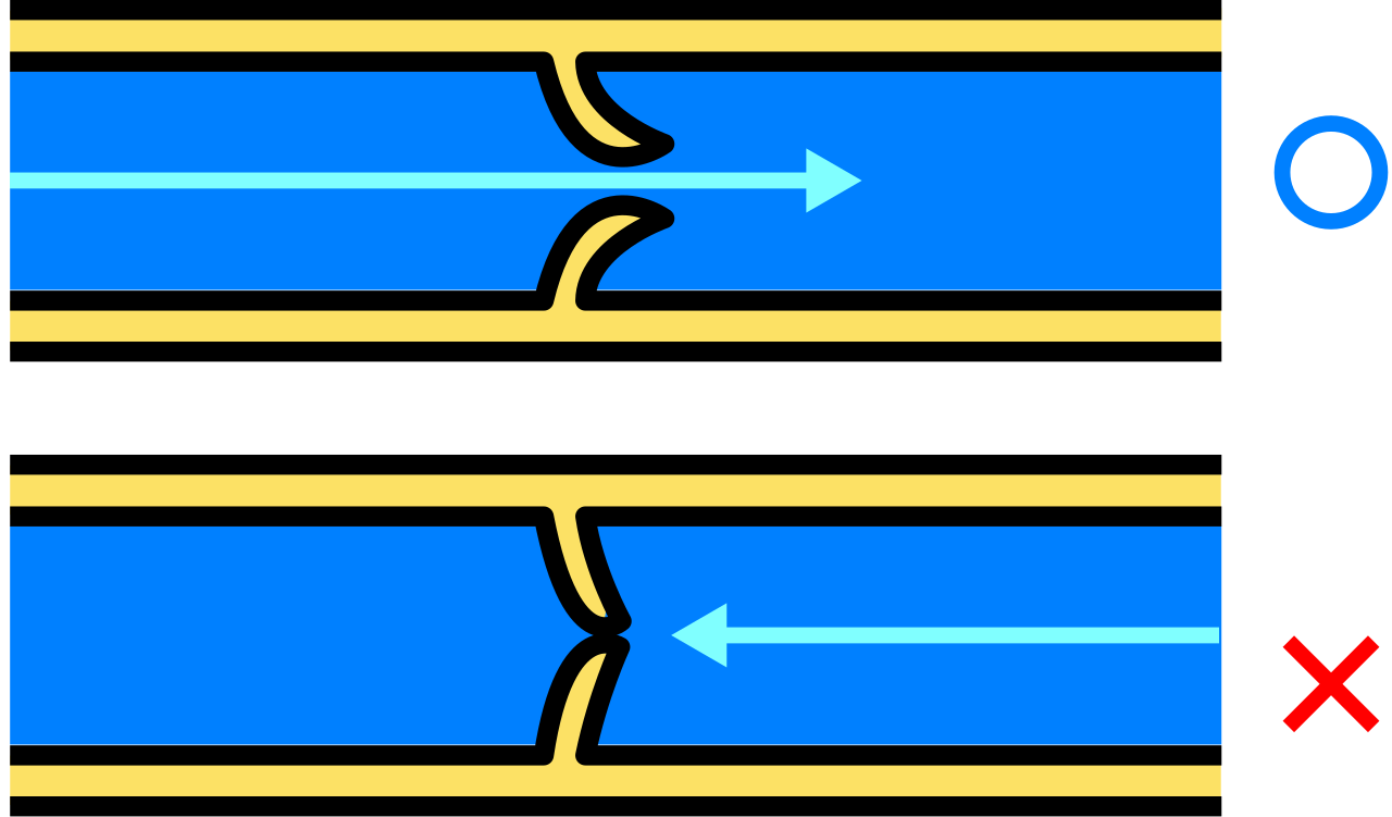
How do we keep the blood moving? We use mechanical compression and movement!
- Antiembolic stockings apply graduated compression to promote venous return and prevent venous stasis.
- Early ambulation enhances the calf muscle pump mechanism to improve venous return.
The SCD Rule of Thumb: Sequential compression devices (SCDs) must be applied prior to surgery to prevent deep vein thrombosis. However, if a clot has already formed, the rules change entirely! Sequential compression devices are contraindicated on an extremity with a confirmed deep vein thrombosis. Why? Because rhythmic squeezing will dislodge the clot, shooting it straight into the lungs as a pulmonary embolism.
Positional Plumbing: Arteries vs. Veins
Because arteries and veins do opposite jobs, we use gravity in opposite ways to help them. Let's compare:
| Vascular Issue | Nursing Intervention | The Physics/Rationale |
|---|---|---|
| Arterial Disease (PAD) | Dependent positioning of the legs (dangling legs downward). | Gravity enhances arterial blood flow to distal tissues by pulling blood down the supply pipes. |
| Venous Disease (PVD/DVT risk) | Elevating the lower extremities above heart level. | Gravity promotes venous return to the heart by draining the pooled blood back down the return pipes. |
General Vascular Rules: We also must avoid physical blockades. Educating clients to avoid crossing their legs at the knees prevents popliteal vessel compression. Furthermore, educating clients to wear loose-fitting clothing prevents mechanical constriction of peripheral blood vessels.
As a nurse, you are the chief systems engineer monitoring the body's dashboard. Your job isn't just to write down numbers; it is to spot trends. Comparing current vital signs against admission baseline measurements is essential for detecting early signs of systemic decompensation. You cannot know a client is crashing if you don't know where they started!
Postoperative clients require frequent vital sign monitoring to detect deviations from preoperative baseline values. Anesthesia, fluid shifts, and surgical trauma put massive stress on the system. If they are bleeding, their drains will show it. Increased drainage output from surgical drains warrants immediate comparison with prior output trends to detect potential hemorrhage.
Early vs. Late Signs of Collapse
When systemic perfusion begins to fail (think shock or massive blood loss), the body runs a strict triage protocol. It sacrifices the less important organs to save the core.
- The Brain Panics First: The brain demands massive amounts of oxygen. A decreased level of consciousness is an early clinical sign of impaired cerebral perfusion. If your client suddenly becomes restless, confused, or lethargic, check their perfusion immediately!
- The Heart Revs Up: To compensate for less blood volume or poor oxygenation, the heart simply pumps faster. Tachycardia often serves as an early compensatory mechanism for decreased cardiac output and poor tissue perfusion.
- The Kidneys Shut Down: The kidneys are the body's fluid regulators. If systemic blood pressure drops, the kidneys clamp down and stop making urine to save water. Therefore, a sudden decrease in urine output indicates a potential reduction in systemic vascular perfusion to the kidneys. Specifically, a urine output of less than 30 milliliters per hour indicates potential inadequate renal perfusion.
- Blood Pressure Drops Last: Many students wait for the blood pressure to fall to identify a problem. That is a dangerous mistake! Hypotension is a late indicator of compromised systemic vascular perfusion. By the time the blood pressure drops, the body’s compensatory mechanisms have entirely exhausted themselves.
Monitoring Fluid Volume
Finally, how do we track if the plumbing system is backing up with fluid? We don't just guess; we use a scale. Comparing daily weights is the most accurate method to monitor systemic fluid volume status changes. One liter of water weighs exactly 2.2 pounds (1 kilogram). Therefore, a sudden weight gain of greater than two pounds in 24 hours suggests fluid retention rather than actual tissue mass gain. No one eats enough calories to gain two pounds of pure fat in a day—that is pure, trapped water.
Summary
There you have it. You are not merely changing dressings or adjusting pillows. You are manipulating gravity to protect the trachea. You are distributing mechanical pressure to save the epidermis. You are optimizing fluid dynamics to perfuse the extremities, and you are constantly scanning the vital dashboard for the earliest signs of systemic failure. Embrace the physics of it all, understand why the body behaves the way it does, and you will be an exceptional, life-saving nurse.