Potential for Complications from Surgical Procedures and Health Alterations
Not sure you’re ready?
Take the ~3-minute readiness diagnostic and see where you stand.
The Physics of Recovery: Mastering Postoperative complications
Welcome to the postoperative world! Surgery, if you really think about it, is just highly controlled trauma. We intentionally alter the human body to fix a problem, but in doing so, we wage war against physics and physiology. We introduce anesthetic drugs that put vital reflexes to sleep, we open blood vessels and lose fluid, and we ask a human being in pain to lay perfectly still.
Nature, however, hates a vacuum and despises stagnant fluids. Our entire job as nurses is to anticipate the physics of what is about to go wrong and intercept it. Let's break down the beautiful, mechanical reality of postoperative complications and how we outsmart them.
When a patient is under general anesthesia and recovering from surgical trauma, the lungs face three massive mechanical threats: collapse, congestion, and invasion.
The Collapsing Balloons
Imagine the lungs as millions of microscopic, wet balloons (alveoli). To keep these balloons inflated, you need deep, continuous pressure. But what happens after surgery? It hurts to breathe! Alveolar collapse (atelectasis) occurs postoperatively due to shallow breathing from incision pain. Because the patient avoids taking deep breaths to protect their sore abdomen or chest, those tiny balloons simply deflate.
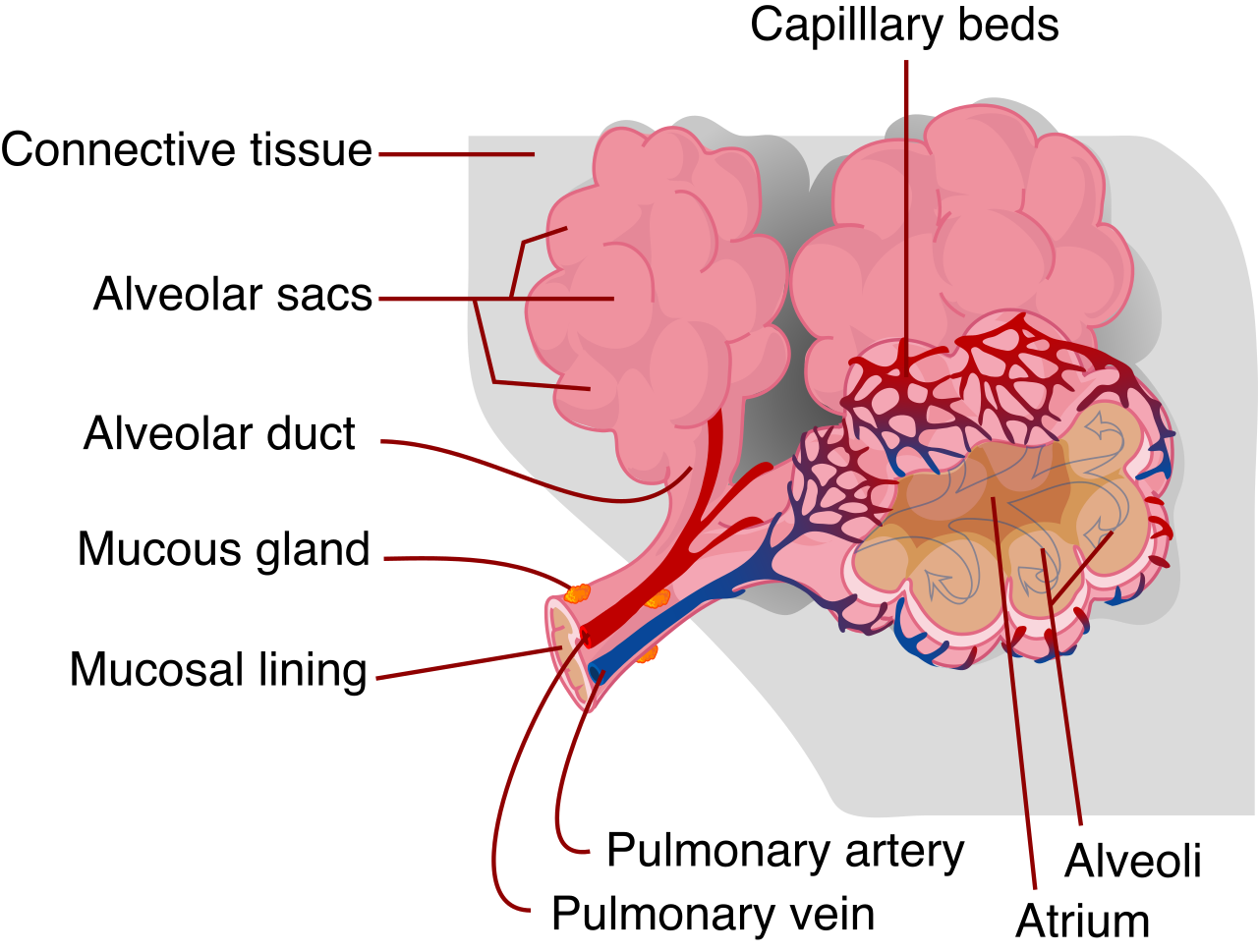
The Timing Principle: Watch the clock. Atelectasis typically manifests 24 to 48 hours after surgery. It is one of the earliest causes of postoperative fever.
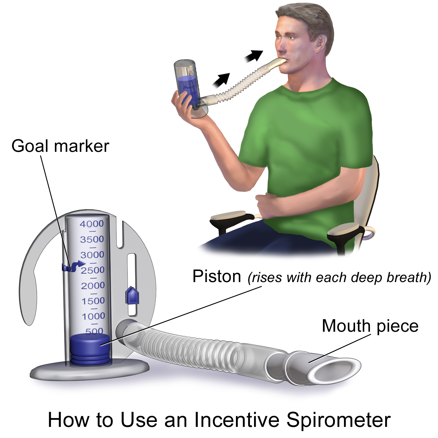
How do we fix this? We give them a mechanical goal. An incentive spirometer promotes sustained maximum inspiration to prevent atelectasis. We aren't just telling them to breathe; we are forcing them to pull a slow, sustained vacuum that physically pops those collapsed alveoli back open.
The Sleeping Brooms
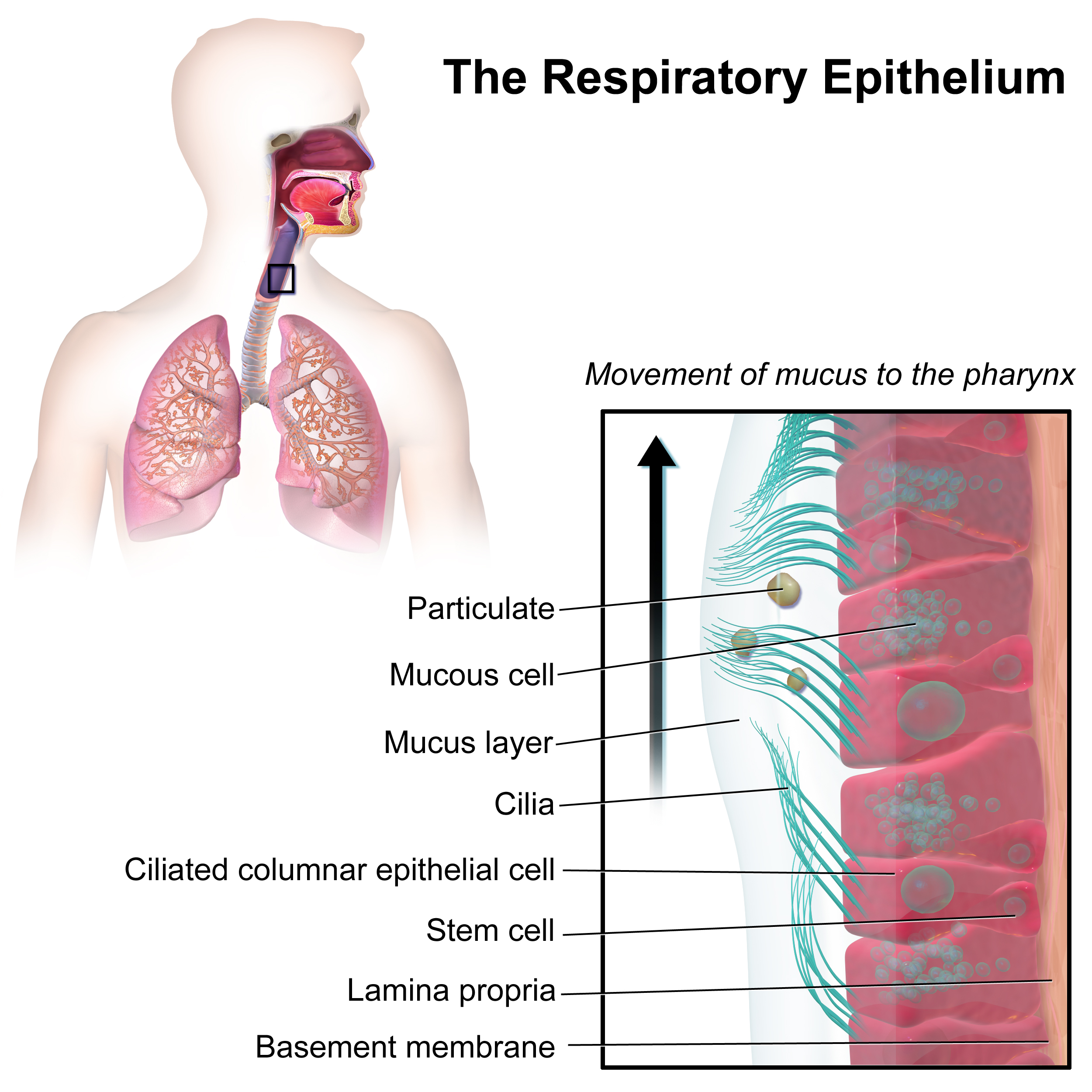
Your respiratory tract is lined with millions of tiny hair-like brooms called cilia, which constantly sweep mucus up and out of the lungs. But anesthetic drugs are powerful suppressants. General anesthesia depresses ciliary action in the respiratory tract. When the brooms stop sweeping, the dust settles. Depressed ciliary action increases the risk of retained respiratory secretions.
Why is this dangerous? Because the lungs are warm, dark, and moist. Retained respiratory secretions create an environment for bacterial growth leading to pneumonia.
The Danger of Gravity: Aspiration
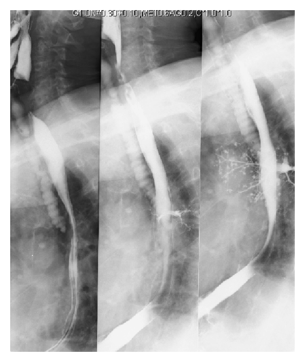
Your trachea and your esophagus are right next to each other. One leads to the lungs, the other to the stomach. Aspiration occurs when gastric contents or secretions enter the tracheobronchial tree. This is a nightmare scenario. Gastric acid destroys delicate lung tissue instantly.
We combat this before surgery even begins. Why do we make patients NPO? Because preoperative fasting reduces the volume and acidity of gastric contents, minimizing the ammunition available if the patient vomits. After surgery, we monitor their neurological recovery. An absent gag reflex indicates a high risk for aspiration, because the body's natural trapdoor mechanism isn't online yet.
If a patient is unconscious, gravity is either your best friend or your worst enemy.
- Placing an unconscious postoperative client in a lateral position facilitates the drainage of oral secretions. Gravity pulls the drool out of the mouth, not down the windpipe.
- Furthermore, placing an unconscious postoperative client in a lateral position prevents the tongue from obstructing the airway.
- Once they are awake but still groggy, elevating the head of the bed decreases the gravitational flow of gastric contents into the esophagus.
How do we know if aspiration happened?
- The Pulse Ox Drop: A sudden drop in oxygen saturation after an episode of emesis strongly suggests an aspiration event. The alveoli are immediately flooded, blocking gas exchange.
- The Anatomical Clue: Because the right mainstem bronchus is wider and steeper than the left, fluids take the path of least resistance. Therefore, decreased breath sounds in the right lower lung lobe indicate potential aspiration of gastric contents.
- The Follow-Up: We continuously rely on pulse oximetry readings to evaluate systemic oxygenation status following interventions to prevent aspiration.
The cardiovascular system is a closed loop of plumbing. It requires volume, and it requires movement. Surgery disrupts both.
The Leaking Tank: Hypovolemia
Blood loss is inevitable in the OR. Surgical blood loss decreases circulating intravascular volume leading to decreased cardiac output. The heart has less blood to pump, so the system pressure drops.
The body instantly recognizes this drop and panics. It activates its emergency override system. Hypovolemic shock triggers the sympathetic nervous system to increase the heart rate, attempting to pump whatever volume is left much faster. Thus, tachycardia is an early clinical sign of hypovolemic shock.
Simultaneously, the body makes a ruthless decision: save the brain and heart, sacrifice the fingers and toes. Hypovolemia triggers peripheral vasoconstriction to shunt blood to vital organs.
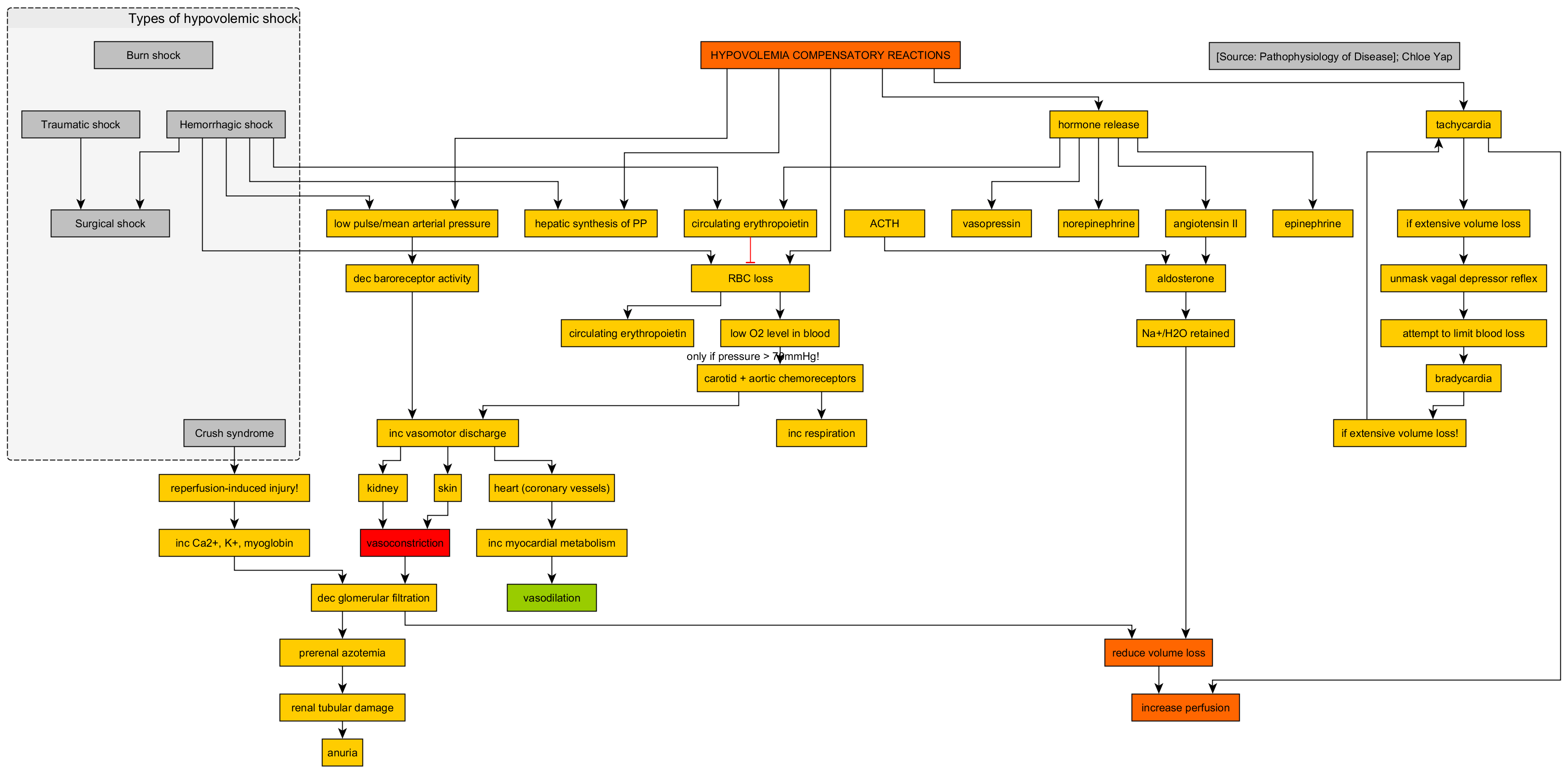
Clinical Trap: Do not wait for the blood pressure to drop to intervene! Hypotension is a late clinical sign of hypovolemic shock. By the time the blood pressure crashes, the body's compensatory mechanisms have already failed.
How do we monitor the internal organs during this? We look at the kidneys! The kidneys require massive blood flow to make urine. Urine output less than 30 milliliters per hour indicates decreased renal perfusion, telling you definitively that the cardiac output is critically low.
The Danger of Stagnation: DVT and PE
Blood is designed to keep moving. If it stops, it clots. Venous stasis from postoperative immobility increases the risk of deep vein thrombosis (DVT).
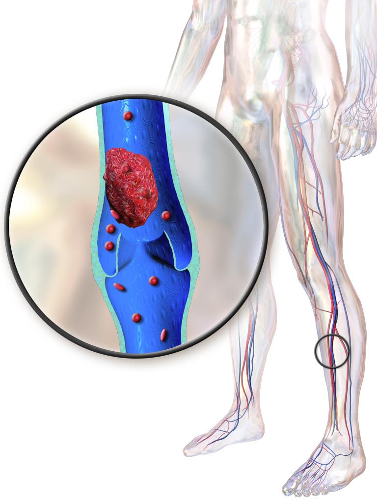
How do we keep the river flowing when the patient is in bed?
- The Calf Pump: Walking is the ultimate cure. Early ambulation stimulates the calf muscle pump to enhance venous return.
- The Vacuum Effect: Remember those deep breaths for the lungs? They help the heart, too! Deep breathing creates negative intrathoracic pressure to draw venous blood toward the heart.
- Mechanical Assistance: We use gear. Anti-embolism stockings provide graduated compression to reduce venous pooling in the legs. We also use machines: Sequential compression devices promote venous return by applying intermittent pressure to the lower extremities. (Just remember, sequential compression devices require periodic removal to assess underlying skin integrity. You cannot leave them on blindly.)
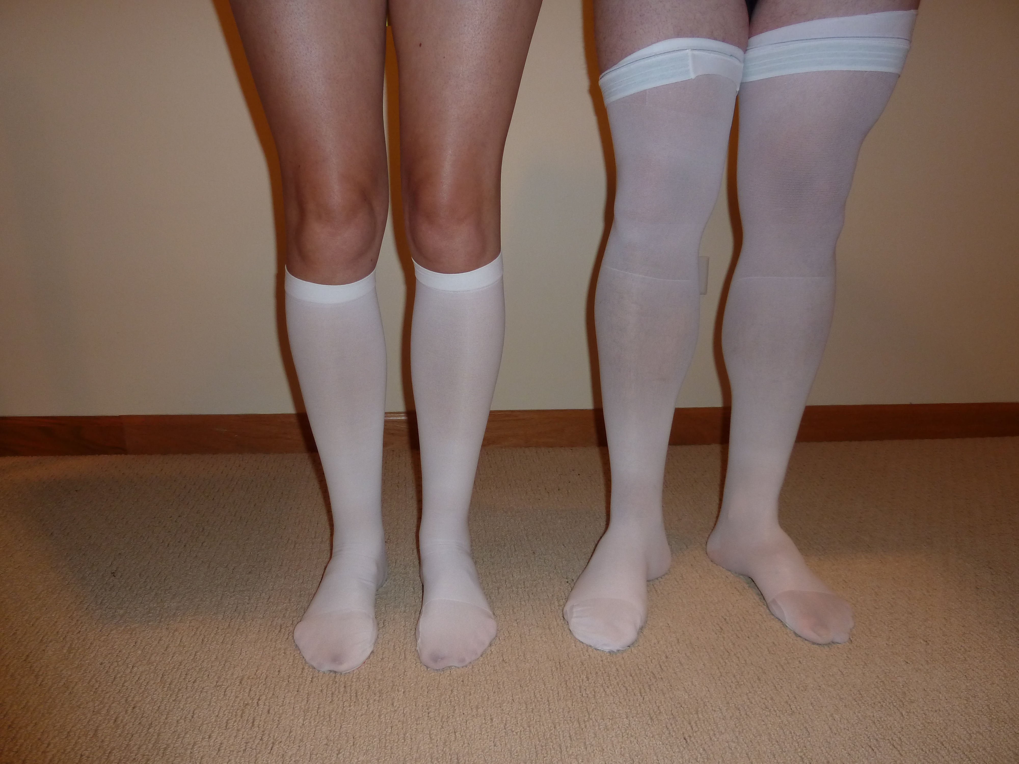
Watch out for accidental tourniquets! You must avoid anything that pinches off the blood flow. Rolling the tops of anti-embolism stockings creates a tourniquet effect, and a tourniquet effect on the lower extremities impairs venous return and increases thrombosis risk. Likewise, placing a pillow directly under the knees creates pressure on the popliteal vein, and this localized pressure on the popliteal vein from pillows or bed adjustments impedes venous return. Keep their legs straight and elevated, not bent over a pillow!
Monitoring and Meds: We use measurement of calf circumference to evaluate the presence of lower extremity edema related to venous stasis. If a clot has already formed, redness and unilateral warmth in the lower extremity indicate a high probability of deep vein thrombosis.
To prevent clots chemically, prophylactic subcutaneous administration of enoxaparin prevents clot formation by inhibiting coagulation Factor Xa.
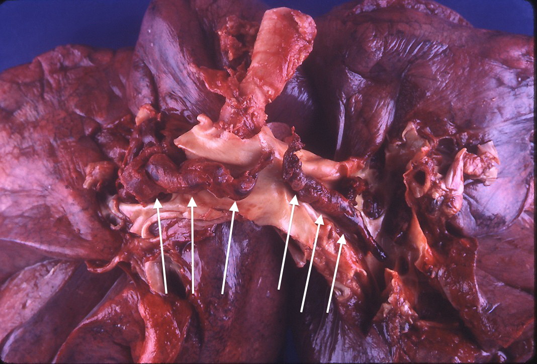
Why do we care so much about a clot in the leg? Because of where it travels. A pulmonary embolism occurs when a thrombus dislodges and occludes the pulmonary artery. This blocks blood from reaching the lungs to get oxygenated. A pulmonary embolism is a potentially fatal complication of deep vein thrombosis.
Surgery tells the brain to shut down non-essential services. The bowel and the bladder go to sleep.
The Lazy Bowel (Paralytic Ileus)
The intestines are dramatic. If you touch them, they quit working. Surgical manipulation of the bowel causes temporary cessation of intestinal peristalsis. This traffic jam is called a paralytic ileus.
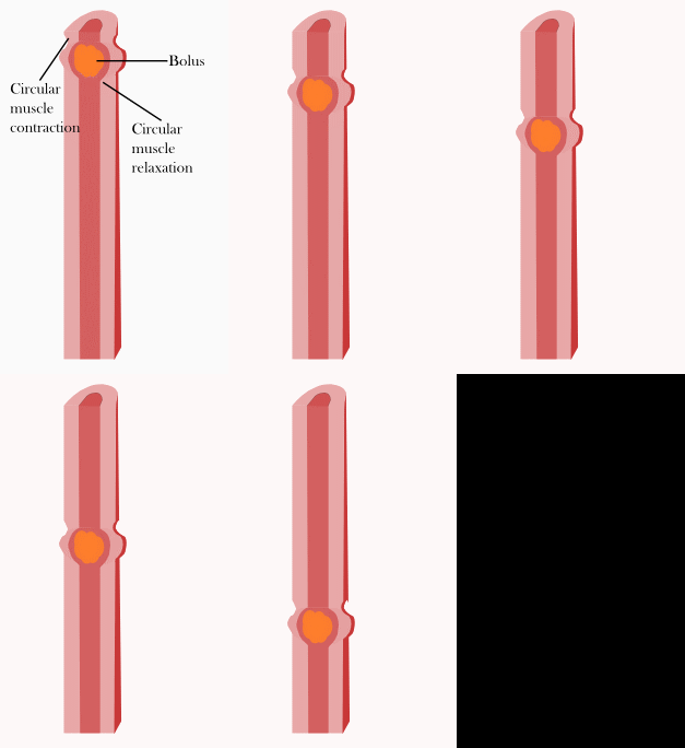
How do you know it's happening? Paralytic ileus presents with an absence of bowel sounds.
How do we fix it? We don't just wait; we move! Early ambulation stimulates gastrointestinal motility to prevent paralytic ileus. Walking physically jostles the abdomen and signals the autonomic nervous system to wake the gut up. You will know you've won because the return of flatus indicates the resolution of paralytic ileus. When they fart, the pipes are open!
The Stubborn Bladder (Urinary Retention)
Just like the bowel, the bladder is paralyzed by the drugs. Anesthesia suppresses the micturition reflex (the brain's signal to pee) and anesthesia decreases bladder tone (the muscle's ability to squeeze).
By definition, postoperative urinary retention is the inability to void 6 to 8 hours after surgery. The kidneys are making urine, but the bladder is holding it hostage. If you suspect this, use your hands: Bladder distention presents as a palpable mass above the symphysis pubis.
We've cut the skin, and now we rely on sutures, staples, and the body's collagen to hold it together. But wounds are under immense mechanical stress.
Dehiscence: The Seams Pull Apart
Wound dehiscence is the separation of surgical incision layers. It doesn't usually happen immediately; it happens when the initial inflammatory phase gives way to early healing, but the tissue isn't strong enough yet. Wound dehiscence typically occurs 5 to 10 days after surgery.
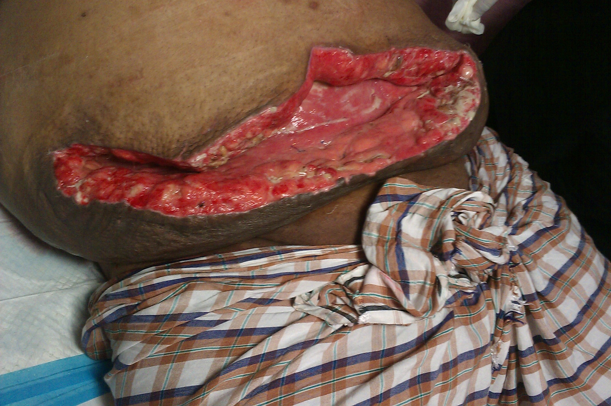
What causes it? Pure mechanical force. Increased intra-abdominal pressure from coughing increases the risk of wound dehiscence. To fight this physics problem, we use counter-pressure. Splinting an abdominal incision with a pillow during coughing reduces mechanical stress on the wound, absorbing the shockwave of the cough.
Evisceration: The Nightmare Scenario
If the layers separate entirely, what's inside comes out. Wound evisceration is the protrusion of internal organs through an open surgical incision.
🚨 CRITICAL RULE: Wound evisceration is a medical emergency requiring immediate surgical intervention. Do not leave the patient. Do not try to shove the organs back in!
Your immediate goal is to keep the exposed bowel alive until the surgeon arrives. Covering an eviscerated wound with sterile saline-soaked dressings prevents tissue necrosis. It keeps the organs wet, viable, and protected from airborne pathogens.
The Invisible Enemy: Infection
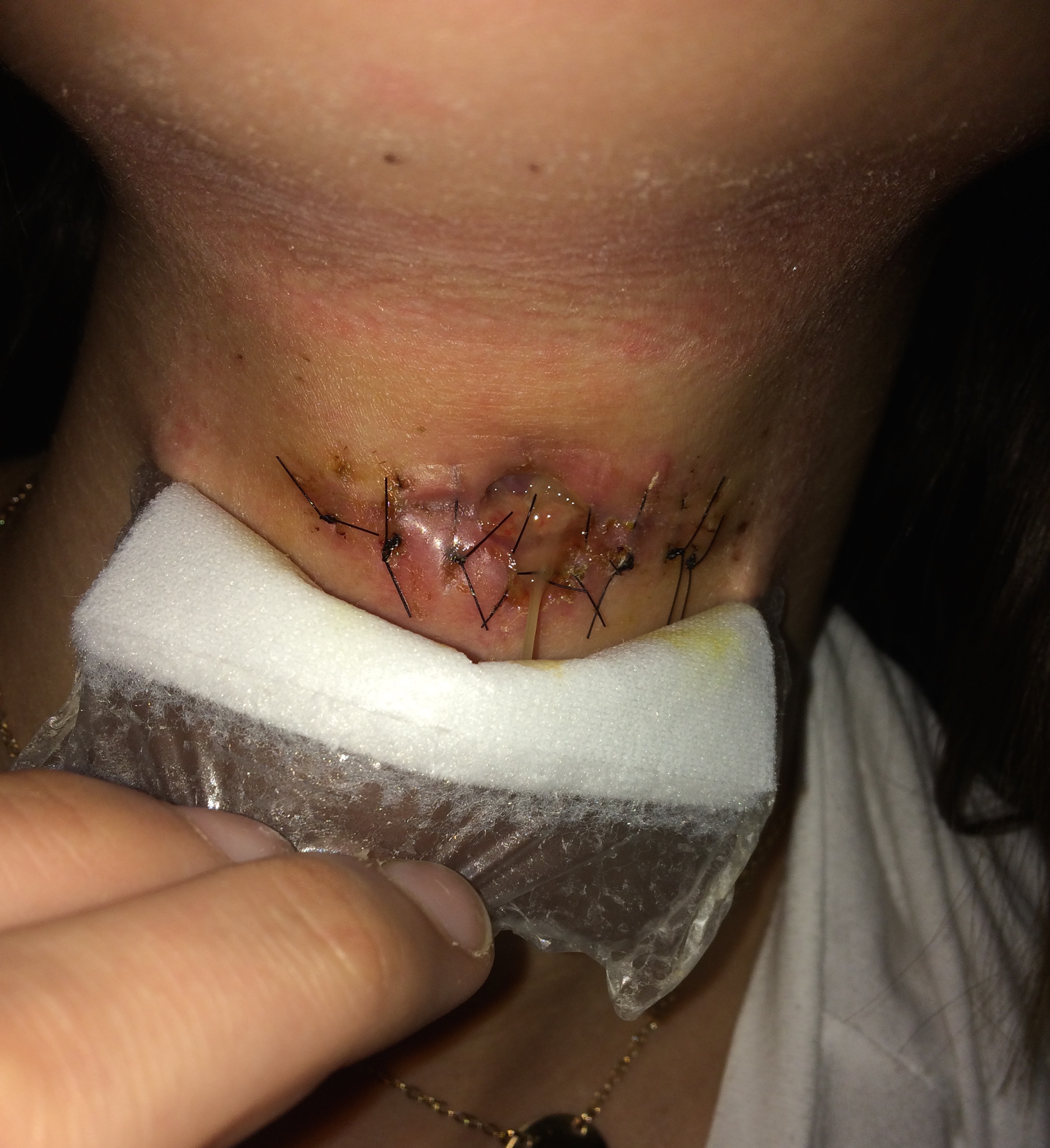
Finally, we must watch for invading armies. Normal postoperative inflammation peaks early, but true infection takes time to brew. Surgical site infections typically manifest 3 to 5 days postoperatively. Watch for purulent drainage, spiking fevers, and red, angry wound edges.
Final Thoughts for the NCLEX
When you look at a postoperative patient, don't just see a list of tasks. See the mechanics. See the collapsed balloons waiting for a spirometer. See the stagnant rivers of blood waiting for the calf pump to fire. See the asleep bowels waiting for a walk down the hallway.
Apply the physics, outsmart the complications, and you will keep your patient alive. Happy studying!