System-Specific Assessments
Not sure you’re ready?
Take the ~3-minute readiness diagnostic and see where you stand.
Welcome to the magnificent machinery of the human body! Nursing is not about simply memorizing a list of arbitrary tasks; it is the fine art of clinical detection. When you walk into a patient's room, you aren't just looking at a person—you are observing a complex, interconnected universe of pumps, pipes, electrical circuits, and gas-exchange bellows.
Our job is to detect the subtle whispers of a system failing before it starts shouting. Early identification of patient deterioration requires prompt notification of the primary healthcare provider. But before you can call for help, you have to know exactly what you are looking at.
Let’s break down the definitive, systems-based approach to patient assessment.
The brain is the body’s master computer, tightly sealed inside a bony vault. Because we can't look at it directly, we have to evaluate its outputs: movement, speech, and reflexes.
The Glasgow Coma Scale (GCS)
When assessing consciousness, we use the Glasgow Coma Scale, which elegantly evaluates a patient's eye opening response, verbal response, and motor response.
- A Glasgow Coma Scale score of 15 indicates a fully alert and oriented patient. Everything is firing beautifully.
- A Glasgow Coma Scale score of 8 or less indicates severe brain injury or coma. (Remember the rhyme: “Less than eight, intubate!”)
Windows to the Brain: Pupils
The eyes are literal extensions of the brain. When we shine a light into them, we are testing the circuitry of the optic and oculomotor nerves.
- Pupil reactivity to light indicates the functional intactness of the oculomotor nerve (Cranial Nerve III).
- We use the acronym PERRLA, which stands for Pupils Equal, Round, Reactive to Light, and Accommodation.
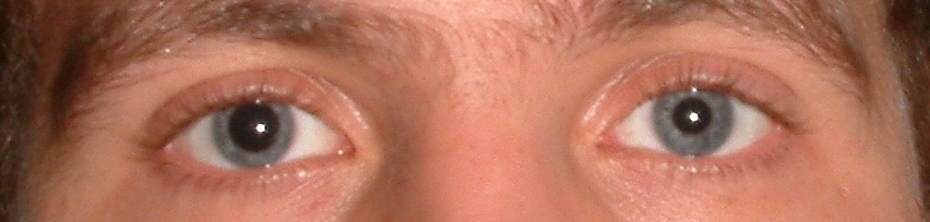
- What if the pressure inside the brain is dangerously high? The swelling brain will physically squish the oculomotor nerve. Because of this, asymmetrical pupils can indicate a severe brain injury or herniation. This is an absolute emergency.
Recognizing Neurological Crises
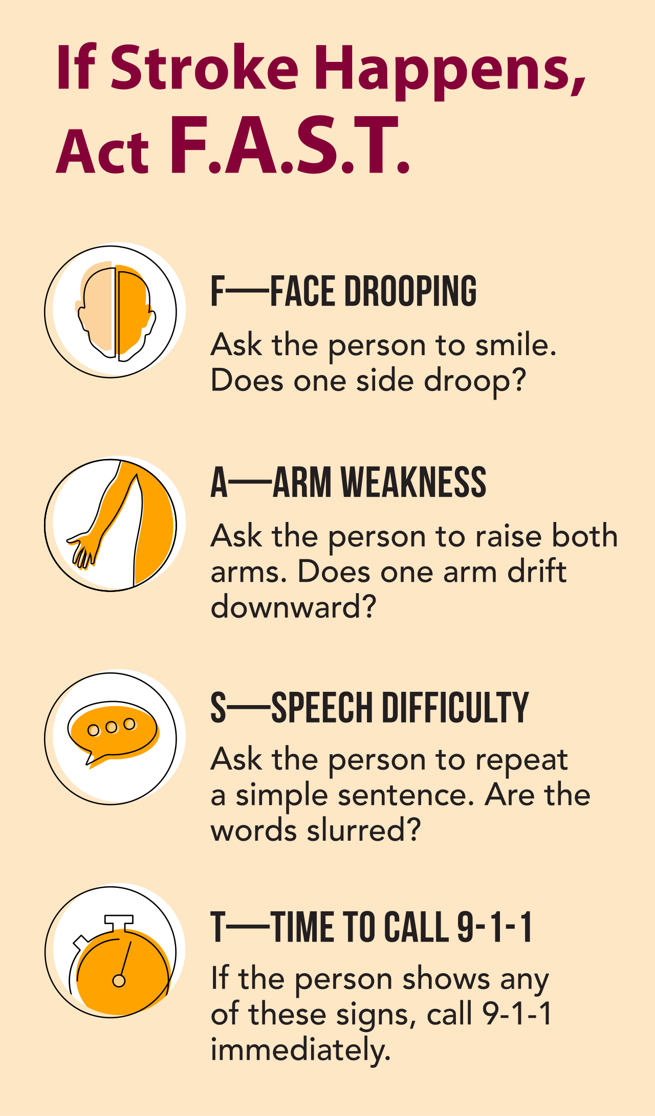
Sometimes, the hardware is damaged focally. Unilateral weakness during a motor assessment strongly suggests a focal neurological lesion like a stroke. To assess this quickly in the field, we use the Cincinnati Prehospital Stroke Scale, which screens for three critical failures:
- The presence of facial droop.
- The presence of arm drift.
- Abnormal speech patterns.
If the brain begins swelling globally, it tries to fight for its own blood supply. This fight manifests as Cushing's triad, which consists of severe bradycardia, irregular respirations, and a widening pulse pressure.
Crucial Warning: Do not confuse Cushing's triad with shock! Shock causes a fast heart rate. Cushing's triad is a late and life-threatening sign of increased intracranial pressure. If you see this, the brain is quite literally running out of room.
Behavioral and Sedation Scales
Sometimes the issue isn't structural damage, but a software glitch caused by chemicals or lack thereof.
- We use the CIWA-Ar clinical scale to evaluate the severity of alcohol withdrawal symptoms.
- In the intensive care unit, we use the Richmond Agitation-Sedation Scale (RASS) to precisely assess the level of sedation and agitation in critically ill patients.
Think of the heart as a mechanical pump and the blood vessels as plumbing. If the pump fails, or the pipes leak, tissue dies.
Assessing the Pump
First, we listen to the pump itself. An apical pulse is assessed by placing the stethoscope at the fifth intercostal space at the left midclavicular line. This is the point of maximal impulse. Because medications like digoxin directly affect the heart's electrical timing, the apical pulse must be auscultated for one full minute prior to administering digitalis medications.
What if the pump is weakening? The presence of an S3 heart sound in an older adult is an early auscultatory sign of heart failure. It sounds like a sloppy, sloshing lub-dub-DUB, indicating blood is crashing into an already overfilled ventricle.
Assessing the Pipes (Perfusion)
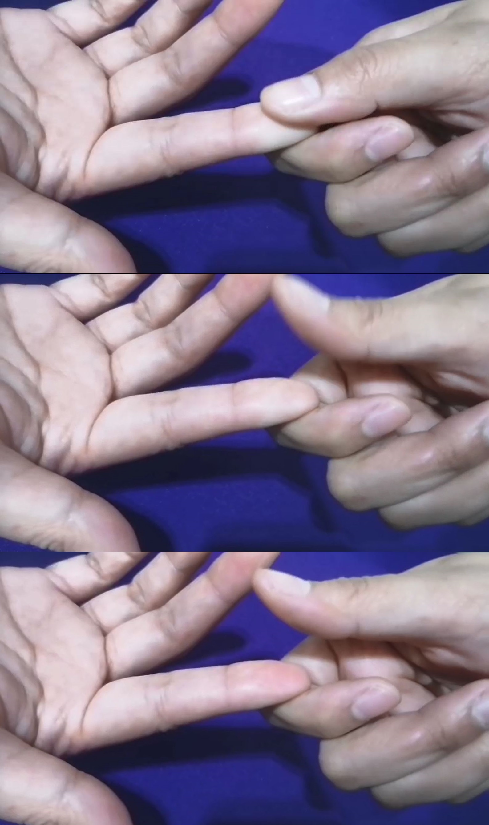
We must ensure blood is reaching the very tips of the fingers and toes. We test this through capillary refill time, where the normal range is less than 3 seconds. If you press a nail bed and the color takes forever to return, a capillary refill time greater than 3 seconds indicates poor peripheral tissue perfusion.
We also feel the pressure waves of the blood—the pulses. Peripheral pulses are graded on a scale:
- 0 indicates an absent pulse.
- 2+ indicates a normal and palpable pulse.
- 3+ or 4+ indicates a bounding pulse (think fluid overload or extreme anxiety).
Fluid Overload and Heart Failure
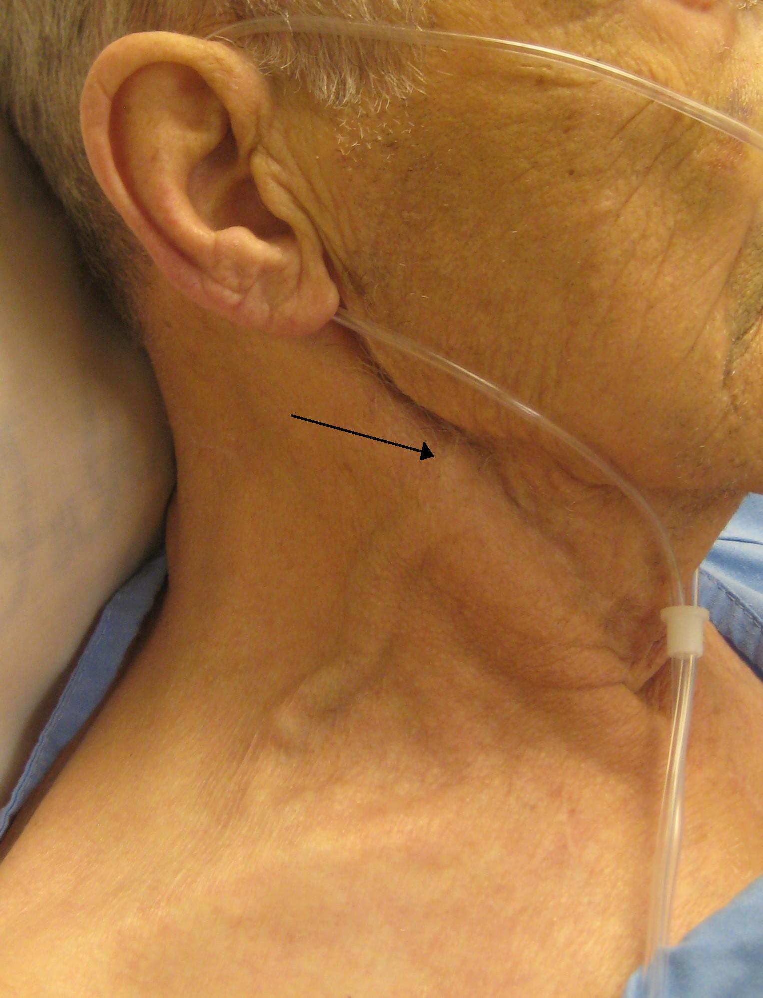
If the right side of the heart is failing, blood backs up into the venous system. Imagine a traffic jam on the highway leading to the heart. This backup becomes visible in the neck. Jugular venous distention (JVD) is properly assessed with the patient positioned supine at a 30 to 45-degree angle. If the neck veins bulge at this angle, the presence of jugular venous distention indicates right-sided heart failure or systemic fluid volume overload.
Gravity will pull that extra fluid down into the tissues, causing edema. Peripheral pitting edema is graded on a scale of 1+ to 4+:
- A 1+ pitting edema describes a 2-millimeter skin depression that disappears rapidly.
- A 4+ pitting edema describes an 8-millimeter skin depression that persists for a long time.
If a patient is bedridden, gravity pulls fluid to their backside, not their feet. Therefore, pitting edema over the sacrum in a bedridden patient indicates systemic fluid volume overload.
Deep Vein Thrombosis (DVT)
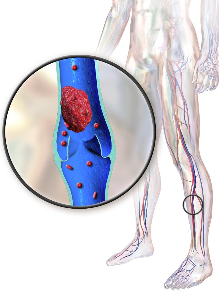
Sometimes the plumbing gets clogged. A deep vein thrombosis is assessed by examining the lower extremities for unilateral swelling, redness, and warmth. Note well: In the old days, nurses flexed the foot to look for calf pain. Stop doing this! Homan's sign is no longer recommended for deep vein thrombosis assessment due to low diagnostic reliability (and the risk of dislodging the clot).
Shock and Kidney Perfusion
If the body is bleeding out or losing fluid, what is the very first thing it does? It panics and pumps faster. Tachycardia is often the first physiological sign of clinical deterioration or shock.
If the pump is failing or volume is low, the kidneys stop making urine to save water. Therefore, a sudden decrease in urine output below 30 milliliters per hour indicates potential decreased cardiac output OR potential acute kidney impairment. Watch that catheter bag; it is a real-time window into cardiac output!
Oxygen is the fuel that keeps the human machine running. Without it, the system crashes rapidly.
The Earliest Sign of Hypoxia
Long before a patient turns blue, their brain will panic from the lack of oxygen. Restlessness or altered mental status is often the earliest behavioral sign of hypoxia.
Respiratory Mechanics
- The normal adult resting respiratory rate is 12 to 20 breaths per minute.
- Bradypnea is defined as a resting respiratory rate of less than 12 breaths per minute.
- Tachypnea is defined as a resting respiratory rate of greater than 20 breaths per minute.
If a patient is struggling to breathe, do not wait for an order to help them mechanically. Elevating the head of the bed is a primary immediate intervention for acute respiratory distress. Let gravity pull the diaphragm down so the lungs can expand!
Auscultating Lung Sounds
Listen to the air moving through the bellows. Abnormal acoustics tell you exactly what is physically wrong inside:
- Crackles auscultated in the lungs indicate the presence of fluid in the alveoli or small airways. (Imagine blowing air through a straw into a glass of milk—that popping sound is a crackle).
- Wheezing auscultated in the lungs indicates narrowing or obstruction of the lower airways. (Like pinching the neck of a balloon as air escapes, making a musical whistle).
- Stridor is a high-pitched respiratory sound indicating dangerous upper airway obstruction. This is a medical emergency. The windpipe is closing!
Oxygen Saturation & Chronic Hypoxia
We measure oxygen using light. Pulse oximetry measures the percentage of arterial hemoglobin saturated with oxygen. However, it relies on blood flow to the fingertips.
- Clinical hypothermia causes severe peripheral vasoconstriction.
- This peripheral vasoconstriction can lead to falsely low pulse oximetry readings on the extremities. If the hands are freezing, the machine will lie to you.
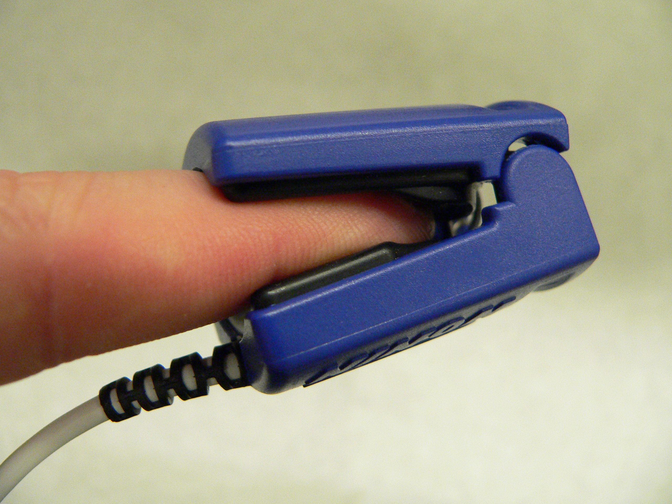
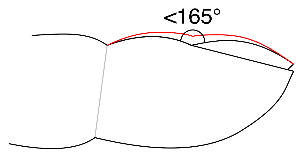
If hypoxia isn't acute but lasts for years (like in COPD), the body structurally changes the fingers to grow more capillaries. Clubbing of the fingers is an assessment finding characterized by an increased angle between the nail base and the skin. Seeing this finger clubbing indicates the presence of chronic systemic tissue hypoxia.
Preventing Pulmonary Collapse
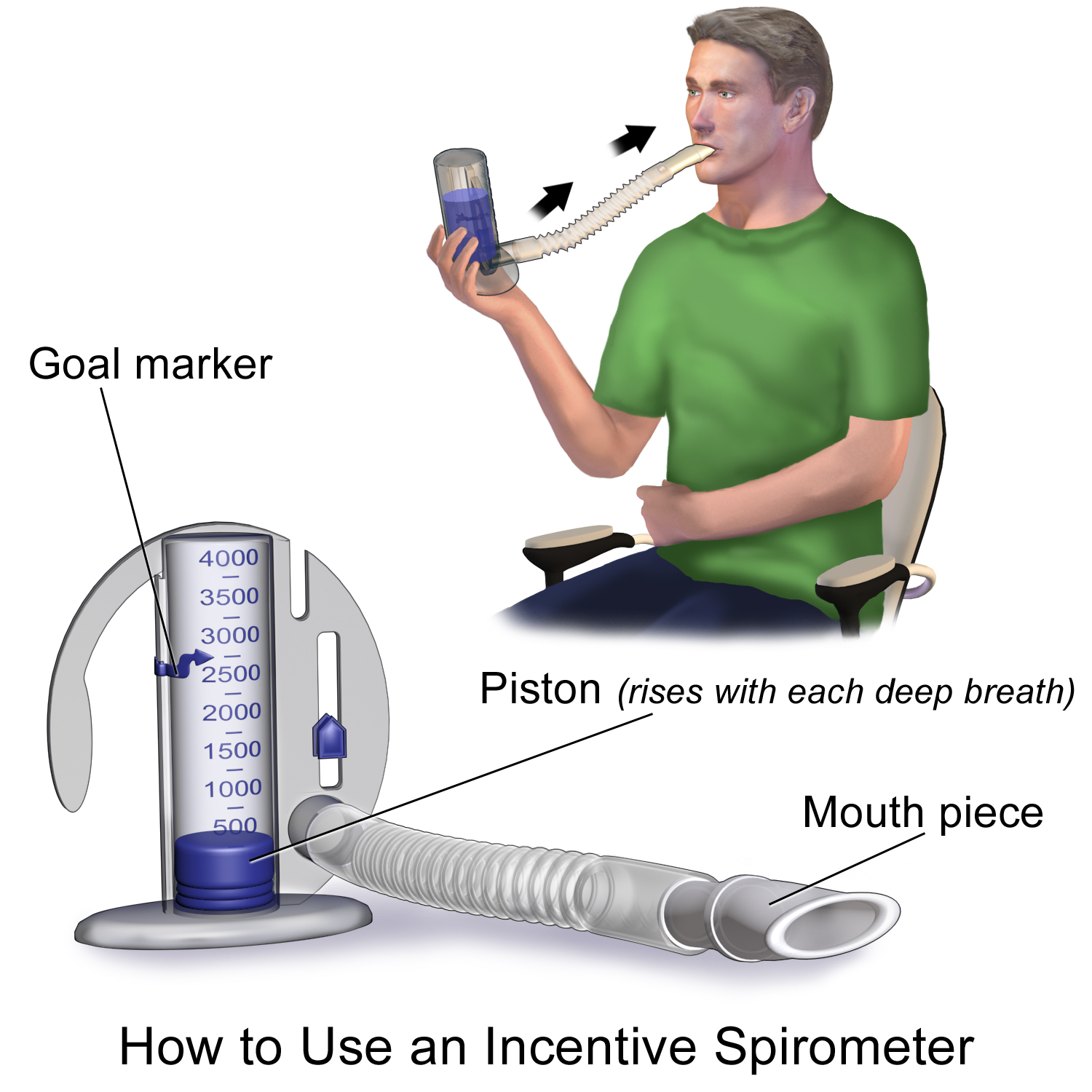
After surgery, it hurts to breathe deeply, so patients take shallow breaths. This leaves the tiny alveoli collapsed. Inadequate lung expansion significantly increases the risk of developing postoperative atelectasis. To fight this, we use a device where incentive spirometry volume measurements evaluate a patient's lung expansion capacity, forcing them to pop those alveoli open.
The skin is our armor. Wound healing requires excellent blood flow, great building materials (protein), and a pristine environment.
Risk Assessments for Pressure Injuries
We use The Braden Scale to assess pressure injury risk. A lower numerical score on the Braden Scale indicates a higher risk of developing pressure injuries. It elegantly evaluates six distinct areas:
- Sensory perception (Can they feel the pain of ischemia?)
- Skin moisture (Is the skin softening from sweat or urine?)
- Patient activity (Are they out of bed?)
- Patient mobility (Can they change positions?)
- Patient nutrition (Do they have fuel to rebuild?)
- Friction and shear (Is the skin tearing on the sheets?)
Assessing Healing Wounds (REEDA)
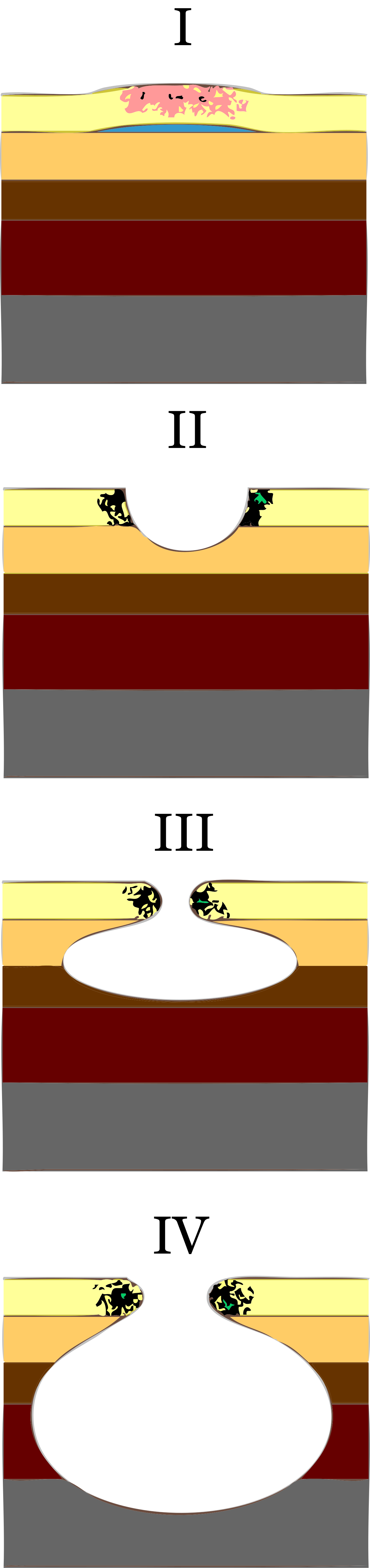
When inspecting a surgical incision or localized wound, the REEDA acronym is used to assess its status.
- R stands for redness.
- E (the first one) stands for edema.
- E (the second one) stands for ecchymosis (bruising).
- D stands for discharge or drainage.
- A stands for the approximation of wound edges (are the edges pulled neatly together?).
The Building Blocks: Albumin
To heal a wound, the body needs protein. Serum albumin levels are used to assess long-term nutritional status related to wound healing.
- The normal range for serum albumin is 3.5 to 5.0 grams per deciliter.
- A serum albumin level below 3.5 grams per deciliter increases the risk of delayed wound healing. No bricks, no wall.
The Great Saboteur: Diabetes Mellitus
Sugar is highly abrasive to tiny blood vessels. Diabetes mellitus impairs peripheral blood flow, making it a major systemic risk factor for delayed wound healing. To know if a patient's blood sugar has been controlled over time, we look at their red blood cells. Hemoglobin A1c blood tests measure the average blood glucose level over the past two to three months.
- Elevated Hemoglobin A1c levels indicate poorly controlled diabetes mellitus.
- This poorly controlled diabetes mellitus increases the risk of microvascular complications and delayed tissue healing.
Pigmentation Considerations
Skin assessment must adapt to the patient's melanin levels.
- Cyanosis in dark-skinned individuals is best assessed by examining the oral mucosa or conjunctiva.
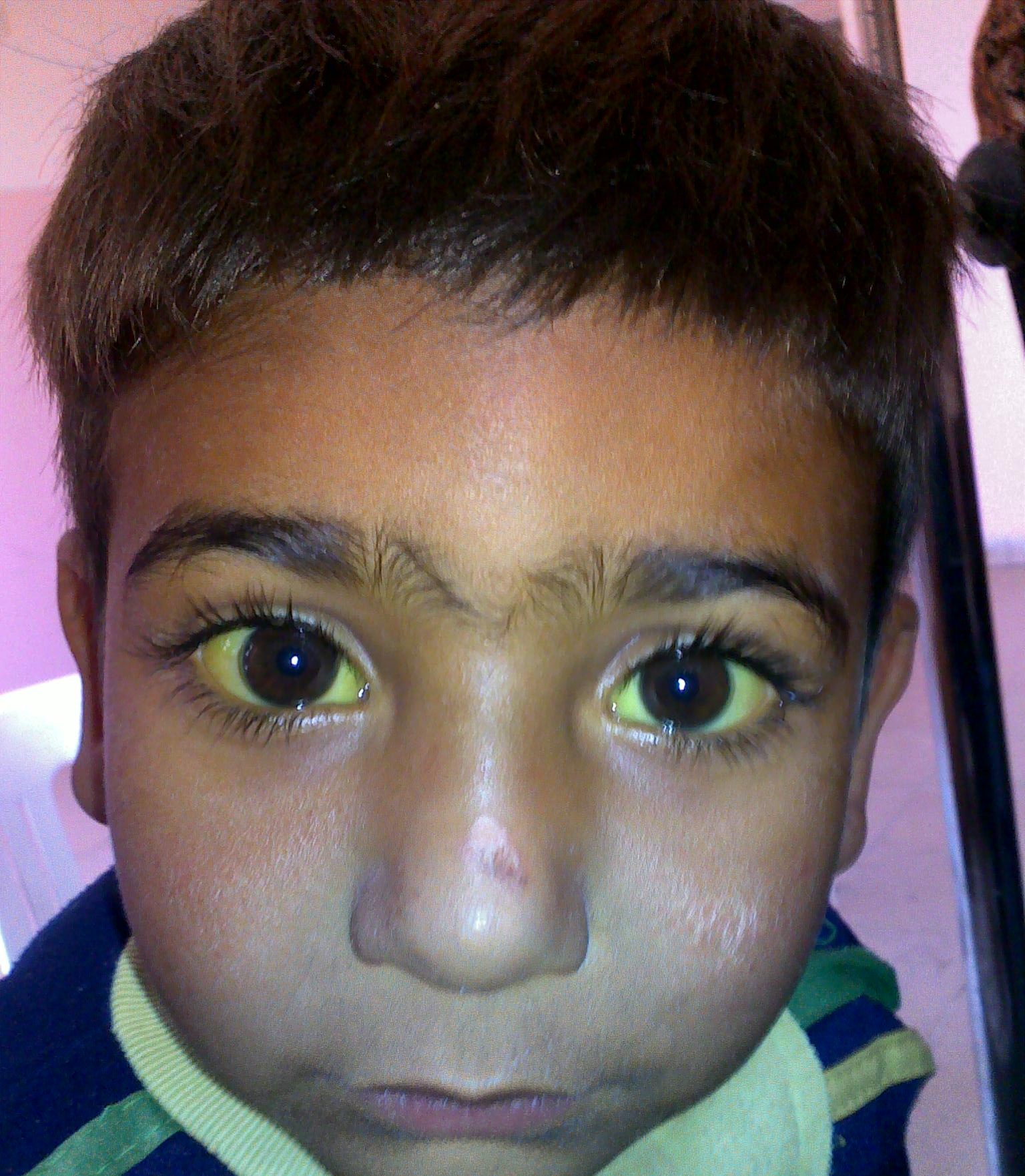
- Jaundice in dark-skinned individuals is best assessed by examining the sclera or the hard palate.
If the nervous system, muscles, or senses fail, gravity wins. Falls are devastating. We proactively measure fall risk using The Morse Fall Scale, where a higher numerical score indicates a higher risk for falls. This scale assigns risk points based on six parameters:
- A patient's history of falling.
- The presence of secondary medical diagnoses.
- The use of ambulatory aids like crutches or walkers.
- The presence of an intravenous line or saline lock (an IV pole is a tripping hazard!).
- Impaired patient gait.
- Impaired or overestimated mental status (patients who think they can walk, but can't).
Assessing Senses and Muscle Strength
You must assess a patient's interaction with the world:
- Visual impairment significantly increases a patient's risk for accidental falls. We test distance vision because if they can't see the obstacle, they will trip. The Snellen chart evaluates distance visual acuity.
- The whisper test screens for high-frequency hearing loss in clinical settings.
- Decreased tactile sensation increases the risk of unrecognized thermal or mechanical injuries. (If a diabetic steps on a tack, they may not feel it). Monofilament testing evaluates the loss of protective tactile sensation in the feet.
We also assess power. Muscle strength is clinically graded on a scale ranging from 0 to 5:
- A muscle strength grade of 5 indicates normal strength against full applied resistance.
- A muscle strength grade of 0 indicates no visible or palpable muscle contraction.
The Postural Threat: Orthostatic Hypotension
When a patient stands, gravity pulls blood to their legs. A healthy vascular system clamps down immediately to push blood back up to the brain. If it fails, the patient faints.
- Assessing for orthostatic hypotension involves measuring blood pressure and heart rate in supine, sitting, and standing positions.
- A drop in systolic blood pressure of at least 20 mmHg upon standing defines orthostatic hypotension.
- Because the brain loses blood pressure and the patient gets dizzy, orthostatic hypotension significantly increases a patient's fall risk upon ambulation.
Finally, we assess the gut.
Swallowing and Aspiration
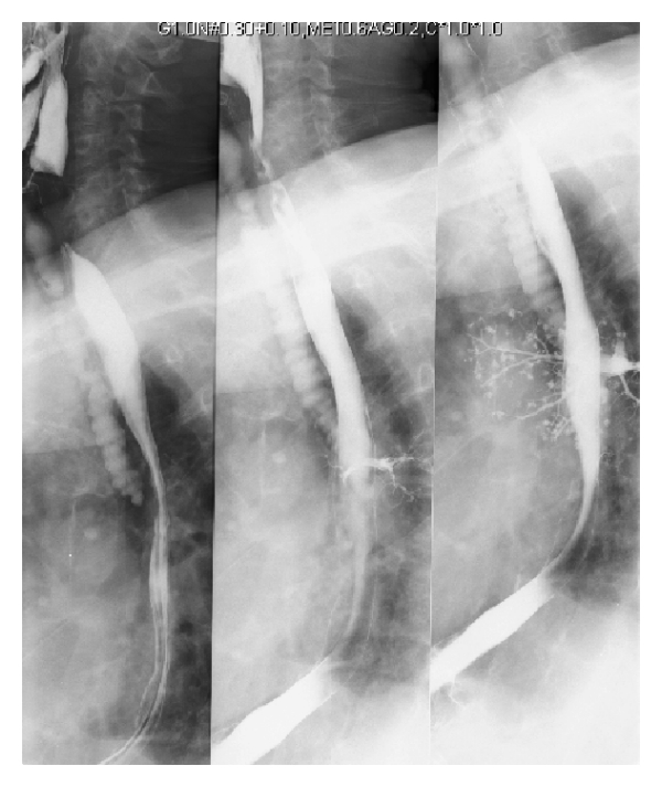
Can the patient safely route food into the esophagus instead of the lungs? Dysphagia is the medical term for difficulty swallowing. It is incredibly dangerous because dysphagia significantly increases a patient's risk of dangerous pulmonary aspiration.
- How do we test the plumbing gates? Aspiration risk is clinically assessed by evaluating the patient's gag reflex and ability to swallow water safely.
Auscultating the Abdomen
The intestines are a muscular tube constantly squeezing fluid and gas.
- Normal bowel sounds occur at a rate of 5 to 30 times per minute.
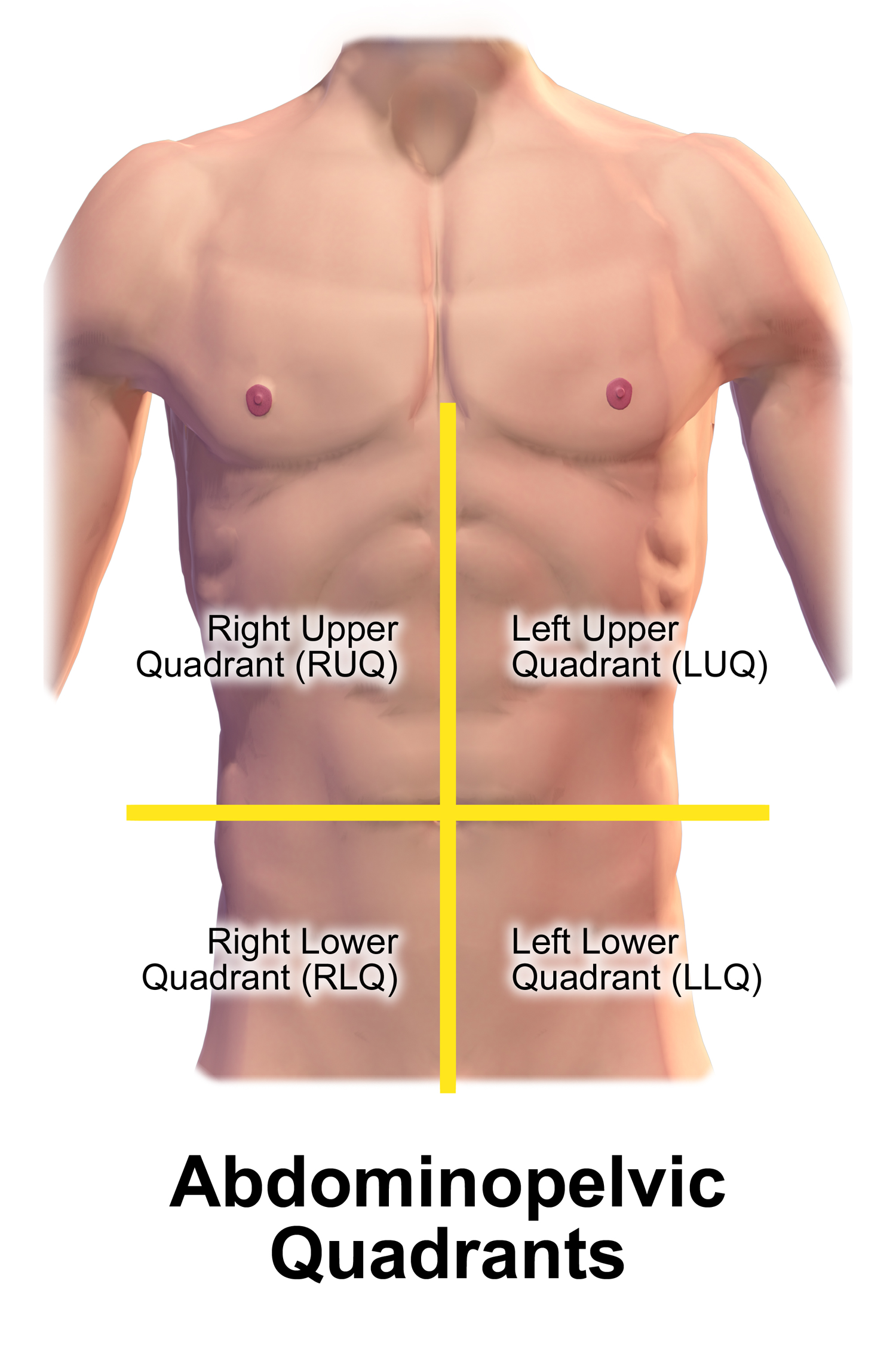
- If the gut has stopped entirely (a paralytic ileus), it goes dead silent. But you cannot rush this assessment. Absent bowel sounds must be confirmed by listening continuously for five minutes in each abdominal quadrant.
Final Thoughts
When you assess a patient, you are a scientific observer. You are connecting an elevated HbA1c to a failed monofilament test, recognizing that poor perfusion leads to falls and delayed wound healing. You are noting restlessness before the pulse oximeter drops. Learn these systems, understand the physics of how they work, and you will not only ace the NCLEX—you will save lives.