Ante/Intra/Postpartum and Newborn Care
Not sure you’re ready?
Take the ~3-minute readiness diagnostic and see where you stand.
Welcome to the fascinating, dynamic, and wonderfully mechanical world of maternal-newborn nursing. If you just memorize a list of clinical signs, you’ll be easily tripped up on your NCLEX-PN. But if you understand how the machinery of the human body actually works during pregnancy, birth, and the postnatal period, the right answers will become completely obvious to you. Nature is an incredible engineer!
Let’s pull back the curtain and look at the antepartum, intrapartum, postpartum, and newborn periods not as isolated textbook chapters, but as one beautiful, continuous physical and emotional process.
Before the baby ever arrives, a complex preparation is happening. We have to look at the mother’s psychological readiness and check under the hood to see how the baby is handling the environment.
The Emotional Blueprint
When a woman discovers she is pregnant, you might expect instant, overwhelming joy. But human psychology is delightfully complex. Maternal ambivalence is a perfectly normal emotional response during the first trimester of pregnancy. Building a human being is a massive biological and lifestyle shift; feeling unsure or conflicted at first is a natural processing mechanism.
As the pregnancy progresses to the end, the mother's focus naturally narrows. Maternal introversion is a common emotional response during the third trimester of pregnancy. She is subconsciously conserving her psychological energy for the monumental task of labor. To truly know if a mother is ready to bring this child home, we must look outside the patient herself. Evaluating the client’s support system is essential for determining emotional preparedness for the postpartum period. Without a physical and emotional safety net, the transition to motherhood becomes infinitely harder.
The Nonstress Test (NST): Listening to the Baby Exercise
How do we know the baby is thriving inside the uterus? We use a nonstress test, which evaluates fetal well-being by monitoring how the fetal heart rate responds to fetal movement. Think about it: when you jog up a flight of stairs, your heart rate naturally accelerates. A healthy fetus should experience the exact same cardiovascular response when it moves around!
To do this, the practical nurse assists by attaching the external fetal monitor to the maternal abdomen. The pregnant client is given a job: she presses a marker button on the fetal monitor to record fetal movements.
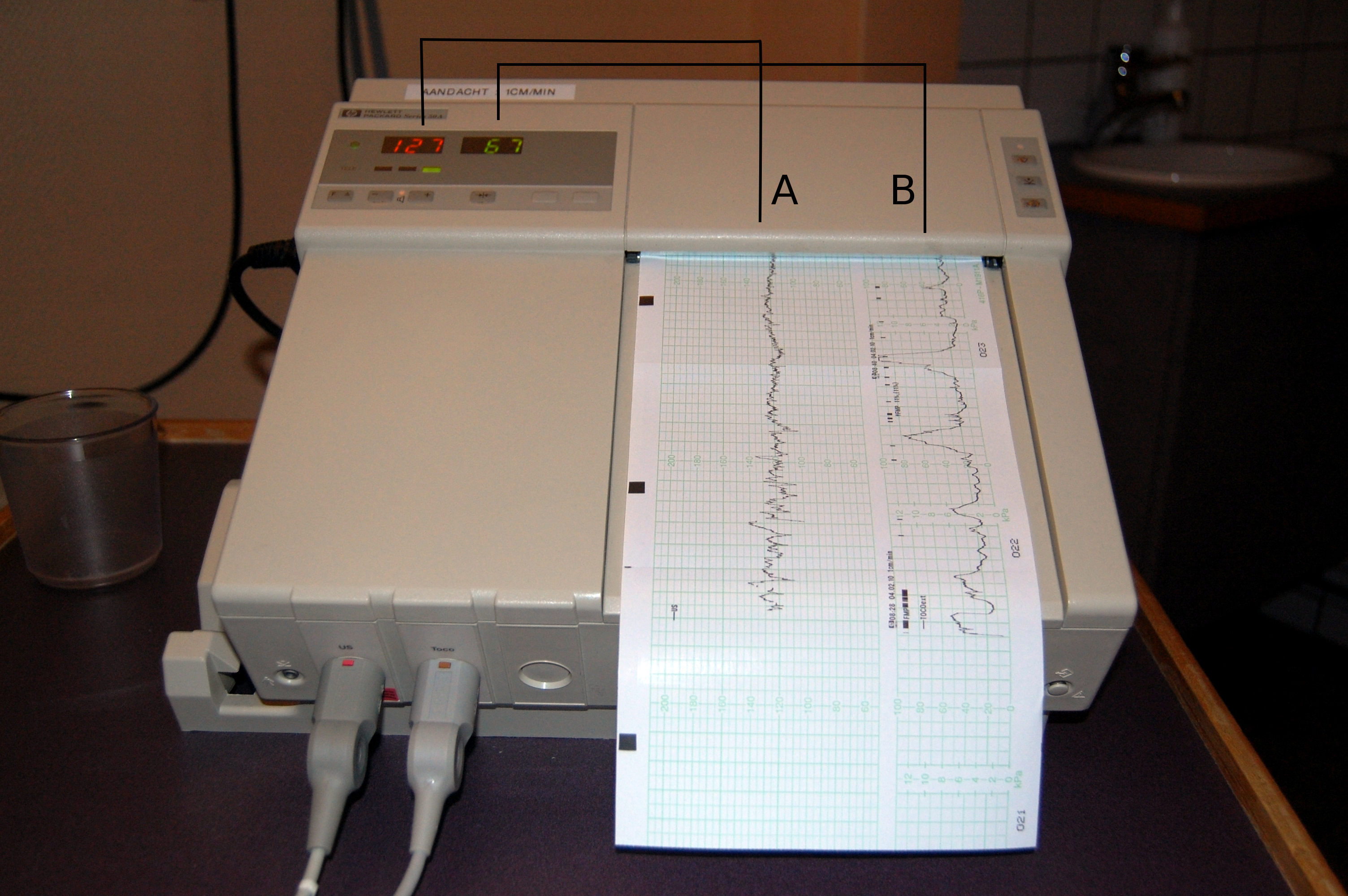
But before we press "start," we must position the mother correctly. Fluid dynamics matter here!
- Placing the pregnant client in a semi-Fowler position during a nonstress test prevents supine hypotension. If she lies flat on her back, the heavy uterus compresses the inferior vena cava, dropping her blood pressure.
- Alternatively, placing the pregnant client in a left lateral position during a nonstress test optimizes placental perfusion, ensuring maximum oxygen delivery to the fetus.

What makes an NST "Good"? A reactive nonstress test is considered a reassuring sign of fetal well-being. For a fetus at 32 weeks gestation or older, a reactive test requires at least two fetal heart rate accelerations within a twenty-minute window.
To qualify as an acceleration, the math must be right: Each fetal heart rate acceleration must peak at least 15 beats per minute above the baseline and must last for at least 15 seconds. Think of it as the "15-by-15 rule."
Labor is an exercise in pressure, hydraulics, and timing.
First, we need to know the resting state of the baby’s heart. The normal baseline fetal heart rate is between 110 and 160 beats per minute. Anything outside of this is a clue that the environment is changing.
Timing the Waves: Contraction Mechanics
To understand labor, the practical nurse must accurately measure the "waves" of uterine muscle activity. Don't mix up frequency and duration; they measure two entirely different things:
- Frequency tells us how often the waves are crashing. The practical nurse assesses the frequency of uterine contractions by timing from the start of one contraction to the start of the next contraction.
- Duration tells us how long a single wave lasts. The practical nurse assesses the duration by timing from the start of a contraction to the end of the same contraction.
The "Squeeze": Reading Fetal Decelerations
During a contraction, the uterus tightens, putting pressure on the baby and the umbilical cord. How the baby's heart rate reacts—and when it reacts—tells us exactly what is happening inside.
| Deceleration Type | Timing & Appearance | What is Happening? | Is it Dangerous? |
|---|---|---|---|
| Early | Aligns exactly with the peak of maternal uterine contractions. | They are a benign physiological response to fetal head compression. The contraction squeezes the baby's head, stimulating the vagus nerve, which harmlessly slows the heart rate. | No. Completely benign. |
| Variable | Appears as an abrupt decrease in fetal heart rate. It looks like a sharp "V" on the monitor. | This indicates umbilical cord compression. Think of stepping on a garden hose—the blood flow is cut off abruptly, causing an immediate, sharp drop in heart rate. | Requires intervention (position change) to unkink the cord. |
| Late | Begins after the peak of a maternal uterine contraction. The heart rate drops late and recovers late. | This indicates impaired uteroplacental blood flow. The placenta is failing. When the contraction peaks, the placenta runs out of oxygen reserves, causing a delayed hypoxic response in the fetus. | Yes. This is an emergency. |
Once the baby and placenta are delivered, the mother’s body must rapidly close off the blood vessels that once fed the placenta. Nature’s solution is a biological tourniquet: the uterine muscle itself.
The Uterine Fundus
The postpartum uterine fundus should feel firm to palpation. Why? Because a firm postpartum uterine fundus minimizes bleeding from the placental attachment site. The muscle fibers physically pinch the bleeding blood vessels shut!
Where should this firm muscle be located?
- The postpartum uterine fundus is expected to be at the level of the maternal umbilicus within twelve hours after delivery.
- From there, it shrinks steadily. The postpartum uterine fundus descends approximately one centimeter per day.
If you palpate the abdomen and feel a soft, squishy uterus, you have a major mechanical failure on your hands. A soft or boggy postpartum uterus indicates inadequate uterine muscle contraction. Because those vessels aren't being pinched shut, a soft or boggy postpartum uterus is a primary indicator of postpartum hemorrhage risk.
Crucial Intervention: What do you do if the biological tourniquet is failing? You wake the muscle up! Fundal massage is the initial nursing intervention for a boggy postpartum uterus.

The Bladder Balloon
Sometimes, the uterus wants to clamp down, but something is physically in the way. Directly beneath the uterus sits the urinary bladder. A distended maternal urinary bladder displaces the postpartum uterus upward and to the right of the midline. If the uterus is pushed up and to the right, it cannot contract properly. Emptying the bladder allows the uterus to return to midline and clamp down.
Lochia: The Cleanup Crew
As the uterus heals, it sheds debris. This vaginal discharge is called lochia, and it follows a predictable, color-coded timeline based on the stages of cellular healing:
- Lochia rubra is a dark red vaginal discharge. It normally occurs during the first three to four days postpartum.
- Lochia serosa is a pinkish-brown vaginal discharge. It normally occurs from day four to day ten postpartum.
- Lochia alba is a whitish-yellow vaginal discharge. It normally occurs from day ten up to six weeks postpartum.
When the baby arrives in the outside world, we have three major priorities: keeping them breathing safely, preventing infection at the umbilical stump, and mastering the fluid dynamics of feeding.
Safe Sleep Mechanics
We must engineer a sleep environment that guarantees an open airway. The supine position (flat on the back) is the only recommended sleep position for a healthy newborn. We do this because placing a newborn in the supine position for sleep reduces the risk of Sudden Infant Death Syndrome (SIDS).
The surrounding environment must follow strict rules:
- The sleep environment must consist of a firm mattress.
- It must be completely free of loose bedding.
- It must be completely free of stuffed toys.
- It must be completely free of bumper pads.
Caring for the Umbilical Cord
The umbilical cord was the baby’s lifeline, but now it is a decaying piece of tissue that needs to dry up and fall off. The umbilical cord stump typically detaches from the newborn within ten to fourteen days after birth.
Until it falls off, we treat it like a "dry dock":
- Caregivers must administer only sponge baths to the newborn until the umbilical cord stump naturally falls off. Submerging it in a tub delays drying.
- The newborn’s umbilical cord stump must be kept completely clean and dry to prevent infection.
- To protect it, caregivers should fold the front of the newborn's diaper below the umbilical cord stump. Why? Because folding the diaper below the umbilical cord stump prevents contamination from newborn urine.

The Thermodynamics and Mechanics of Infant Feeding
Babies grow rapidly and require constant fuel. Healthy newborns require feeding eight to twelve times within a twenty-four-hour period. How do we know they are getting enough? Look at the output! A healthy newborn produces six to eight wet diapers per day by the end of the first week of life.
Breastfeeding Mechanics: A baby does not "chew" milk out of the breast; they create a powerful vacuum seal. Therefore, a proper breastfeeding latch requires the infant's mouth to cover the mother's entire nipple and requires the infant's lips to flange outward against the mother's breast. Once pumped, breast milk can be safely stored at room temperature for up to four hours.

Formula and Bottle Physics: If using formula, prepared infant formula can be safely stored in the refrigerator for up to twenty-four hours.
Now, let's talk about thermodynamics—specifically, heating the milk. Heating infant formula in a microwave creates uneven hot spots. Because a baby cannot tell you the milk is burning them until it is too late, uneven hot spots in microwaved infant formula pose a severe burn risk to the infant's mouth. Instead, infant formula should be warmed by placing the bottle in a container of warm water. Always verify safety: caregivers should test the temperature of warmed infant formula by sprinkling a few drops on the inner aspect of the caregiver's wrist.
Finally, let's look at the fluid dynamics of bottle feeding:
- The infant should be held in a semi-upright position during bottle feeding. Gravity is your friend here; holding the infant semi-upright reduces the risk of formula entering the Eustachian tubes, which prevents painful ear infections.
- The infant's bottle must be tilted during feeding to keep the nipple completely filled with milk. If there is air in the nipple, the baby swallows air. Keeping the bottle nipple filled with milk prevents the infant from swallowing excess air.
- Because babies are imperfect little machines, some air always gets in. Therefore, caregivers should burp the infant frequently during feedings to expel swallowed air.

Study the reasons behind these facts. Understand the mechanics of the uterus clamping down, the physics of a baby's latch, and the fluid dynamics of the placenta during a contraction. When you step into your NCLEX-PN, you won't just be remembering facts—you'll be seeing the beautifully logical machinery of human life in action. Good luck!